-
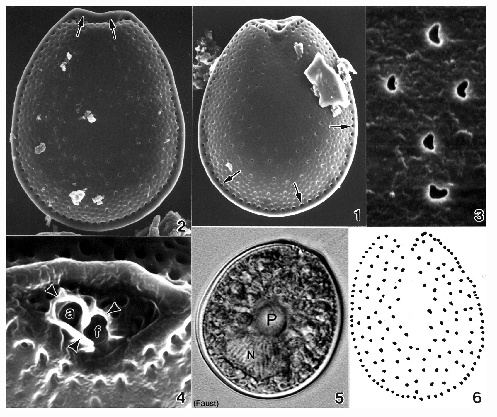
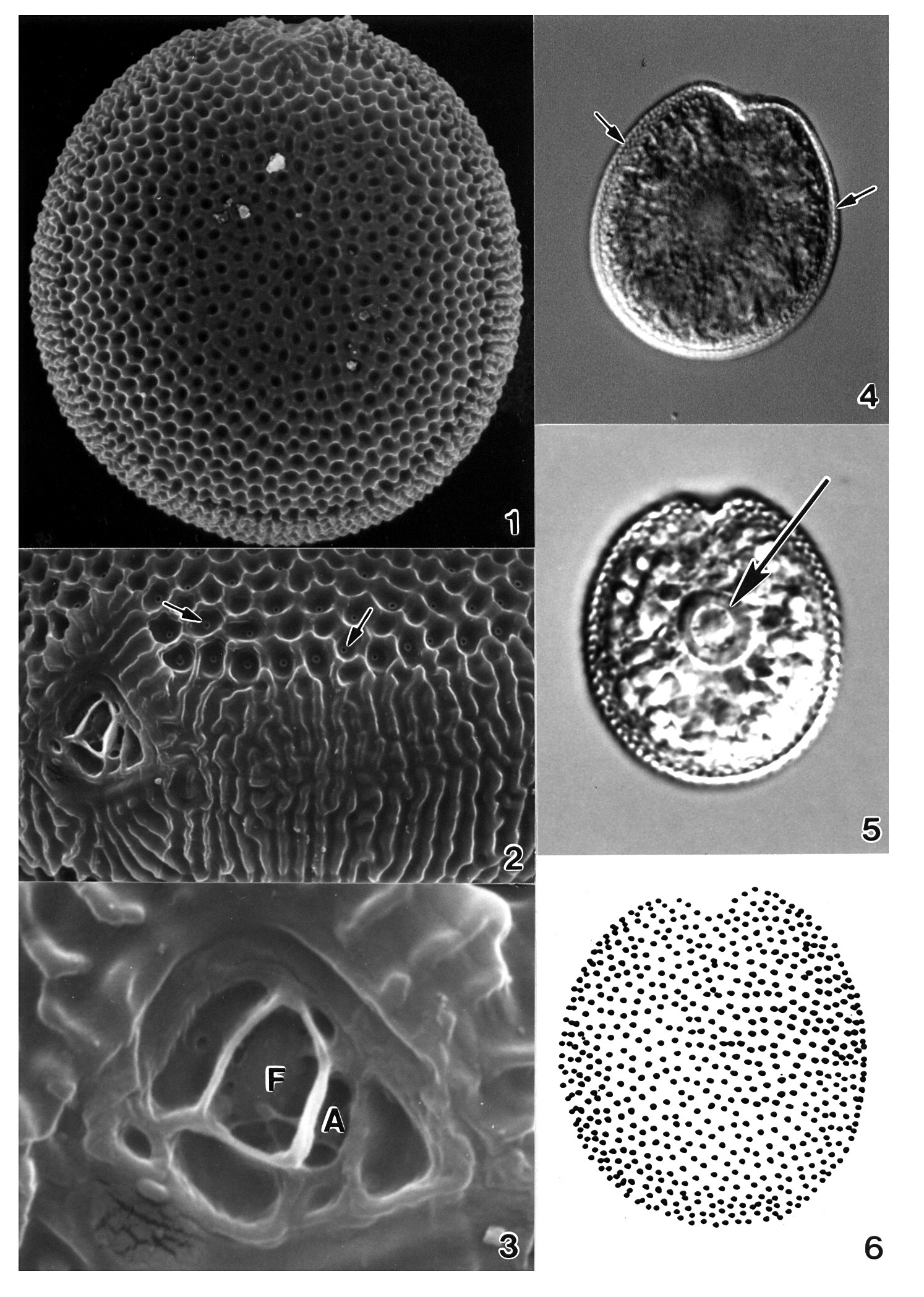
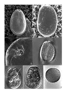
Plate 43. Prorocentrum lima. Figs. 1-3. SEM. Fig. 1. Right valve. Cells oblong to ovate with narrowed anterior. Marginal pores and scattered surface pores present; valve center devoid of pores. Intercalary band smooth and wide. Fig. 2. Left valve; bacteria attached (arrows). Fig. 3. Periflagellar area: shallow, broad, V-shaped depression on right valve. Flared periflagellar collar encircles auxiliary (a) pore (arrow); larger flagellar pore (f) adjacent (after Faust 1991). Figs. 4-7. LM. Fig. 4. Thecal pore arrangement. Fig. 5. Right valve with central pyrenoid (arrow). Fig. 6. Left valve and posterior nucleus (n). Fig. 7. Triple-layered resting cyst. (Figs. 1,2,4-7 after Faust 1993c)
-
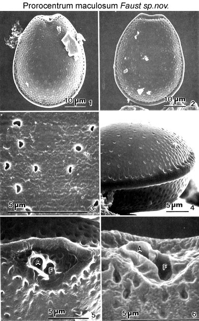
Figs. 1-6. . Prorocentrum maculosum sp. nov. Fig.1. Appearance of the right valve, including the periflagellar area. The valve surface is rugose with scattered poroids and the centre of the valve is devoid of poroids. The cell margin is surrounded by a row of marginal pores. Fig.2. In left valve view. The anterior end is flat to slightly concave and the centre of this valve is devoid of pores. Fig.3. Unevenly distributed kidney-shaped to circular or oblong valve pores are present on the valves. Fig.4. Distinct ridge appears as a flange around the cell, with a row of equally spaced marginal pores. Fig.5. The periflagellar area, set in a V-shaped depression, is a broad triangle with a raised margin. Flagellar pore (F) and auxiliary pore (A) are about equal in size and are surrounded by a curved and flared apical collar. Fig.6. The apparently protuberant apical collar viewed from the side.
EMu: Holotype SEM negative # 40012; SEM stub #140; Field # 66-87; Accession # 407159; Catalog # 60; Figure # 1.
-
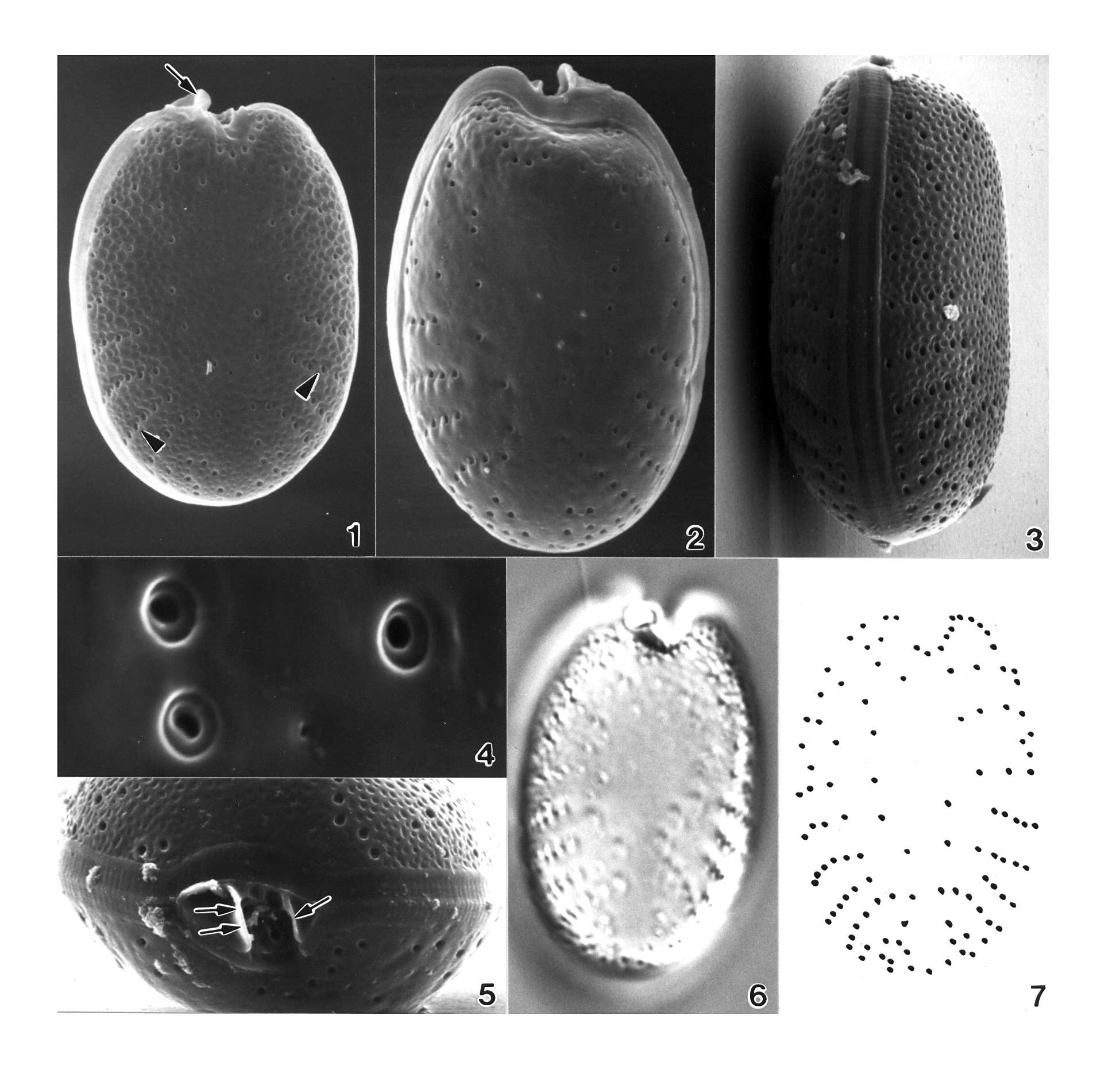
Plate 44. Prorocentrum maculosum. Figs. 1-4. SEM. Fig. 1. Right valve: cell broadly ovate, narrowing apically. Valve surface rugose with scattered poroids; valve center devoid of poroids. Marginal pores evenly spaced (arrows). Fig. 2. Left valve: anterior end flat to slightly concave with raised apical ridge (arrows). Valve margins appear as a flange around cell. Fig. 3. Valve poroids: unevenly distributed on valve surface; circular to oblong or kidney-shaped. Fig. 4. Periflagellar area: broad V-shaped depression on right valve. Apical ridge (raised margin) on left valve. Flagellar (f) and auxiliary (a) pores surrounded by protuberant periflagellar collar (arrowheads); equal in size. Fig. 5. LM. Right valve: central pyrenoid (P) and large posterior nucleus (N) (M.A. Faust). Fig. 6. Line drawing: valve poroid and marginal pore arrangement (Figs. 1-4,6 after Faust 1993b)
-
Plate 45. Prorocentrum mexicanum. Figs. 1-5. SEM. Fig. 1. Right valve: cell oval. Periflagellar collar curved and prominent (arrow). Trichocyst pores radially arranged (arrowheads). Fig. 2. Left valve. Apical area excavated (M.A. Faust). Fig. 3. Lateral view: cell ovate to convex; intercalary band broad and transversely striated. Cell surface rugose. Fig. 4. Trichocyst pores round with smooth edge, within deep furrowed depressions. Fig. 5. Periflagellar area: small, V-shaped shallow depression. Prominent curved periflagellar collar (double arrows) adjacent to auxiliary pore; protuberant periflagellar plate (single arrow) opposite and adjacent to flagellar pore. Fig. 6. LM. Right valve: radial pore arrangement visible (M.A. Faust). Fig. 7. Line drawing: trichocyst pore arrangement. (Figs. 1,3-5,7 after Faust 1990b)
-
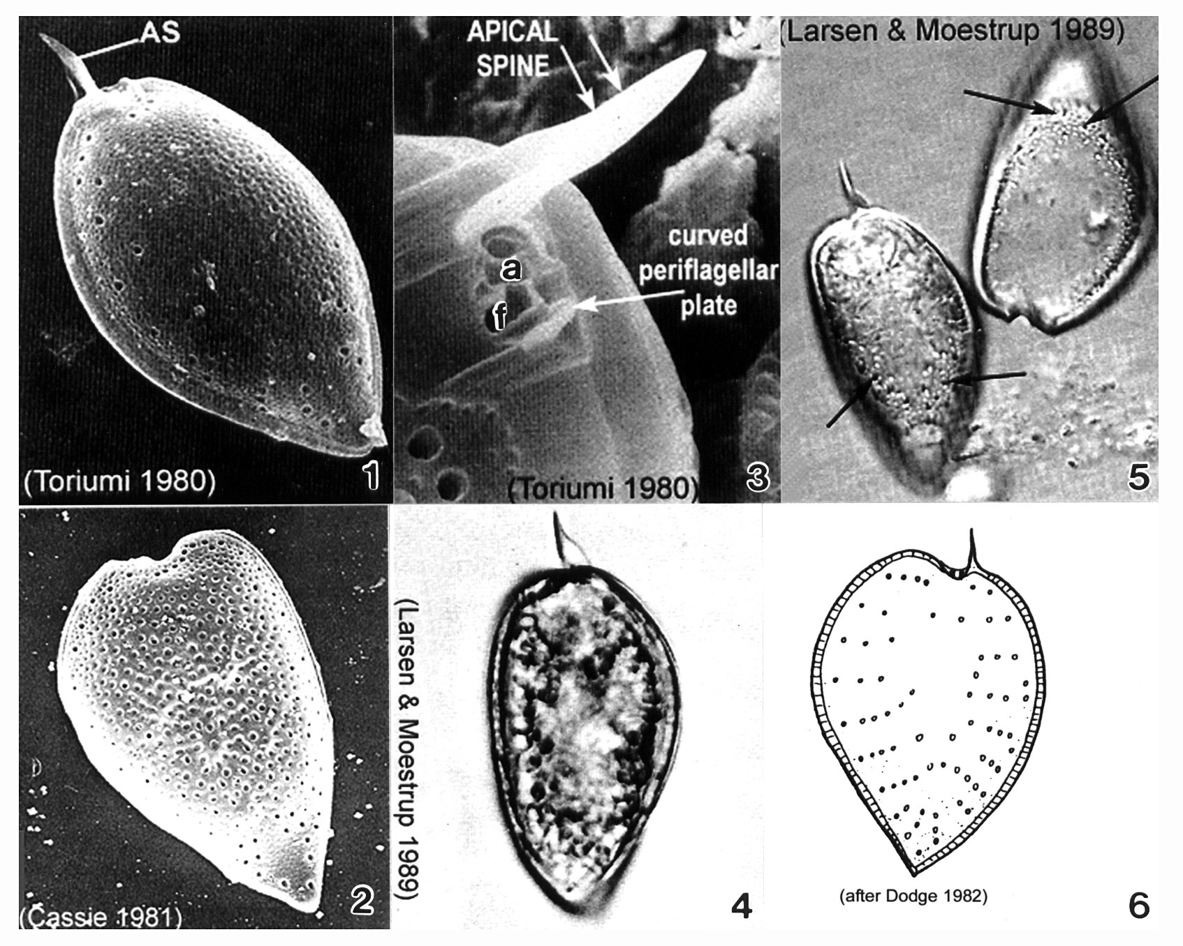
Plate 46. Prorocentrum micans. Figs. 1-3. SEM. Fig. 1. Right valve: cell tear-drop shaped; rounded anteriorly, pointed posteriorly, broadest in the middle. Apical spine (AS) winged. Rugose thecal surface. Intercalary band smooth and wide. Fig. 2. Heart-shaped cell. Apical spine missing. Fig. 3. Periflagellar area: small, shallow triangular depression on right valve. Flagellar (f) and auxiliary (a) pores present; curved periflagellar plate adjacent to f. Large winged AS directly opposite. Figs. 4-5. LM: Left valve. Winged AS visible. Fig. 5. Empty theca with visible trichocyst pores (arrows). Fig. 6 Line drawing: trichocyst pore arrangement.
-
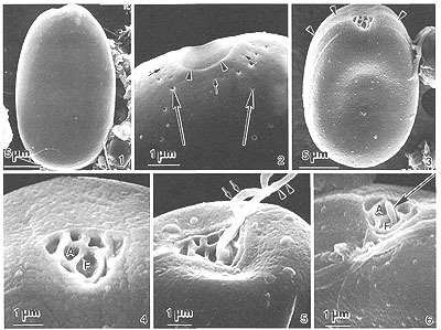
Figs. 1-6. . Prorocentrum norrisianum sp. nov. FIG.1. Cells are ovate in valve view; the cell surface is smooth with scattered pores. FIG.2. Left valve margin at the anterior end is flat or inclined with a distinctly marked collar (arrowheads). Large pores (large arrows) and small pores (small arrows) are present on the valve surface. Large pores are round with a smooth edge; small pores are indented. FIG.3. Cell's anterior end is flat. The intercalary band is smooth (arrowheads). FIG.4. Periflagellar area is V-shaped, located on the right valve; it has a flagellar (F) and an auxiliary (A) pore, unequal in size. The two flagella are not present. FIG.5. Flagella, the longitudinal (arrowheads) and transverse (arrows), emerge from the flagellar pore. FIG.6.A short, tubular, peduncle-like structure (arrow) protrudes from the flagellar pore (F). The auxiliary pore (A) is to the left of the flagellar pore.
EMu: Holotype SEM negative #115034; SEM #115; Field # 3; Accession # 407164; Catalog # 1415; Figure # 1.
-
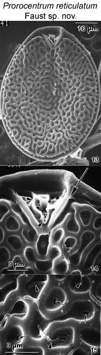
Figs. 13-15. . Prorocentrum reticulatum sp. nov. FIG.13. Right valve view of periflagellar area. Cells are oval. The valve surface is reticulated. FIG.14. A network of ridges with alternating depres¬sions ornaments the valve surface. The anterior end of the left valve is flat. The periflagellar area is set in a narrow V-shaped depression with raised margin. A narrow apical collar (arrow¬heads) wraps around the flagellar pore (F) and auxiliary pore (A). The longitudinal flagellum emerges from the flagellar pore (arrow). FIG.15. Reticulae are a network of ridges with round to oval depressions, and at the center a narrow, kidney-shaped opening (arrows). Some bacteria (arrowheads) are attached to the valve surface.
EMu: HOLOTYPE SEM negative #209017; SEM Stub #209; FIELD # 1041-96; ACCESSION # 202408;CATALOG # 1425; FIGURE #13.
-
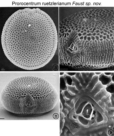
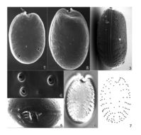
Figs. 21-24. . Prorocentrum ruetzlerianum sp. nov. Fig. 21. The body is ovoid and covered with pentagonal areolae. FIG. 22. The cell is round in side view and convex in the middle of the valve, where each deep depression (areola) has a small circular opening (arrow). Scale bars = 200 µm. FIG.23. Intercalary band is broad, transversely rugose, the sinuous rugae being unique to this species. FIG 24. The periflagellar area is flat, has a triangular orientation set into a shallow, V-shaped depression of the right valve, and composed of platelets of unequal size and shape. Flagellar pore (f), and auxiliary pore (a). The two flagella are not present. Scale bars = 200 µm.
EMu: SEM NEGATIVE # 23012; SEM STUB # 23; FIELD # 78-87; ACCESSION # 407159; CATALOG # 30; Figure # 21.
-
Plate 48. Prorocentrum ruetzlerianum. Figs. 1-3. SEM. Fig. 1. Right valve: cell round to ovoid, covered with pentagonal areolae. Cell surface rugose. Fig. 2. Anterio-lateral view. Each areola with small circular pore at its base (arrows). Intercalary band broad, transversely rugose. Fig. 3. Periflagellar area: small, shallow, unornamented depression on right valve; large flagellar (f) pore and smaller auxiliary (a) pore. Figs. 4-5. LM (M.A. Faust). Right valve: striated valve margins (small arrows); large central pyrenoid (large arrow). Fig. 6. Line drawing: areolae arrangement. (Figs. 1-3,6 after Faust 1990b)
-
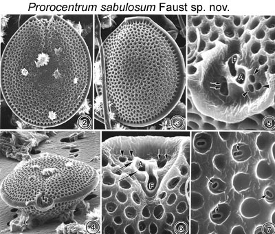
Figs. 2-7. . Prorocentrum sabulosum sp. nov. FIG. 2. Cell is in right lateral view, including the periflagellar area. The valve surface is areolated. Cells are oval in valve view. FIG. 3. Cell is in left lateral view. The anterior end is flat to slightly concave. The cell margin is smooth. FIG. 4. Apical view, cells are ellipsoid, the apical area exhibits a rounded lip, and both left and right valves are excavated. Foreign material is on cell surface. FIG. 5. The periflagellar area, set in a V-shaped depression, is a broad triangle with raised margin. The flagellar pore (F) is larger than the auxiliary pore (A). A large pore is adjacent to the flagellar pore (arrows), and three smaller pores are adjacent to the auxiliary pore (arrowheads). FIG. 6. A narrow apical collar wraps around the flagellar and auxiliary pores (large arrows) viewed from the side. The position of the large pore (small arrows) and small pores (arrowhead) are from the side. FIG.7. Aeolae are round to oval with a smooth margin. Some areolae have oblong-shaped trichocyst pores (arrows).
EMu: SEM NEGATIVE # 23012; SEM STUB # 23; FIELD # 78-87; ACCESSION # 407159; CATALOG # 30; Figure # 21.
-
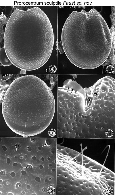
Figs. 8-13. Prorocentrum sculptile sp. nov. FIG.8. Cell shape is broadly ovate with a rounded, indented, anterior area in left valve view. FIG.9. Anterior end on the cell in right valve view is a deep-sculptured indentation. FIG.10. The posterior-lateral view is ellipsoid. The intercalary band is smooth. FIG.11. The valve surface has shallow depressions of variable shapes, round to oblong with smooth margins. Trichocyst pore openings that are round, similar in size, and at times open or closed are situated in some of the depressions (arrows). FIG.12. The periflagellar area on the right valve has a deep, V-shaped, narrow, 7-8-µm-long curved depression and an inclined, thin, apical structure (arrows). The transverse flagellum (arrowheads) is shown. FIG.13. Ejected trichocysts are situated in furrowed depressions on the valve surface (arrows).
EMu: HOLOTYPE SEM NEGATIVE #133051; SEM stub # 133; Field # 556-92; Accession # 407166; Catalog # 92; Figure # 8.
-
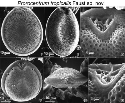
Figs. 7-12. Prorocentrum tropicalis sp. nov. FIG.7. Cells are broadly oval in valve view. The valve surface is rugose with scattered poroids. The cell margin has a ledge. The anterior end of the left valve is flat to slightly concave. FIG. 8. The right valve of a newly divided cell, the margin is narrow. FIG, 9. Periflagellar area on right valve at the anterior end of a cell; a ridge appears around the cell. The ridge is granular and horizontally striated (arrowheads). FIG. 10. Apical view of a cell. Flagella are not shown. FIG. 11. Periflagellar area is abroad triangle with a raised margin, unornamented. Flagellar pore (F) and auxiliary (A) pore are unequal in size. The valve surface is rugose with evenly distributed poroids; at the center of a poroid, a small round dome is situated. FIG. 12. A flexible, short, tubular, peduncle-like structure (arrow) emerges from the flagellar pore.
EMu: Holotype SEM negative #159090; SEM #133; Field # 556-92; Accession # 407166; Catalog # 1420; Figure # 9.
-

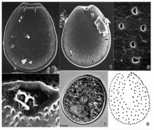
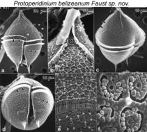
Figs. 1.SEM of Protoperidinium belizeanum sp. nov. (a) Ventral view. Cell is pyriform with an apical horn and two antapical spines. Apical plate 1' is meta. Cingulum is equatorial and ascending. Cingular wall reticulated (arrowheads). Postcingular plates narrow, qua¬drangular and wide (arrows), (b) Apical pore complex (APC) (X) includes an apical horn and a flange (arrows). Apical horn partially covered with biodetritus. (c) Epitheca excavated ventrally. Location of apical intercalary plate 3a (arrowheads), (d) Hypotheca is round. Cin¬gular list is prominent (arrowheads). Postcingular plates narrow (short arrows). Sulcus deep bordered by narrow list. Antapical spines are two, each with three fins, associated with antapical plates 1" and 2". (e) Thecal surface with three distinct ornamentation: 1) retic¬ulated pattern of ridged hexagonal depressions with a knob at network junctions (arrowheads), 2) depressions with a central rimmed pore (large arrows), and 3) vertical striation of paired particles above intercalary band (short arrows), (f) Depressions with a central rimmed pore (arrows) surrounded by small oblong particles Miniscule pore within the rimmed pore (arrowhead).
EMu: SEM NEGATIVE # 155141; SEM STUB # 155; FIELD # 631-93; ACCESSION # 407168; CATALOG # 1598; FIGURE # 3.
-
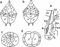
FIG. 2. Line drawing of Protoperidinium belizeanum sp. nov. Thecal plates are (a) ventral view, (b) dorsal view, (c) apical view, and (d) antapical view, (e) Details of the sulcal platelets.
EMu: SEM NEGATIVE # 155141; SEM STUB # 155; FIELD # 631-93; ACCESSION # 407168; CATALOG # 1598; FIGURE # 3.
-
Peniscola, Valencia, Spain
-
Peniscola, Valencia, Spain