NMNH Prorocentrum reticulatum type specimen
Description:
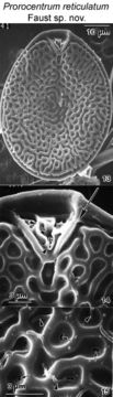
Figs. 13-15. . Prorocentrum reticulatum sp. nov. FIG.13. Right valve view of periflagellar area. Cells are oval. The valve surface is reticulated. FIG.14. A network of ridges with alternating depres¬sions ornaments the valve surface. The anterior end of the left valve is flat. The periflagellar area is set in a narrow V-shaped depression with raised margin. A narrow apical collar (arrow¬heads) wraps around the flagellar pore (F) and auxiliary pore (A). The longitudinal flagellum emerges from the flagellar pore (arrow). FIG.15. Reticulae are a network of ridges with round to oval depressions, and at the center a narrow, kidney-shaped opening (arrows). Some bacteria (arrowheads) are attached to the valve surface. EMu: HOLOTYPE SEM negative #209017; SEM Stub #209; FIELD # 1041-96; ACCESSION # 202408;CATALOG # 1425; FIGURE #13.
Included On The Following Pages:
- Life (creatures)
- Cellular (cellular organisms)
- Eukaryota (eukaryotes)
- SAR (Stramenopiles, Alveolates, Rhizaria)
- Alveolata (alveolates)
- Dinophyceae
- Prorocentrales
- Prorocentraceae
- Prorocentrum
- Prorocentrum reticulatum
- Dinoflagellata (dinoflagellates)
This image is not featured in any collections.
Source Information
- license
- cc-by-nc-sa-3.0
- copyright
- National Museum of Natural History, Smithsonian Institution
- original
- original media file
- visit source
- partner site
- NMNH Marine Dinoflagellates
- ID