-
-
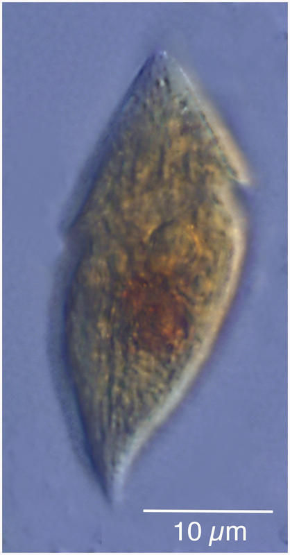
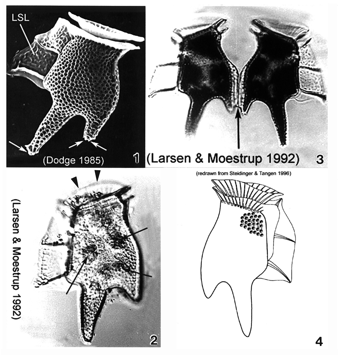
Plate 19. Dinophysis tripos. Fig. 1. SEM: lateral view. Cell large, oblong and heavily areolated. Hypothecal projections with toothed posterior ends (arrows). Left sulcal list (LSL) large, wide and reticulated. Figs. 2,3. LM: lateral view. Fig. 2. Anterior cingular list (ACL) projected anteriorly obscuring low epitheca (arrowheads). Narrow cingulum. Chloroplasts visible (arrows). Fig. 3. Paired cells. Hypothecal projection on dorsal margin sometimes seen with a narrow list (arrow) connecting two daughter cells during cell division. Fig. 4. Line drawing.
-
Heterotrophic dinoflagelate from the Etang de Thau
-
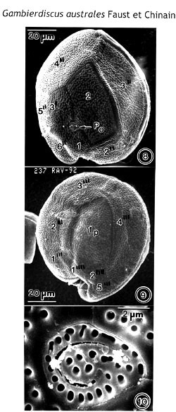
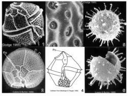
Figs. 8-10. Scanning electron micrographs of Gambierdiscus australes (RAV-92), sp. nov. Figs. 8, 9. Cells round to ellipsoid. The cell surface is smooth with scattered small pores. Fig. 8. Epithecal view. The Po plate is oriented ventrally. Fig. 9. Hypothecal view. The Ip plate, long and narrow, occupies 30% of the hypotheca width. Fig. 10. The Po plate is a broadly ellipsoid plate, with fish-hook-shaped apical opening surrounded by 31 pores.
EMu: Holotype SEM negative # 237047; SEM stub # 237; Field # RAV-92; Catalog # 1526; Figure #8.
-
-
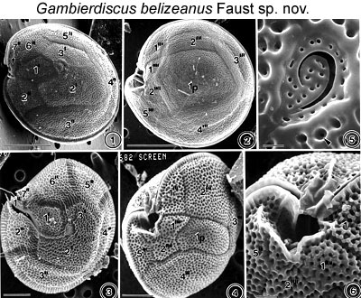
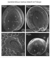
Figs. 1-6. Scanning electron micrographs of the surface morphology of Gambierdiscus toxicus and Gambierdiscus belizeanus are shown. FIGS.1-2. Scanning electron micrographs of the surface morphology of Gambierdiscus toxicus Adachi et Fukuyo. FIG.1. Cell in epithecal view. FIG.2. Cell in hypothecal view. Cell shape is round, compressed and ellipsoidal. Cell surface is smooth with scattered small pores. Thecal plate is large and quadrangular. FIGS.3-6. Gambierdiscus belizeanus sp. nov. FIG.3. Cell in epithecal view slightly damaged. Cell surface areolated and plates partially separated. FIG. 4. Cell is in hypothecal and ventral view. Plate is narrow. FIG.5. Apical pore complex is triangular with a fish-hook-shaped apical pore. A round pore is present in the areolae (arrowhead). FIG.6. Cingulum deep, ascending into a deep sulcal hollow.
EMu: Holotype SEM NEGATIVE # 132003B; SEM STUB # 152; FIELD # 682-93; ACCESSION # 407167; CATALOG # 798; FIGURE # 3.
-
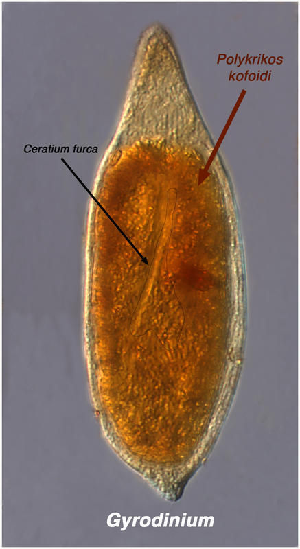
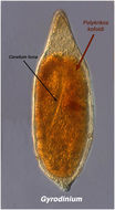
This probably is Gyrodinium eating Ceratium
-
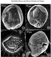
Figs. 11-14. Scanning electron micrographs of Gambierdiscus pacificus (HO-91), sp. nov., and Gambierdiscus belizeanus. Figs. 11-13. Gambler discus pacificus, sp. nov. Fig. 11, 12. Cells are round to ellipsoid. The Cell surface is smooth with scattered small pores. Fig. 11. Epithecal view. The Po plate is oriented ventrally. Fig. 12. The Ip plate, short and narrow, occupies 20% of the hypotheca width. Postcingular plates 2'" and 4'" are wide. Fig. 13. The Po plate is four-sided plate with a narrow fish-hook-shaped apical opening surrounded by 31 pores. Fig. 14. Gambierdiscus belizeanus. Cell in hypothecal view. The cell surface is areolated. The Ip plate, narrow and pentagonal, is wedged between very wide postcingular plates 2'", and 4'". The cingulum, deeply excavated, is ascending into a deep sulcal hollow.
EMu: Gambierdiscus pacificus
Holotype SEM negative # 241006; SEM stub # 241; Field # HO-91; Catalog # 1528; Figure #11.
-
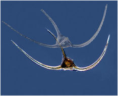
Gyrodinium ate a Polykrikos which had eaten a Ceratium. Specimen from the Etang de Thau.
-
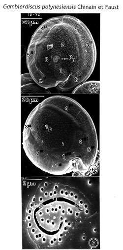
Figs. 5-7. Scanning electron micrographs of Gamblerdiscus polynesiensis (TB-92), sp. nov. Figs. 5, 6. Cells are round to ellipsoid. Cell surface is smooth with small scattered pores. Fig. 5. Epithecal view. The PO plate is oriented ventrally. Fig. 6. Hypothecal view. The Ip plate, broad and pentagonal, occupies 60% of the of hypotheca width. Postcingular plates 2'", 3'" and 4'" are narrow. The cingulum, deep, is ascending into a deep sulcal hollow. Fig. 7. The Po plate is triangular with fish-hook-shaped apical opening surrounded by 44 pores.
EMu: Holotype SEM negative # 242010; SEM stub # 242; Field # TB-92; Catalog # 1522; Figure #5.
-
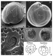
Arctic Dinophysis norvegica (empty!)from the Chukchi Sea in summer 2011.
-
Figs. 1-4. Scanning electron micrographs illustrate the surface morphology of Gambierdiscus toxicus (GTT-91). Fig. 1. In epithecal view. The cell shape is round, and the apical pore plate (Po) oriented ventrally. Fig. 2. In hypothecal view. Posterior intercalary plate (Ip) broad and pentagonal, centrally located, occupying about 1/3 of cell's width. Fig.3. Po plate ellipsoid with a fish-hook-shaped apical pore surrounded by rows of 28 evenly distributed pores. Fig. 4. Cell in central view, shape compressed and bordered by a cingular list. The cell surface is smooth with small scattered pores.
NOTE: This is the apotype of the genus Gambierdiscus. It is an important toxic species. I would like to add this species to the dinoflagellate ‘Types’ since the SEM plate of G. toxicus is the only record. Adachi and Fukuyo (1979) described G. toxicus sp. nov. only in line drawing to illustrate the morphology of plates.
-
Ceratium arcticum specimens from the Chukchi Sea.
-
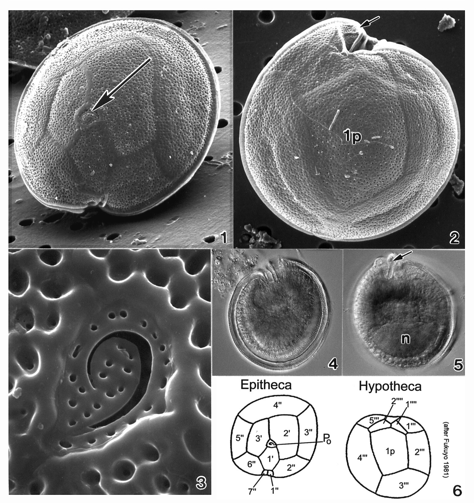
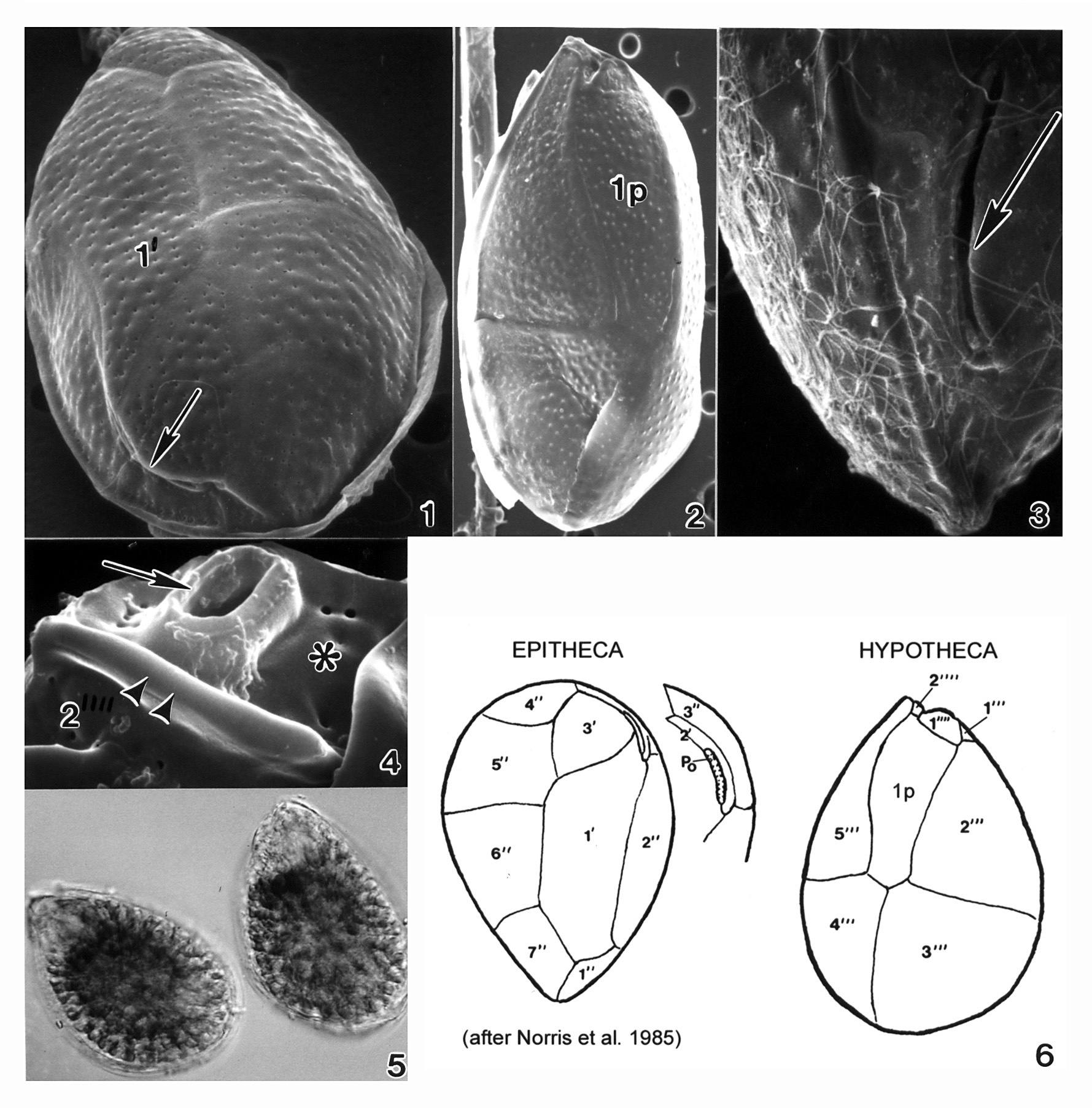
Plate 20. Gambierdiscus toxicus. Figs. 1-3. SEM. Fig. 1. Epitheca: cell round to ellipsoid; anterior-posteriorly compressed. Cell surface smooth with small scattered pores. Apical pore complex located at the apex (arrow). Fig. 2. Hypotheca: 1p plate large and pentagonal. Sulcal region deeply excavated (arrow). Fig. 3. Apical pore plate with characteristic fishhook shaped apical pore. Fig. 4. LM. Epitheca: cingulum and sulcal region in focus. Fig. 5. LM. Hypotheca: sulcal ridge (arrow); large nucleus (n). Fig. 6. Line drawing: thecal plate arrangement.
-
-
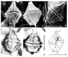
Plate 21. Gonyaulax polygramma. Figs. 1-3. SEM. Fig. 1. Ventral view: cell large, elongate and quadrilateral. Epitheca with prominent apical horn (arrow). Cingulum left-handed, displaced 1.5 X its width; sulcus widens posteriorly. Longitudinal ridges on thecal surface with reticulations in between. Fig. 2. Lateral ventral view: transverse (TF) and longitudinal (LF) flagella present. One antapical spine (arrow). Fig. 3. Dorsal view: hypotheca truncate with straight sides. Three antapical spines (arrows): one large and two small. Figs. 4-5. LM. Fig. 4. Ventral view: reticulations evident; one long antapical spine (arrow). Fig. 5. Dorsal view: prominent longitudinal ridges. Fig. 6. Line drawing.
-
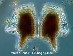
Dinophysis from the SW Pacific (Biosope Cruise)
-
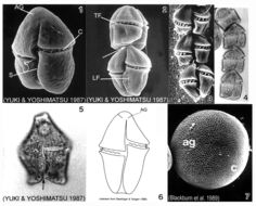
Plate 23. Gymnodinium catenatum. Figs. 1-3. SEM: ventral view. Fig. 1. Cell small, elongate-ovoid with slight dorso-ventral compression. Conical apex; rounded and notched antapex. Cingulum (C) excavated; sulcus (S) long. Distinctive horse-shoe shaped apical groove (AG) encircles apex. Fig. 2. Two cell chain; attachment point visible (arrow). Premedian cingulum displaced 2X its width. Longitudinal (LF) and transverse (TF) flagella visible. Fig. 3. Chain cells with anterior-posterior compression. Terminal cell slightly longer. Thecal surface rugose to smooth (Blackburn et al. 1989). Figs. 4-5. LM. Fig. 4. Chain-formation (Yuki and Yoshimatsu 1987). Fig. 5. Single cell. Conical epitheca with concave to flat apex. Bilobed hypotheca (arrow). Fig. 6. Line drawing. Fig. 7. SEM: cyst with microreticulations. ag=apical groove; c=cingulum
-
-
Plate 29. Lingulodinium polyedrum. Figs. 1-3. SEM. Fig. 1. Ventral view: cells angular and polyhedral-shaped. Thick plates well defined and coarsely areolate. Epitheca with shoulders and nearly flattened apex. Hypotheca with straight sides and flattened antapex (arrow). Cingulum deep and displaced 1-2 X its width. Sulcus widens posteriorly. Fig. 2. Apical view: first apical plate (1') long and narrow. Apical pore plate (Po) with raised inner elliptical ridge. Cingulum with lists (arrowheads). Strong ridges along sutures outline thecal plates. Fig. 3. Thecal areolae with large trichocysts (arrow)(Lewis and Burton 1988). Fig. 4. Line drawing. Figs. 5-6. SEM: resting cysts. Fig. 5. Cyst sperical with numerous tapering spines. Fig. 6. Cyst theca after excystment.
-
A heterotrophic dinoflagellate found with Phaeocystis on which it likely feeds.
-
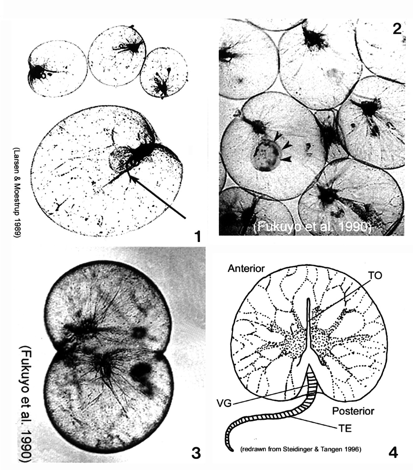
Plate 30. Noctiluca scintillans. Figs. 1-3. LM. Fig. 1. Cells large, balloon-shaped, nearly spherical, and colorless. A single flagellum housed in the ventral groove (arrow). Fig. 2. Cytoplasmic strands extend from nucleus (near the groove) to cell perifery. Engulfed cell (arrowheads). Fig. 3. Asexually dividing cell. Fig. 4. Line drawing. Deep and wide ventral groove (VG) houses the tooth (TO), an extension of the cell wall. Striated tentacle (TE).
-
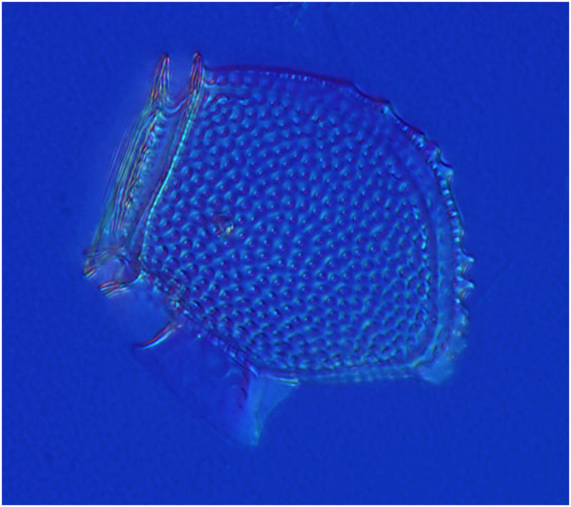
Plate 31. Ostreopsis heptagona. Figs. 1-4. SEM. Fig. 1. Epithecal view: cells broadly oval, oblong and pointed. Long curved apical pore plate, Po, off-center (arrow). Plate 1' heptagonal and distinctive. Fig. 2. Hypothecal view: plate 1p pentagonal and dorso-ventrally elongate. Fig. 3. Po long, narrow and curved. Narrow mucilage strands cover cell surface. Fig. 4. Ventral view: location of ventral opening (arrow), ventral plate (asterisk), and rigid plate (asterisk) within cingulum. Fig. 5. LM. Two cells. Fig. 6. Line drawing: thecal plate arrangement.
-
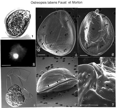
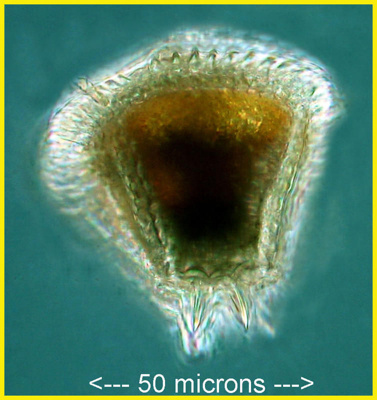
FIGS. 1-7. Ostreopsis labens sp. nov. FIGS. 1-3. Light microscope views. Scale bar = 25 μm. Cells contain chloroplasts and a spherical posterior nucleus (n). FIG.1. Cell is in epithecal view. FIG. 2. Location of nucleus stained with DAPI. FIG.3. Hypothecal plates partially separated with numerous pores. Cell with an engulfed prey organism (arrowhead); red color not detected on a black and white print. FIGS. 4-7. Cells viewed with SEM. FIG. 4. Cell is broadly ovoid in epithecal view. Note the curved, long apical pore (Po) located off-center (arrow). Scale bar =10 µm. FIG. 5. Cell is in hypothecal view. Cell is smooth with scattered pores (arrows). Scale bar =10µm. FIG. 6. Cell is slightly convex in lateral view. Note lipped, equatorial cingulum (arrowheads). Scale bar =10 µm. FIG. 7. Antapical plate 1"is with a slightly curved list (arrowhead). The sulcus narrow, recessed and hidden adjacent to plate 2". The ventral opening (arrow) is situated on the ventral surface adjacent to a ridged plate (Rp) (asterisk). Scale bar = 5 μm. EMu: : HOLOTYPE SEM NEGATIVE # 170058; SEM STUB # 170; FIELD # 745-94; ACCESSION # 410840; CATALOG # 984; FIGURE # 4