-
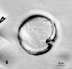
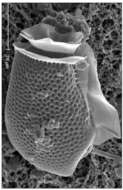
Fig 6: Alexandrium tamarense anterior part of theca showing outline of 1' plate
-
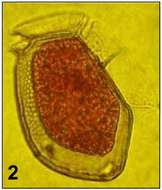
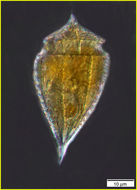
Fig 7: Image of Alexandrium tamarense
-
Fig 1: Prorocentrum micans Schematic diagram (ventral view) redrawn from Tomas et al. 1997.
-
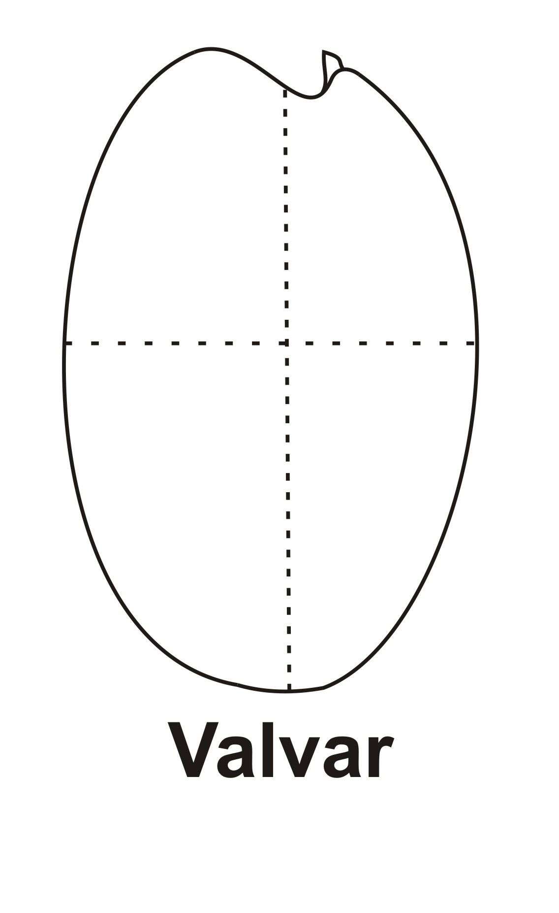
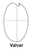
Fig 1: Prorocentrum rhathymum Schematic diagram (valvar view) redrawn (and edited) from Cortés-Altamirano & Sierra-Beltran 2003.
-
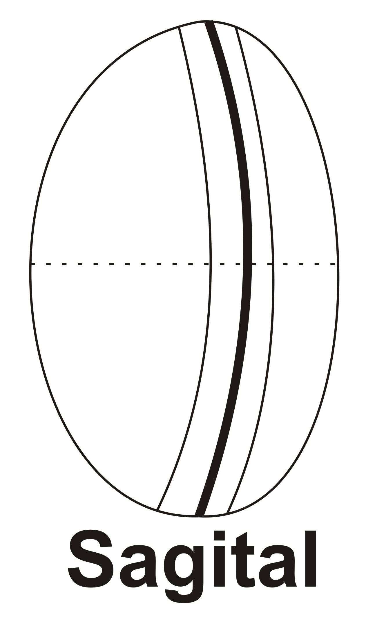
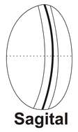
Fig 2: Prorocentrum rhathymum Schematic diagram (sagital view) redrawn from Cortés-Altamirano & Sierra-Beltran 2003.
-
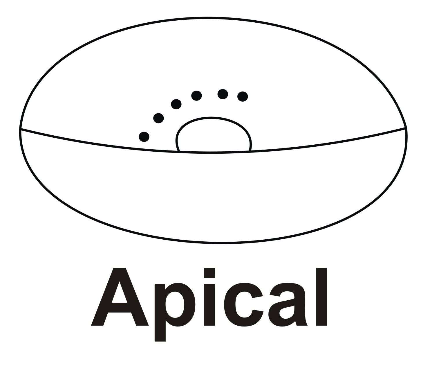
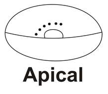
Fig 3: Prorocentrum rhathymum Schematic diagram (apical view) redrawn (and edited) from Cortés-Altamirano & Sierra-Beltran 2003.
-
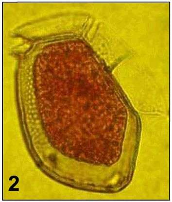
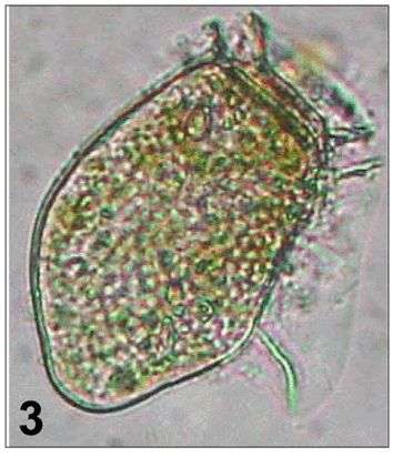
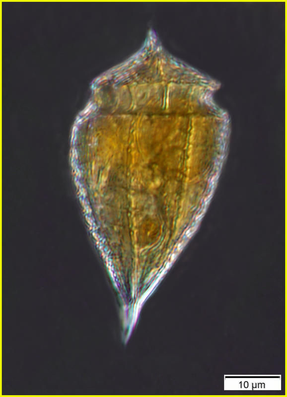
Fig 1: Dinophysis acuta Schematic drawing of a cell in lateral view
-
Fig 2: Dinophysis acuta Lugol's preserved cell showing shrinkage of cell contents
-
Fig 3: Dinophysis acuta Live cell in lateral view
-
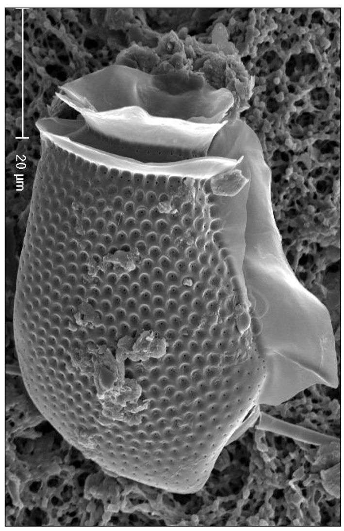
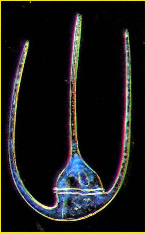
Fig 4 Eletron micrograph of a D. acuta cell showing details of pore structure and sulcal lists
-
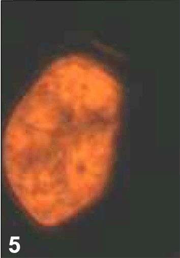
Fig 5 Epifluorescence image under blue light
-
Specimen from the Ionian Sea found at 75 m depth. Lugol's-fixed.
-
Uit: www.nies.go.jp/biology/ mcc/strainlist_a.htm
Ecomare
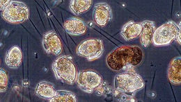
Alexandrium; Alexandrium.
-
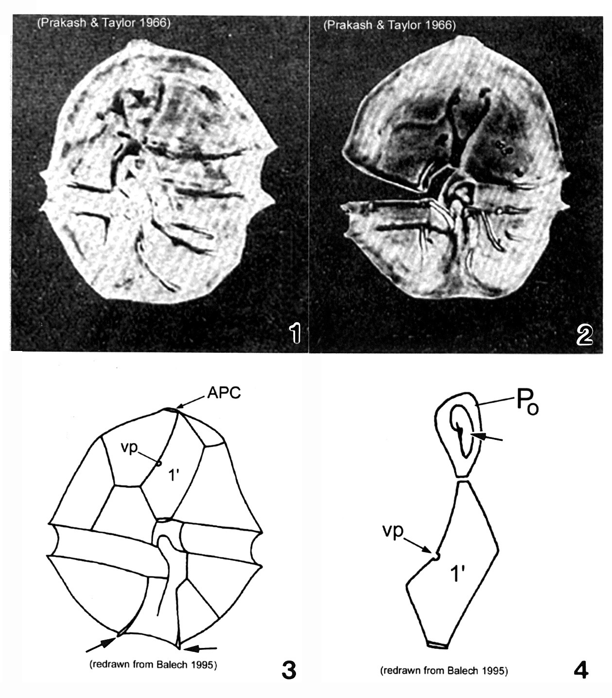
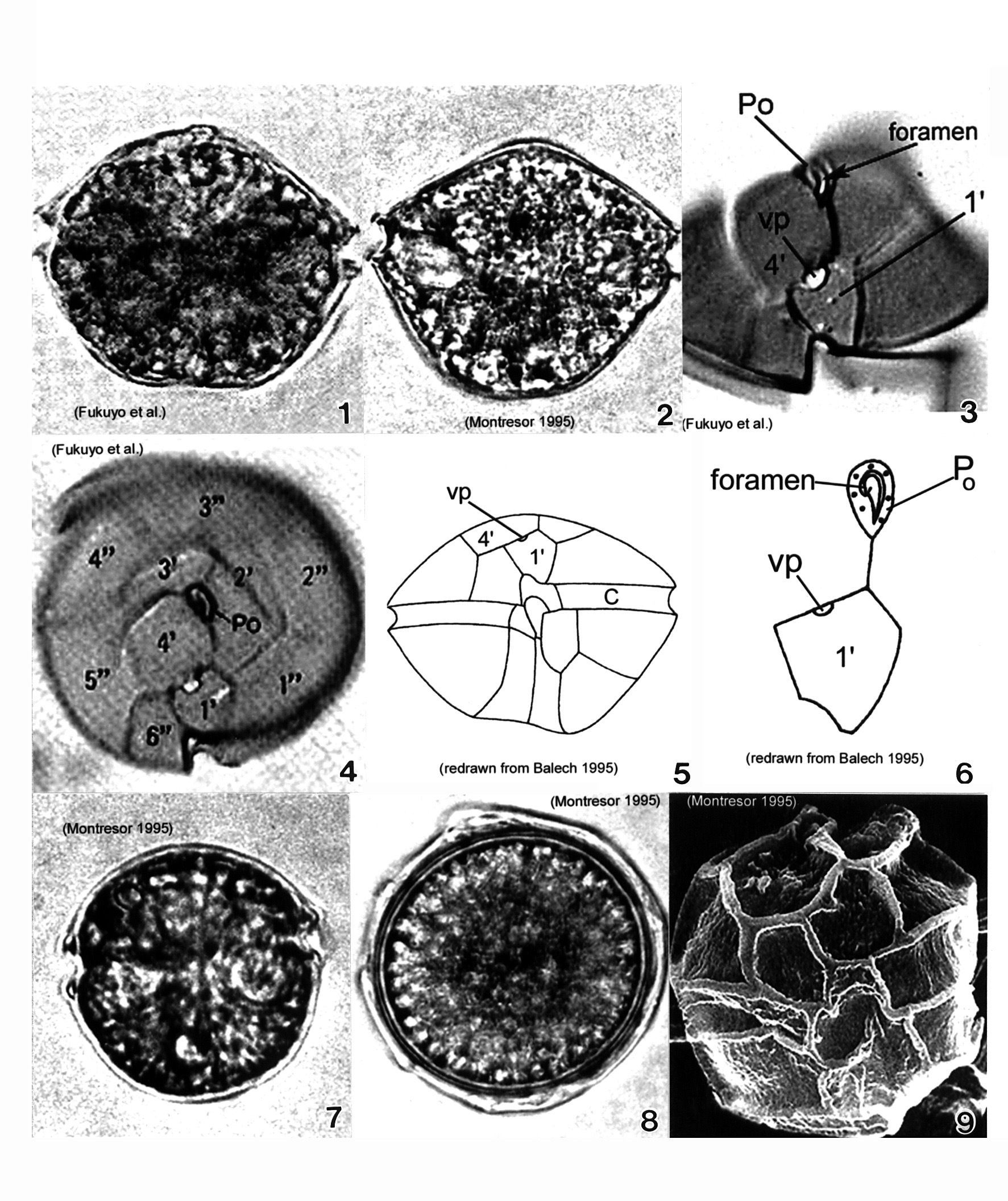
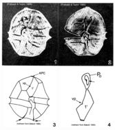
Plate 1. Alexandrium acatenella. Figs. 1-2. LM: ventral view of empty thecae. Cell small to medium, longer than wide, angular to round. Conical epitheca with shoulders; larger than hypotheca. Figs. 3-4. Line drawings. Fig. 3. Ventral view: 1' plate bears ventral pore (vp). Hypotheca with two antapical spines (arrows). Fig. 4. Po comes in direct contact with 1' plate. APC: comma-shaped foramen (arrow).
-
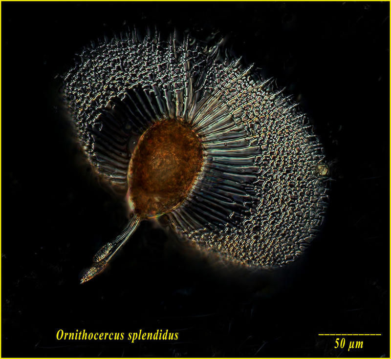
He thought it splendid. Figures from the original description of Ornitheroceras splendidus by Franz Schütt in 1893. The paper is available on the Classic Taxonomic Monographs page.
-
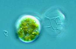
Histioneis hyalina (probably). From Station 52 of the Tara Oceans Expedition
-
University of Liverpool http://www.liv.ac.uk/
Ecomare
David J.S. Montagnes; David J.S. Montagnes.
-
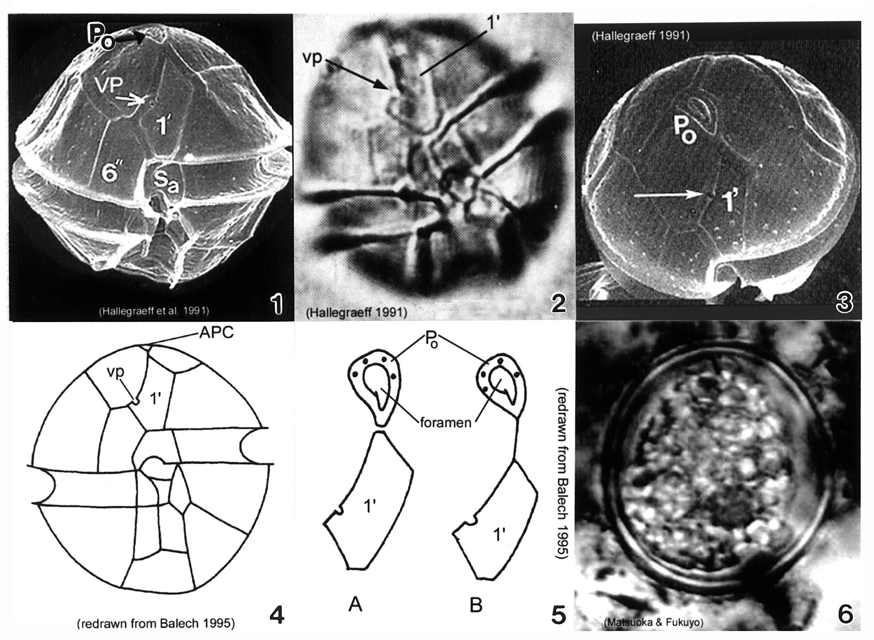
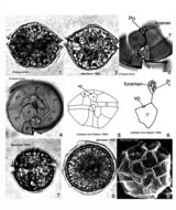
Plate 3. Alexandrium minutum. Fig. 1. SEM: ventral view. Cell small and ellipsoidal. Epitheca conical, larger than hypotheca. Hypotheca short and wide; antapex obliquely flattened. Intercalary bands present. Cingulum deep, lipped; displaced 1X its width. Sulcus shallow (sa=anterior sulcal plate). Apical pore plate (Po) in direct contact with 1' plate. Fig. 2. LM: ventral view. Ventral pore (vp) present on 1' plate. Fig. 3. SEM: apical view. Po large, narrow and oval; indirectly connected to 1' plate. Vp present (arrow). Figs. 4-5. Line drawing. Fig. 4. Ventral view. 1' plate slender and rhomboidal. Fig. 5. Po connection to 1' plate: a. direct; b. indirect via thin suture. Fig. 6. LM: cyst circular in apical view.
-
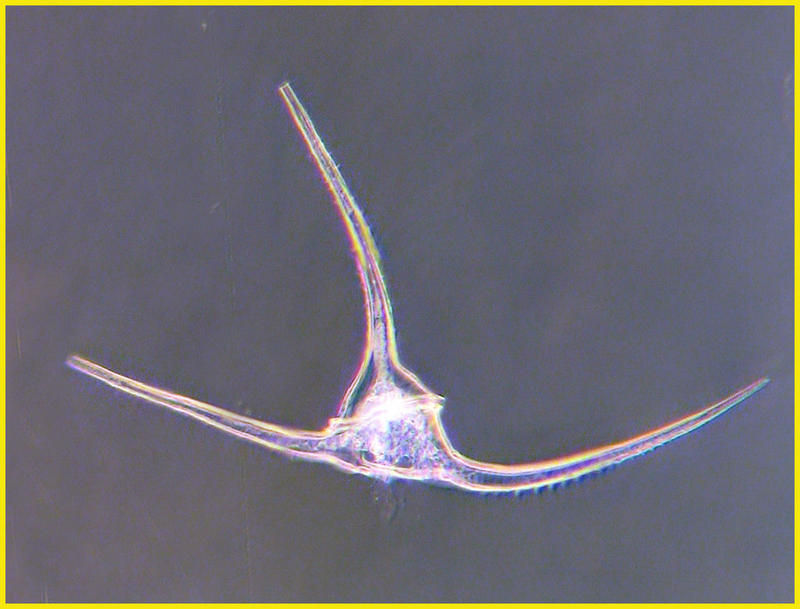
Oxytoxum nanum
-
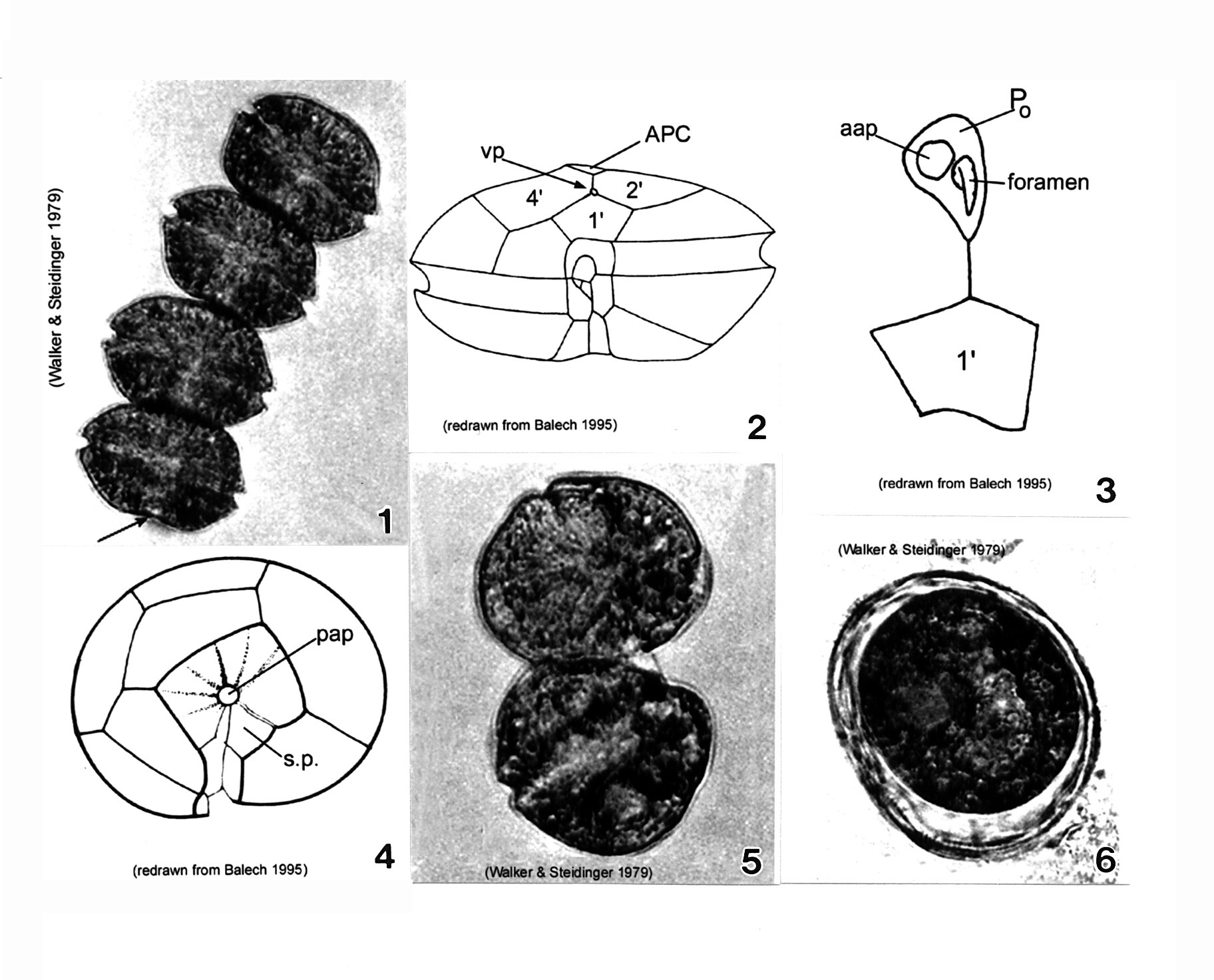
Plate 4. Alexandrium monilatum. Fig. 1. LM: four-cell chain. Cells large, wider than long, flattened anterio-posteriorly. Antapex slightly concave (arrow). Figs. 2-4. Line drawings. Fig. 2. Ventral pore (vp) depicted (Florida specimens) at anterior margin of 1' plate where it comes in contact with plates 2' and 4'. Cingulum (C) deeply excavated, wide, descending; displaced one time its width. Fig. 3. Apical pore plate (Po) does not come in contact with 1' plate. Anterior attachment pore (aap) large, round and dorsally situated in the APC. Foramen comma-shaped. Fig. 4. Antapical view: posterior sulcal plate (sp) large, rhomboid and concave with radial markings. Posterior attachment pore (pap) large and centrally located. Figs. 5-6. LM. Fig. 5. Two isogamous gametes fusing at oblique angles. Fig. 6. Mature resting cysts: dark and round, with a triple layered wall.
-
-
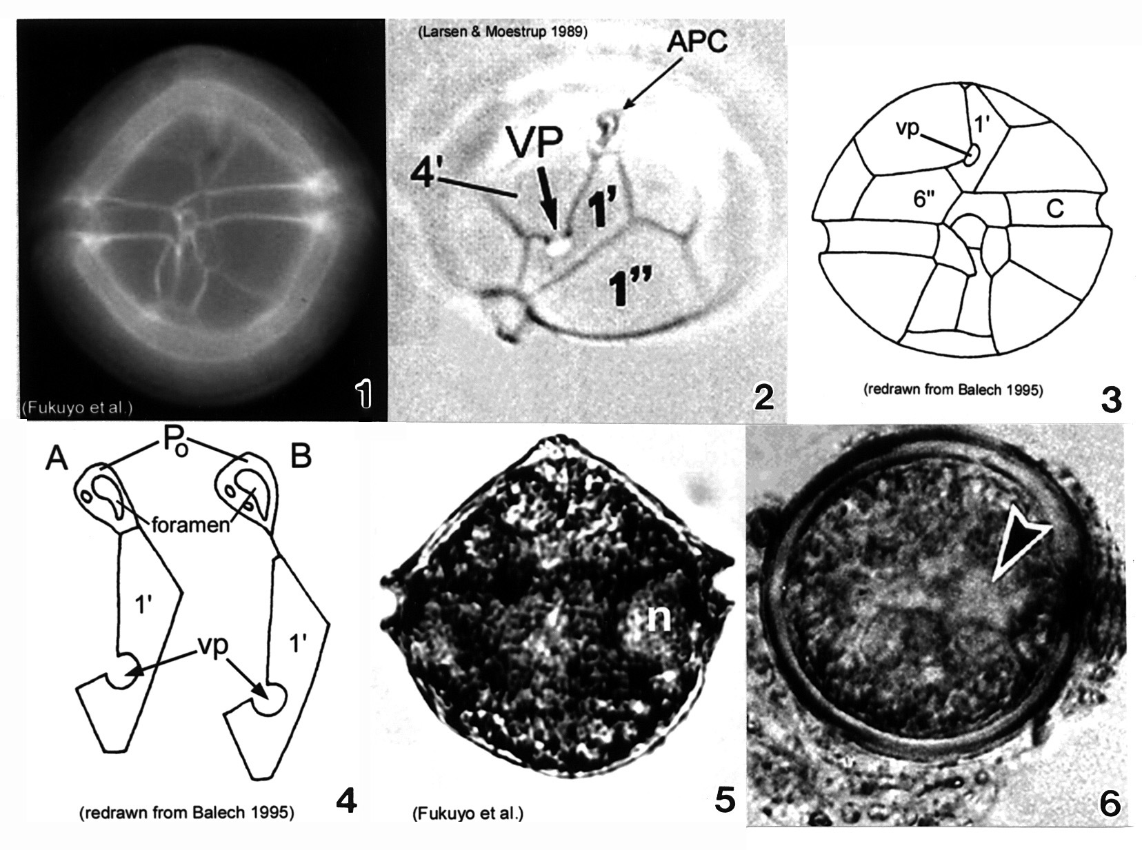
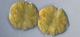
Plate 5. Alexandrium ostenfeldii. Figs. 1-3. LM. Fig. 1. Ventral view. Cell large and nearly spherical. Cingulum deeply excavated. Epitheca broad and convex-conical. Hypotheca hemispherical with an obliquely flattened antapex. Fig. 2. Epitheca: apical view. Ventral pore (vp) large and distinct. First apical plate (1') forms a 90 degree angle at the point where vp and 4' plate come in contact. Apical pore complex (APC) with comma-shaped foramen. Figs. 3-4. Line drawings. Fig. 3. Ventral view: 6'' plate wider than high. Cingulum (C) slightly excavated. Fig. 4. APC and 1' plate: a. Po in direct contact with 1'; b. Po in indirect contact with 1' via thin suture. Fig. 5. LM: vegetative cell. Small equatorial nucleus (n). Fig. 6. LM: temporary cyst large and spherical, covered in mucilage. Nucleus visible (arrowhead)(Mackenzie et al. 1996).
-
-
Plate 6. Alexandrium pseudogonyaulax. Figs. 1-4. LM. Fig. 1. Ventral view. Cell broadly pentagonal; wider than long. Epitheca short and dome-shaped. Hypotheca longer than epitheca. Cingulum shallow and barely displaced. Fig. 2. Dorsal view. Antapex obliquely concave. Fig. 3. Epitheca: ventral view. Apical pore plate (Po) with comma-shaped foramen. 1' plate pentagonal with large wide ventral pore (vp) on 4' plate margin. Fig. 4. Epitheca: apical view. 1' plate does not come in contact with Po. Po oval and longitudinal on apex. Figs. 5-6. Line drawings. Fig. 6. Po and 1' plate not in contact. Fig. 7. LM: isogamous gametes smaller and rounder than vegetative cells. Fig. 8. LM: round resting cyst. Fig. 9. SEM: paratabulate cyst.