-
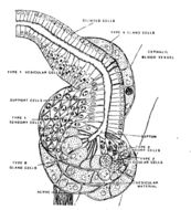
Diagram of the cerebral organ of Amphiporus lactifloreus in frontal section; the top of the diagram is anteriorFrom: Fine structure of the cerebral organs in hoplonemerteans (Nemertini), with a discussion of their function Helen M. Amerongen Zoomorphology (1987) 10: 145-159
-
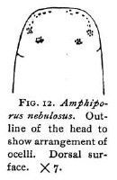
Outline of the head to show the arrangement of ocelli. Dorsal surface.Coe, W. R. (1901). The Nemerteans of the Expedition. Proceedings of the Washington Academy of Sciences, Vol. 3, 1-110.
-
Outline of anterior portion of body to show the arrangement of ocelli. Coe, W. R. (1901). The Nemerteans of the Expedition. Proceedings of the Washington Academy of Sciences, Vol. 3, 1-110.
-
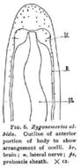
Zygonemertes albida: Outline of anterior portion of body to show arrangement of ocelli.
-
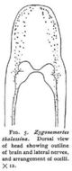
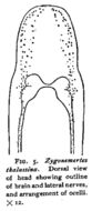
Dorsal view of anterior portion of body showing outline of brain and lateral nerves, and arrangement of ocelli.Coe, W. R. (1901). The Nemerteans of the Expedition. Proceedings of the Washington Academy of Sciences, Vol. 3, 1-110.
-
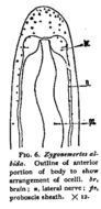
Zygonemertes thalassina: Dorsal view of head showing outline of brain and lateral nerves, and arrangement of ocelli x12
-
Photomicrograph of a longitudinal section through the posterior end of a female that was apparently depositing cleaving... Photomicrograph of a longitudinal section through the posterior end of a female that was apparently depositing cleaving embryos into an egg string. The single arrowhead marks the caudal end of the epidermis. Scale bar, 100 um.
-
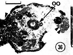
Photomicrograph of a longitudinal section through the posterior end of a female that was fixed while laying an egg string...Photomicrograph of a longitudinal section through the posterior end of a female that was fixed while laying an egg string. The single arrowhead marks a subterminal constriction, and the double arrowheads point to a mass of spermatozoa. Cellular debris occurring toward the end of the worm probably represents artifactual damage incurred when the worm was removed from its egg string. Ruthenium red/sodium cacodylate primary fixative. 00, oocyte. Scale bar, 100 um.
-
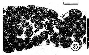
A section of an egg string containing blastulae (arrowhead). Note that extraembryonic cells are lacking within the egg string...A section of an egg string containing blastulae (arrowhead). Note that extraembryonic cells are lacking within the egg string. Ruthenium red - sodium cacodylate primary fixative. Scale bar, 100 um.
-
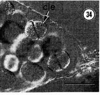
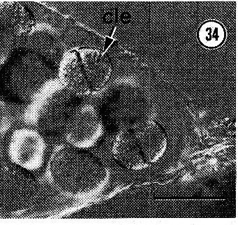
The distal end of an egg string with zygotes undergoing first cleavage (cle). Scale bar, 100 um.
-
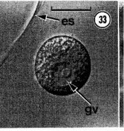
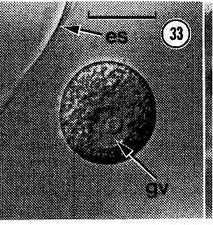
The peripheral region of a newly deposited egg string (es) showing an oocyte with an intact germinal vesicle (gv)...the peripheral region of a newly deposited egg string (es) showing an oocyte with an intact germinal vesicle (gv). Scale bar, 50 u.m.
-
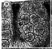
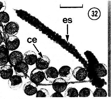
An egg string (es) containing numerous developing embryos. ce, crab egg. Scale bar, 0.5 mm.
-
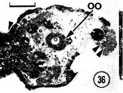
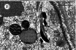
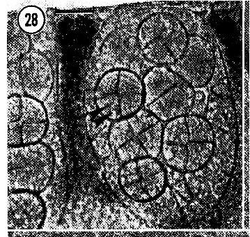
Spermatozoa (sp) tightly appressed to an intraovarian oocyte (00). ic, intestinal cell. Scale bar, l um.
-
Spermatozoa (sp) tightly appressed to an intraovarian oocyte (00). ic, intestinal cell. Scale bar, l um.
-
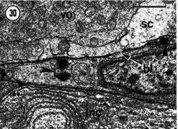
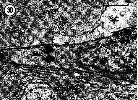
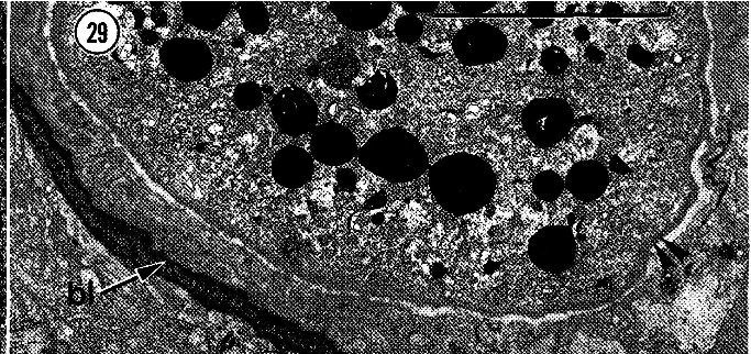
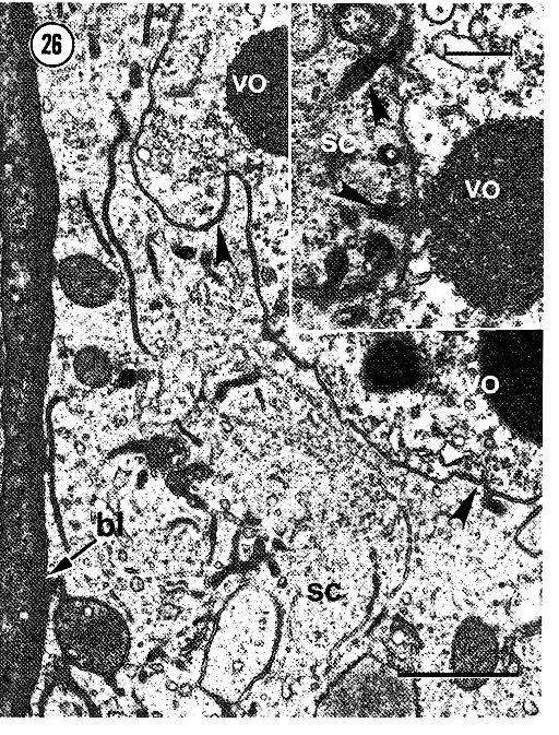
Spermatozoa (arrowhead) occurring in the connective tissue compartment directly outside an ovary. hi, basal lamina; inc...Spermatozoa (arrowhead) occurring in the connective tissue compartment directly outside an ovary. hi, basal lamina; inc, lateral nerve cord; sc, somatic cell; VO, vitellogenic oocyte. Scale bar, 1 u.m.
-
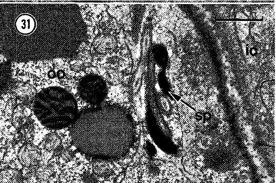
A fertilized zygote within the lumen of an ovary. The double arrowheads point to an electron-dense egg membrane....A fertilized zygote within the lumen of an ovary. The double arrowheads point to an electron-dense egg membrane. hi, basal lamina. Scale bar, 10 u.m.
-
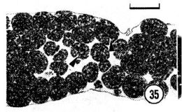
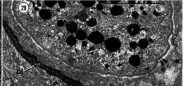
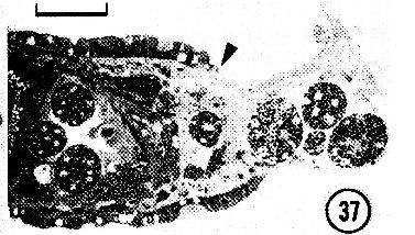
Photomicrograph of a compressed female worm with cleaving embryos in its ovaries (double arrowheads). Scale bar, 50 u.m.
-
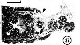
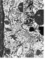
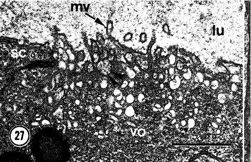
The apical surface of a vitellogenic oocyte (va) that is surrounded by somatic cells (se) with basally located tubular elements.The apical surface of a vitellogenic oocyte (va) that is surrounded by somatic cells (se) with basally located tubular elements. The double arrowheads mark a putative endocytotic vesicle. lu, lumen of ovary; mv, microvilli. Scale bar, 1 u.m.
-
The border between a vitellogenic oocyte (vo) and a somatic cell (se) with numerous tubular elements. Some of the tubules...The border between a vitellogenic oocyte (vo) and a somatic cell (se) with numerous tubular elements. Some of the tubules seem to fuse with the oolemma (arrowhead). hi, basal lamina. Scale bar, 1 um. Inset: several tubules (arrowheads) apparently in the process of fusing to the oolemma of a vitellogenic oocyte (vo). se, somatic cell. Scale bar, 0.5 um.
-
Basal infoldings and the vesicular to tubular elements (double arrowheads) that apparently become incorporated in the somatic...Basal infoldings and the vesicular to tubular elements (double arrowheads) that apparently become incorporated in the somatic cell (se) . Note the numerous dense particles directly outside the basal lamina (hi). Scale bar, 1 um.
-
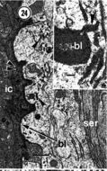
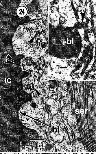
The border between the ovary and intestine. Numerous electron-dense particles (double arrowheads) occur in the narrow...The border between the ovary and intestine. Numerous electron-dense particles (double arrowheads) occur in the narrow extracellular space between the basal laminae (hI) of theovary and intestine. The single arrowheads mark tubular elements occurring within the basal region of a somatic cell. ie, intestinal cell; ser, smooth endoplasmic reticulum. Scale bar, 1 um. Inset: higher magnification view of a tubular vesicle (arrowhead) attached to the inner surface of the basal lamina (hI). Scale bar, 100 nm.
-
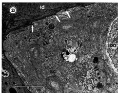
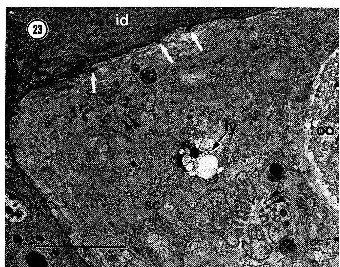
An adintestinal somatic cell (sc). The double arrowheads mark peculiar clusters of nuclear bodies, and the single arrows point..An adintestinal somatic cell (sc). The double arrowheads mark peculiar clusters of nuclear bodies, and the single arrows point to infoldings of the basal lamina. id, intestinal diverticulum; ly, lysosome; 00, oocyte. Scale bar, 10 urn.
-
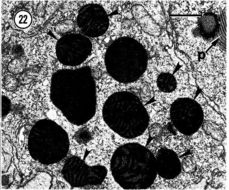
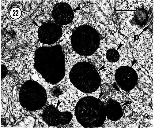
Large yolk bodies with plaques (p) in their internal regions (arrowheads). Scale bar, 1 um.
-
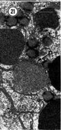
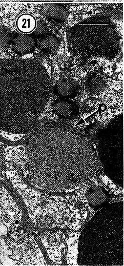
Plaquelike inclusions (p) that seem to fuse with electron-dense material during yolk formation. Scale bar, 1 um.