-
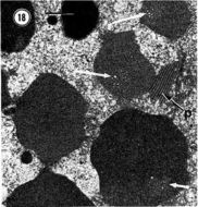
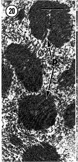
Stacks of plaque like inclusions (p) apparently in the process of forming ovoid bodies. Scale bar, 1 um
-
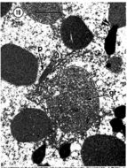
A membrane-bound yolk granule (gr) and nascent plaquelike bodies (p) being formed in the vicinity...A membrane-bound yolk granule (gr) and nascent plaquelike bodies (p) being formed in the vicinity of the rough endoplasmic reticulum. The double arrowheads mark an apparent fusion between a granule and lipid droplet. Scale bar, I J.Lm.
-
Stages in yolk formation. The arrows mark vesicular components of developing yolk bodies. p, plaque. Scale bar, 1 um.
-
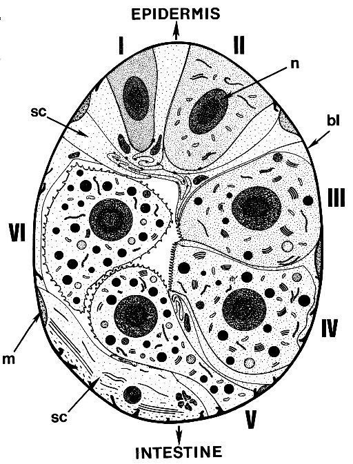
A diagram of an ovary showing various stages of oogenesis...A diagram of an ovaryshowing variousstagesof oogenesis andthe relationship between germinal and somaticcells in the ovarian epithelium. I, oogonium or a young previtellogenic oocyte; II, a previtellogenic oocyte; III, an early vitellogenic oocyte; IV, a late vitellogenic oocyte; V, a yolk-filled oocyte in the process of being moved intothe lumenof the ovary;VI, a primaryoocytein the ovarian lumen. hi, basallamina; m, myofilaments; n, nucleus; SC, somatic cell.
-
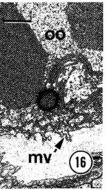
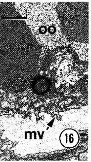
Stubby microvilli (mv) arising from a primary oocyte (00) in the lumen of the ovary. The apical surface of the ovarian...Stubby microvilli (mv) arising from a primary oocyte (00) in the lumen of the ovary. The apical surface of the ovarian epithelium is depicted at the bottom of the micrograph. Note that the oocyte lacks well developed extracellular coats or surrounding follicle cells. Scale bar, 1 urn.
-
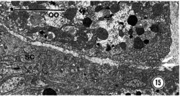
15. A primary oocyte (00) in the lumen of the ovary. The double arrows point to patches of dense material that may represent...A primary oocyte (00) in the lumen of the ovary. The double arrows point to patches of dense material that may represent parts of a poorly developed vitelline envelope. sc somatic cell. Scale bar, 10 um.
-
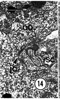
A junctional complex (jc) adjoining an oocyte (00) and its neighboring somatic cell (sc). Scale bar, 1 um
-
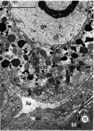
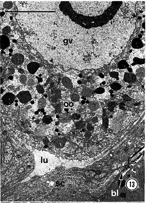
primary oocyte (00) in the lumen of the ovary (lu). bl, basal lamina; gv, germinal vesicle; sc, somatic cell. Scale bar, 10 urn.
-
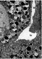
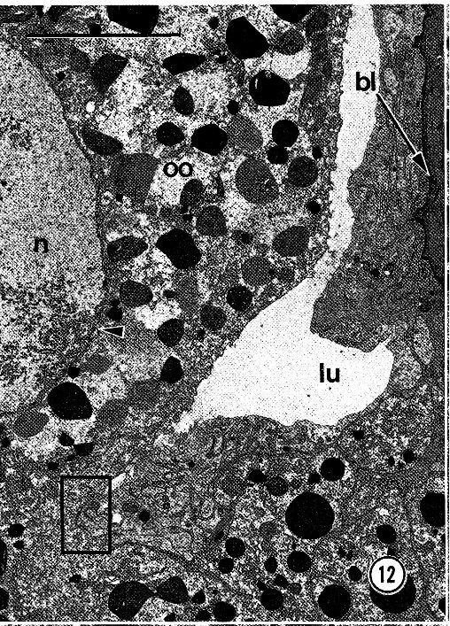
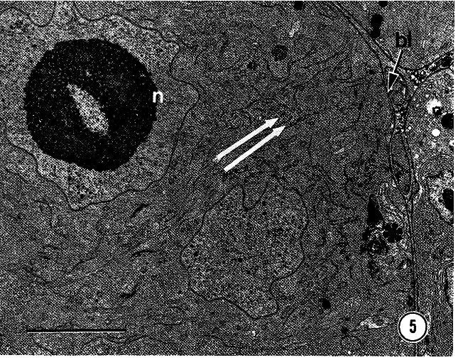
An oocyte (00) in the process of being moved into the ovarian lumen (lu). Membranous lamellae (arrowhead) can be seen attached..An oocyte (00) in the process of being moved into the ovarian lumen (lu). Membranous lamellae (arrowhead) can be seen attached to the inner surface of the nuclear envelope. bl, basal lamina; n, nucleus. The box outlines the region shown at higher magnification in Fig. 14. Scale bar, 10 micrometers.
-
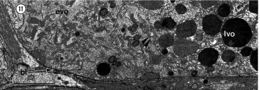
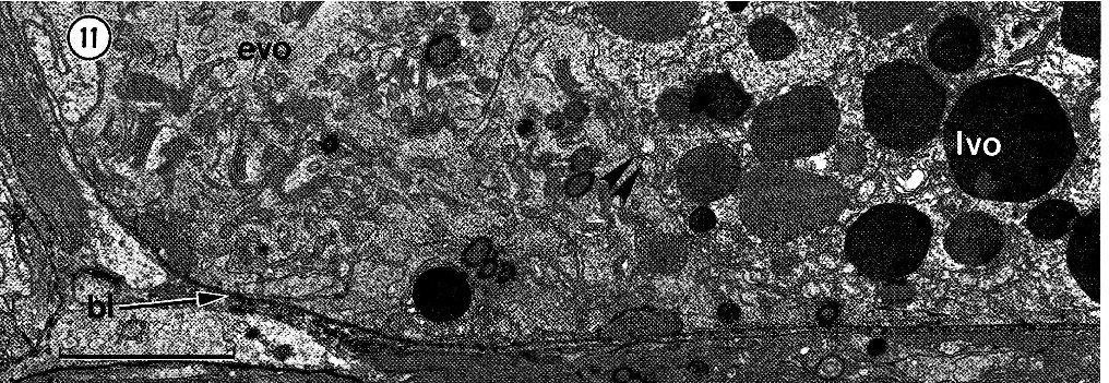
Oocytes at early (evo) and late stages of vitellogenesis (Ivo). The double arrowheads mark the border between two oocytes...Oocytes at early (evo) and late stages of vitellogenesis (Ivo). The double arrowheads mark the border between two oocytes. bl, basal lamina. Scale bar, 5 micrometers.
-
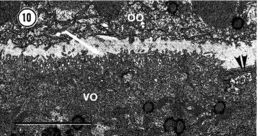
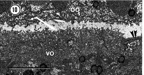
Vitellogenic oocyte (vo) that abuts the ovarian lumen. The double arrowheads point to apical processes of somatic cells...Vitellogenic oocyte (vo) that abuts the ovarian lumen. The double arrowheads point to apical processes of somatic cells, and the single arrow marks microvilli arising from the oocyte. 00, oocyte in lumen. Scale bar, 5 micrometers.
-
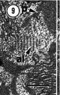
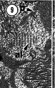
A rare example of annulate lamellae (aI) in a vitellogenic oocyte. p, plaque. Scale bar, 1 micrometer.
-
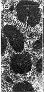
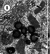
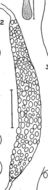
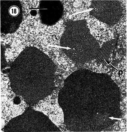
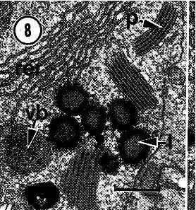
Various inclusions within a vitellogenic oocyte...Various inclusions within a vitellogenic oocyte. l, lipid; p, plaque; rer, rough endoplasmic reticulum; vb, vesicular body. Scale bar, 1 micrometer.
-
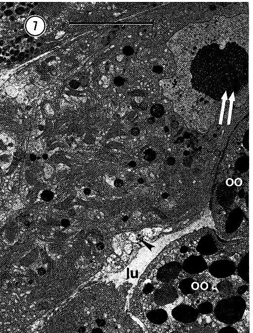
The ovarian epithelium with two vitellogenic oocytes. The double arrows mark the nucleus of one of the oocytes...The ovarian epithelium with two vitellogenic oocytes. The double arrows mark the nucleus of one of the oocytes, and the single arrow points to somatic cell processes. lu, lumen of ovary; 00, oocyte in lumen. Scale bar, 10 micrometers.
-
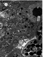
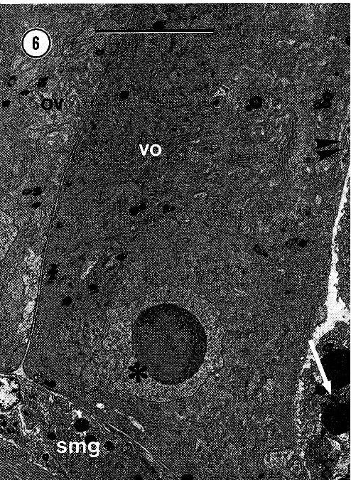
Two closely appressed ovaries (ov). The nucleus of a young vitellogenic oocyte (vo) is marked by an asterisk...Two closely appressed ovaries (ov). The nucleus of a young vitellogenic oocyte (vo) is marked by an asterisk in the right-hand ovary. The double arrows point to somatic cell processes bordering the lumen of the ovary, and the single arrow points to a primary oocyte in the ovarian lumen. smg, submuscular glands. Scale bar, 10 micrometers.
-
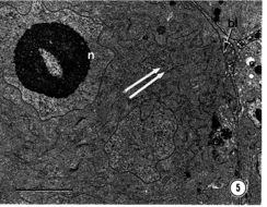
Two previtellogenic oocytes with numerous mitochondria and cisternae of the rough engoplasmic reticulum (double arrows)...Two previtellogenic oocytes with numerous mitochondria and cisternae of the rough engoplasmic reticulum (double arrows). The asterisk marks a basally located portion of a somatic cell. hi, basal lamina; n, nucleus of oocyte. Scale bar, 5 urn.
-
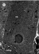
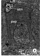
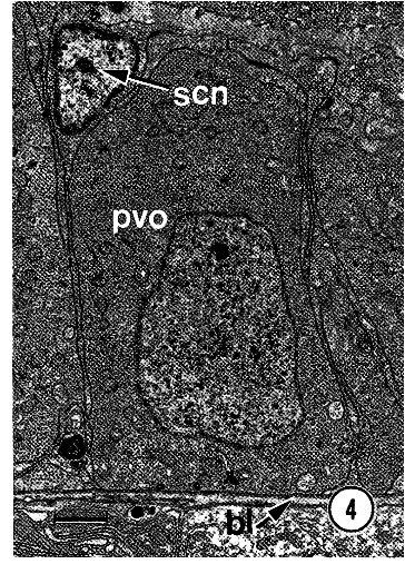
A previtellogenic oocyte (pvo). hi, basal lamina; sen, somatic cell nucleus. Scale bar, 1 micrometer
-
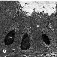
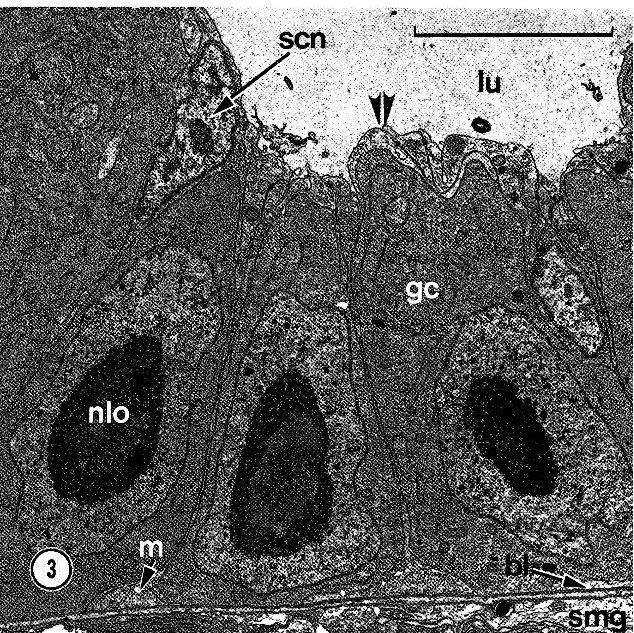
Transmission electron micrograph (TEM) of three small germinal cells (ge) in the subepidermal region of the ovarian epithelium..Transmission electron micrograph (TEM) of three small germinal cells (ge) in the subepidermal region of the ovarian epithelium. The double arrowheads mark apical processes of somatic cells that cover the germinal cells. hi, basal lamina; iu, lumen of ovary; m, myofilaments; nlo, nucleolar body; scn, somatic cell nucleus; smg, submuscular glands. Scale bar, 5 micrometers.
-
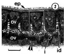
Photomicrograph of a slightly oblique frontal section of a gravid worm...Photomicrograph of a slightly oblique frontal section of a gravid worm, showing prominent globules of ingested crab yolk within the intestine (i) and intestinal diverticula (id). The double arrowheads mark a region similar to that shown in Fig. 3; the rectangle outlines the area depicted in diagrammatic form in Figure 17. ep, epidermis; OV, ovary. Scale bar, 100 urn
-
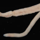
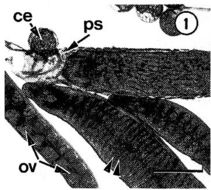
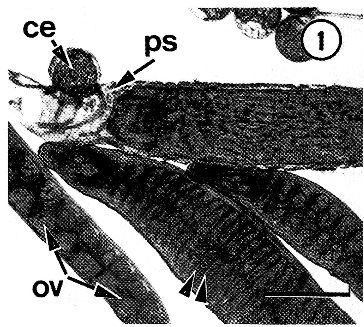
Photomicrograph of compressed gravid specimens of Carcinonemertes epialti...Photomicrograph of compressed gravid specimens of Carcinonemertes epialti. One of the worms is pictured in a parchmentlike sheath (ps). The arrowheads mark diverticula of the intestine that are separated by an ovary (ov). ee, crab egg. Scale bar, 0.5 mm.
-
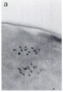
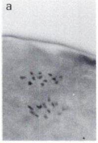
Polar body formation in egg of control Carcinonemertes epialti....Polar body formation in egg ofcontrol Carcinonemertes epialti. Chromosomes nearest edge of the cell are those ofthe polar body. Some cells showed 12, other cells showed 13 chromosomes during meiotic divisions. Magnification: 1500X.
-
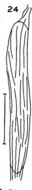
Outline of basis of C. epialti, as seen in section. Scale 0.02 mm.
-
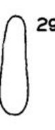
Mucous sheath of C . epialti from Euphylax dovii. Scale 0 .5 mm.
-
Mucous sheath of C. epialti from egg mass of Euphylax dovii at Payta, Peru. Scale 0 .5 mm.