-
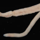
Holotype: worm #144, frontal section
-
Paratype, worm #142, transverse section
-
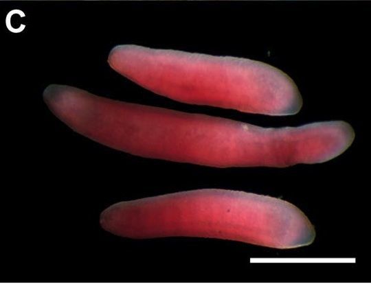
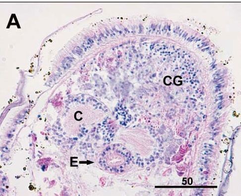
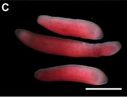
Ovicides jasoni from A. alayseae, formalin preserved specimens. The longest specimen is 3.2 mm in length, bar scale = 1.0 mm.
-
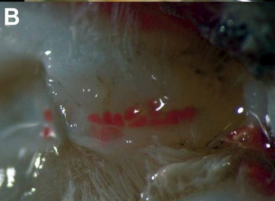
Worms ensheathed on the axilla of A. alayaseae
-
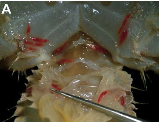
Juvenile worms in situ on the sternum, pleon, and pleopods of Austinograea alayseae, Lau Basin, Tui Malila vent site, 21 May...Juvenile worms in situ on the sternum, pleon, and pleopods of Austinograea alayseae, Lau Basin, Tui Malila vent site, 21 May 2005, Dive #144
-
Slightly oblique transverse section through paratype (1592-185 worm 1 slide 2) showing the arrangement of the submuscular...Slightly oblique transverse section through paratype (1592-185 worm 1 slide 2) showing the arrangement of the submuscular glands in interspersed rows around the circumference of the worm.
-
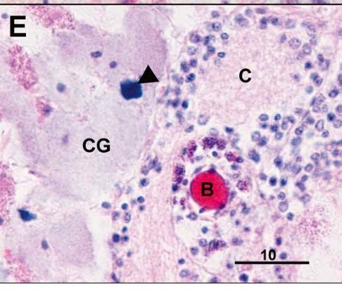
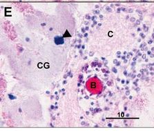
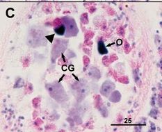
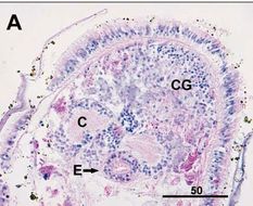
Paratype (worm 1592-182 worm 1b) with large, weakly basophilic cephalic glands (CG) with darkly basophilic granule (arrow)...Paratype (worm 1592-182 worm 1b) with large, weakly basophilic cephalic glands (CG) with darkly basophilic granule (arrow) anterior to cerebrum.
-
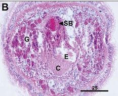
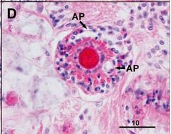
Transverse section through the stylet bulb of paratype (worm 1592-185 worm 4b). Note the presence of the two accessory stylet...Transverse section through the stylet bulb of paratype (worm 1592-185 worm 4b). Note the presence of the two accessory stylet pouches (AP) lateral to the basis.
-
Paratype (worm 1592-182 worm 17) with weakly basophilic cephalic glands (CG) in the region of an ocellus. Note the presence...Paratype (worm 1592-182 worm 17) with weakly basophilic cephalic glands (CG) in the region of an ocellus. Note the presence of the darkly basophilic granule associated with the cephalic gland.
-
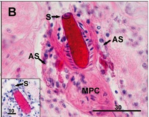
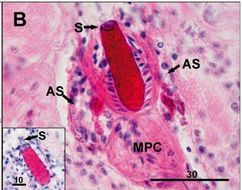
Paratype (worm 1592-182-slide 1-worm 1) with eosinophilic basis, two accessory stylets (AS) and armed stylet possessing a hub...Paratype (worm 1592-182-slide 1-worm 1) with eosinophilic basis, two accessory stylets (AS) and armed stylet possessing a hub (S); inset showing dagger-like stylet (1592-182 slide 1 worm 17).
-
Holotype (worm 1592-96-slide 2 - worm 17) with weakly developed cephalic glands (CG) anterior to the cerebrum; frontal section.
-
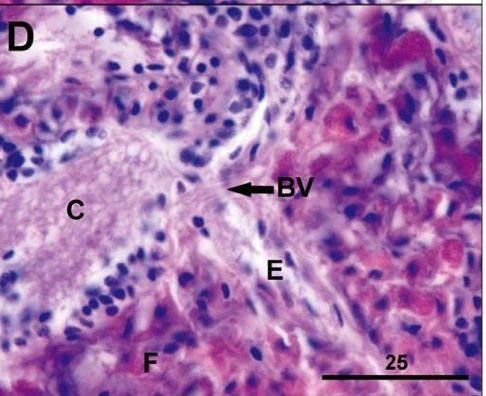
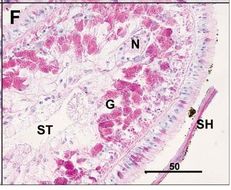
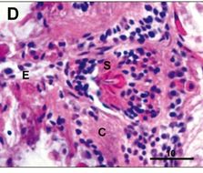
Paratype (slide 2, worm 9). Anterior loop of the primary blood vessel (BV) dorsal to the esophagus (E) and adjacent to the...Paratype (slide 2, worm 9). Anterior loop of the primary blood vessel (BV) dorsal to the esophagus (E) and adjacent to the cerebrum (C). Note the large number of eosinophilic submuscular glands in the esophageal region.
-
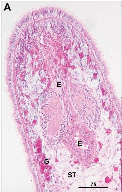
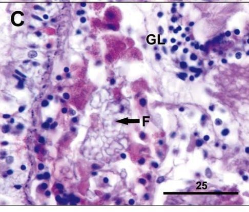
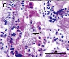
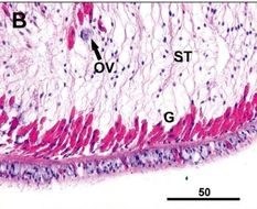
Paratype (same slide, worm 8). The well-developed, vesiculated frontal organ (F) is anterior to the glial cells (GL) of the...Paratype (same slide, worm 8). The well-developed, vesiculated frontal organ (F) is anterior to the glial cells (GL) of the cerebrum.
-
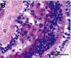
Paratype (same slide, worm 5). Anterior of worm showing weakly basophilic frontal organ (F) developed as a field anterior...Paratype (same slide, worm 5). Anterior of worm showing weakly basophilic frontal organ (F) developed as a field anterior to cerebrum (C) on both sides of the esophagus (E).
-
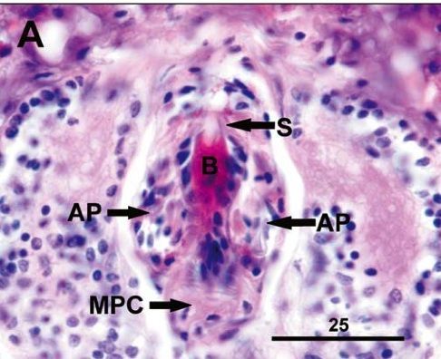
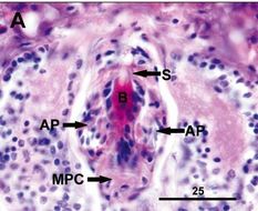
Holotype (Carcino B.l. (1), slide 3, worm 1). Stylet bulb region of worm showing pyriform stylet (S) on the basis (B) with two..Holotype (Carcino B.l. (1), slide 3, worm 1). Stylet bulb region of worm showing pyriform stylet (S) on the basis (B) with two accessory stylet pouches (AP) anterior to the middle proboscis chamber (MPC).
-
Portion of the stylet in the anterior proboscis chamber showing the stylet hub, a portion of the esophagus.
-
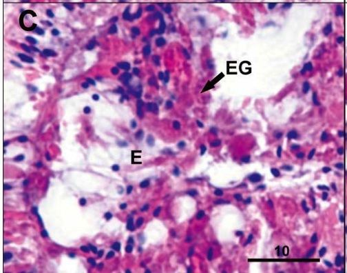
Eosinophilic glands (EG) lining the anterior esophagus.
-
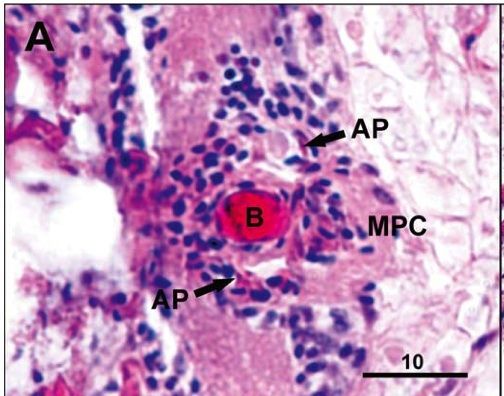
Stylet bulb region of worm showing two accessory stylet pouches (AP) adjacent to the basis.
-
Medial section of body with single row of submuscular glands (G) and a presumptive ovum undergoing resorption (Ov).
-
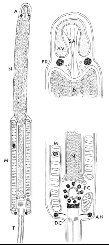
Schematic diagram of the Malacabdella grossa spermatozoon and its acrosomal region and neck region.From: The spermatozoon of the nemertine Malacobdella grossa Bjorn Afzelius J. Submicro. Cytol., 3, 181-192, 1971 (A) Acrosome (AN) Annulus (AV) Acrosomal vesicle (DC) Distal cenriole (M) Mitochondrial sheath (N) Nucleus (PC) Proximal centriole (PR) Post-acrosomal ring (SA) Subacrosomal material (T) TailWith these considerations as a background a fine structural investigation was started using the sperm of the parasitic nemertine Malacobdella grossa (O. F. Müller). The sperm of this species can be characterized as advanced as evidenced by the studies of Retzius (1904) and Franzén (1956). The mode of fertilization is incompletely known. (A) Acrosome (AN) Annulus (AV) Acrosomal vesicle (DC) Distal cenriole (M) Mitochondrial sheath (N) Nucleus (PC) Proximal centriole (PR) Post-acrosomal ring (SA) Subacrosomal material (T) TailWith these considerations as a background a fine structural investigation was started using the sperm of the parasitic nemertine Malacobdella grossa (O. F. Müller). The sperm of this species can be characterized as advanced as evidenced by the studies of Retzius (1904) and Franzén (1956). The mode of fertilization is incompletely known.From: The spermatozoon of the nemertine Malacobdella grossa Bjorn Afzelius J. Submicro. Cytol., 3, 181-192, 1971(A) Acrosome (AN) Annulus (AV) Acrosomal vesicle (DC) Distal cenriole (M) Mitochondrial sheath (N) Nucleus (PC) Proximal centriole (PR) Post-acrosomal ring (SA) Subacrosomal material (T) TailWith these considerations as a background a fine structural investigation was started using the sperm of the parasitic nemertine Malacobdella grossa (O. F. Müller). The sperm of this species can be characterized as advanced as evidenced by the studies of Retzius (1904) and Franzén (1956). The mode of fertilization is incompletely known.
-
-
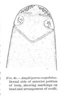
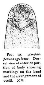
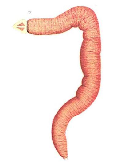
Dorsal view of anterior portion of body showing markings on the head and the arrangement of ocelli.Coe, W. R. (1901). The Nemerteans of the Expedition. Proceedings of the Washington Academy of Sciences, Vol. 3, 1-110.
-
-
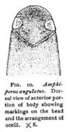
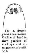
Outline of head to show position of markings and arrangement of ocelli.Coe, W. R. (1901). The Nemerteans of the Expedition. Proceedings of the Washington Academy of Sciences, Vol. 3, 1-110.