
Toxoplasma gondii(tŏk'sə-plāz'mə gŏn'dē-ī') is anobligate,intracellular,parasiticprotozoanthat causes the diseasetoxoplasmosis.[1]
Found worldwide,T. gondiiis capable of infecting virtually allwarm-bloodedanimals.[2]In humans, it is one of the most common parasites;[3]serologicalstudies estimate that up to a third of the global population has been exposed to and may be chronically infected withT. gondii, although infection rates differ significantly from country to country.[4]Although mild, flu-like symptoms occasionally occur during the first few weeks following exposure, infection withT. gondiigenerally produces no symptoms in healthy human adults.[5][6]However, in infants,HIV/AIDSpatients, and others withweakened immunity, infection can cause serious and occasionally fatal illness (toxoplasmosis).[5][6]
Infection in humans and other warm-blooded animals can occur
AlthoughT. gondiican infect, be transmitted by, andasexually reproducewithin humans and virtually all other warm-blooded animals, the parasite cansexually reproduceonly within theintestinesof members of thecat family (felids).[8]Felids are therefore defined as thedefinitive hostsofT. gondii, with all other hosts defined as intermediate hosts.
T. gondiihas been shown to alter the behavior of infectedrodentsin ways thought to increase the rodents' chances of beingpreyedupon by cats.[9][10][11]Because cats are the only hosts within whichT. gondiican sexually reproduce to complete and begin its lifecycle, such behavioral manipulations are thought to beevolutionary adaptationsto increase the parasite'sreproductive success,[11]in one of the manifestations the evolutionary biologistRichard Dawkinsattributes to the "extended phenotype". Although numerous hypotheses exist and are being investigated, the mechanism ofT. gondii–induced behavioral changes in rodents remains unknown.[12]
A number of studies have suggested subtle behavioral or personality changes may occur in infected humans,[13]and infection with the parasite has recently been associated with a number ofneurological disorders, particularlyschizophrenia.[10]However, evidence forcausalrelationships remains limited.[10]
Toxoplasma gondii is an apicomplexan protozoan parasite that infects most species of warm-blooded animals, including humans, and can cause the disease toxoplasmosis. Serologic prevalence data indicate that toxoplasmosis is one of the most common of humans infections throughout the world. A high prevalence of infection in France has been related to a preference for eating raw or undercooked meat, while a high prevalence in Central America has been related to the frequency of stray cats in a climate favoring survival of oocysts and soil exposure. The overall seroprevalence in the United States among adolescents and adults, as determined with specimens collected by the third National Health and Nutrition Examination Survey (NHANES III) between 1988 and 1994, was found to be 22.5%, with a seroprevalence among women of childbearing age (15 to 44 years) of 15%.
The only known definitive hosts for Toxoplasma gondii are members of family Felidae (domestic cats and their relatives). Unsporulated oocysts are shed in the cat’s feces. Although oocysts are usually only shed for 1-2 weeks, large numbers may be shed. Oocysts take 1-5 days to sporulate in the environment and become infective. Intermediate hosts in nature (including birds and rodents) become infected after ingesting soil, water or plant material contaminated with oocysts. Oocysts transform into tachyzoites shortly after ingestion. Thes tachyzoites localize in neural and muscle tissue and develop into tissue cyst bradyzoites. Cats become infected after consuming intermediate hosts harboring tissue cysts. Cats may also become infected directly by ingestion of sporulated oocysts. Animals bred for human consumption and wild game may also become infected with tissue cysts after ingestion of sporulated oocysts in the environment. Humans can become infected by any of several routes:
In the human host, the parasites form tissue cysts, most commonly in skeletal muscle, myocardium, brain, and eyes; these cysts may remain throughout the life of the host. Diagnosis is usually achieved by serology, although tissue cysts may be observed in stained biopsy specimens. Diagnosis of congenital infections can be achieved by detecting T. gondii DNA in amniotic fluid using molecular methods such as PCR.
Toxoplasma gondii is 'n eukariotiese sporosoo wat die katsiekte toksoplasmose veroorsaak.
Die genus Toxoplasma is monotipies. Dit wil sê dat T. gondii die enigste spesie in hierdie genus is.
Die eintlike gasheer van hierdie obligate parasiet is lede van die familie Felidae waartoe die huiskat behoort. Die ongesporuleerde oösiste word in die kat se uitwerpsel uitgeskei. Dit duur 1 tot 5 dae voor die siste sporuleer en besmetlik raak.
Die natuurlike sekondêre gashere is voëls en knaagdiere wat grond, water of plantmateriaal wat besmet is drink of vreet. In die sekondêre gasheer vorm die oösiste tagisoïete wat in die senuwee- en spierweefsel innestel en bradisoïet-siste vorm. Katte vreet hierdie vleis en raak besmet.[1] 'n Deel van die bradisoïete vorm gametosiete wat manlik of vroulik is en seksueel in die kat voortplant.
Mense kan op verskeie maniere besmet raak:
Daar is navorsing wat toon dat die parasiet die gedrag van die besmette sekondêre gasheer beïnvloed, byvoorbeeld dat geïnfekteerde rotte 'n verminderde afkeur vir die kat se urine ondervind, en ook makliker as prooi gevang word deur katte.
Toxoplasma gondii is 'n eukariotiese sporosoo wat die katsiekte toksoplasmose veroorsaak.
Die genus Toxoplasma is monotipies. Dit wil sê dat T. gondii die enigste spesie in hierdie genus is.
Toxoplasma gondii és una espècie de protozou paràsit causant de la toxoplasmosi, una malaltia en general lleu, però que es pot complicar fins a esdevenir fatal, especialment en els gats i en els fetus humans.[1] El gat és el seu hoste definitiu, encara que altres animals homeoterms com els humans també poden allotjar-lo. Per exemple, ha estat aïllat a dofins, llúdries marines,[2] morses,[3] linxs,[4] isards,[5] oques salvatges[6] i diferents tipus de bestiar.[7] També infecta les gavines,[8] les cigonyes i les aus rapinyaires.[9] S'ha observat una alta prevalença de T. gondii entre els pollastres de corral que viuen sobre sòls contaminats i la carn dels exemplars infectats és una font comuna d'adquisició del paràsit.[10] A més, es creu que en aquests animals la presència del paràsit facilita la coccidiosi cecal provocada per un altre protozou patogen, Eimeria tenella, existint in vitro una interacció entre els dos microorganismes.[11] Rattus norvegicus és el rosegador amb l'índex infectiu més elevat i un element d'importància en la disseminació del protozou.[12]
T. gondii es considera l'única espècie vàlida del gènere Toxoplasma. Estudis epidemiològics moleculars, però, han posat de manifest l'existència de com a mínim quatre llinatges clonals en el paràsit, caracteritzats pel seu grau de virulència en múrids de laboratori.[13]
Fou descobert l'any 1908 per diferents investigadors a la sang del rosegador africà Ctenodactylus gundi i a la d'un conill del Brasil, encara que el seu cicle vital complert no es conegué fins 60 anys més tard.[14] La primera prova serològica per detectar-lo va ser creada el 1948.[15]
Aquest protozou és present als cinc continents i s'estima que infecta a un 25-30% de la població mundial. La prevalença de la infecció varia molt entre països i també entre les diferents comunitats que habiten una mateixa regió. La seroprevalença més baixa es dóna a Nord-amèrica, al Sud-est asiàtic, a l'Europa del Nord i al Sàhara. A l'Europa central i del sud la prevalença és moderada, sent la més alta la corresponent a l'Amèrica del Sud i l'Àfrica tropical.[16]
És un paràsit que ha creat diferents rutes potencials de transmissió entre diferents espècies d'hoste i també entre una mateixa espècie. Si T. gondii s'adquireix durant la gestació pot ser transmès verticalment pels taquizoïts al fetus a través de la placenta, ocasionant una toxoplasmosi congènita.[17] Els efectes lesius del protozou sobre el fetus són especialment importants en el cas que la infecció materna tingui lloc en el tercer trimestre d'embaràs,[18] No totes les primoinfeccions per T. gondii en dones gestants comporten una toxoplasmosi neonatal. Segons dades de l'any 2007, un 61% d'elles no transmeten la infecció al fetus. Pel que fa a la resta de casos el 26% dels nounats pateixen una infecció subclínica i el 13% presenta signes clínics aguts (7%) o moderats (6%). La prevalença d'aquestes primoinfeccions varia molt en funció de la zona geogràfica en particular, oscil·lant entre 1 i 14 casos/1.000 embarassos.[19] La hidrocefàlia, les calcificacions cerebrals i els danys oculars són patologies greus pròpies de la infecció congènita pel protozou. La toxoplasmosi també és una de les causes de part preterme, possiblement infradiagnosticada a molts països per manca de programes de detecció.[20] La infecció materna pel paràsit origina nombrosos avortaments espontanis i altera la normal proporció entre els dos sexes de la descendència humana.[21] T. gondii és l'agent patogen responsable de molts casos de retinocoroïditis en nens[22] i adults.[23] Rares vegades, aquest protozou provoca hepatitis agudes.[24] En persones immunodeprimides desencadena infeccions oportunistes molt serioses[25] i que sovint afecten al SNC, com ara encefalitis,[26] meningitis,[27] o abscessos cerebrals,[28] ja que el microorganisme té un marcat neurotropisme i és capaç de travessar la barrera hematoencefàlica.[29] És una causa excepcional de granulomes al tronc de l'encèfal[30] o de mielitis transversa en individus sense problemes d'immunitat.[31] S'ha relacionat la infecció latent pel paràsit amb la gènesi de diversos trastorns neuropsiquiàtrics,[32][33][34] encara que no existeixen evidències concloents fonamentades en estudis poblacionals que confirmin aquest supòsit.[35] Un grup d'investigadors, emprant tècniques proteòmiques i d'espectrometria de masses, ha demostrat en un model murí que la infecció crònica per T. gondii ocasiona canvis neuroinflamatoris que alteren profundament la composició proteínica dels processos sinàptics.[36] Es creu que en animals com ara els ximpanzés[37] o les rates[38] el paràsit és la causa de modificacions del comportament tendents a facilitar la seva propagació.
La transmissió horitzontal d'aquest protozou involucra tres estadis del seu cicle vital. Per exemple, ooquists infecciosos es poden adquirir de medis contaminats (en especial l'aigua)[39] o poden ingerir-se quists tissulars o taquizoïts continguts en la carn o les vísceres de molts animals.
S'ha comprovat que l'aigua és una font important de toxoplasmosis en països tropicals i subtropicals que utilitzen aigües de superfície sense purificar pel consum humà. El brot millor documentat, però, de toxoplasmosi aguda en humans provocat per aigua contaminada fou el de la població de Victoria (Vancouver Island, Canadà), l'any 1995. Estudis epidemiològics retrospectius demostraren que la causa del brot va ser la presència d'ooquists a la xarxa municipal de distribució d'aigua potable.[40]
La transmissió pot ocórrer també per mitjà de taquizoïts presents en productes sanguinis, teixits per trasplantament o llet sense pasteuritzar[41] i formatges artesanals fets amb llet crua. Segons un estudi realitzat amb dades de diversos països europeus corresponents al període 2010-2014, l'índex de mortalitat causat per aquest protozou entre els receptors de trasplantaments en general és d'un 17%, observant-se una menor supervivència en malalts T. gondii seropositius, trasplantats hepàtics i receptors de trasplantament al·logènic de cèl·lules mare hematopoiètiques.[42] En el passat, el consum de carn crua o poc cuita de porc o de xai era la principal via de transmissió als humans. Avui dia, i des de l'increment de les immunodeficiències adquirides, la facilitació de la mobilitat poblacional i l'augment del consum i tràfic de la carn d'animals salvatges (bushmeat), es desconeix quina modalitat de transmissió global és la més important epidemiològicament.[43] Les estimacions de l'OMS indiquen que a Europa es produeixen cada any més d'un milió d'infeccions per T. gondii transmeses per via alimentària.[44] A l'Estat espanyol, la seroprevalença en dones embarassades oscil·la entre el 11 i el 28%, depenent del territori i l'any d'estudi, mentre que la incidència de toxoplasmosi gestacional és del 1,9‰. A hores d'ara la infecció per aquest paràsit no és una malaltia de declaració obligatòria.[45] Les dades d'un estudi de l'any 2010 mostraven una seropositivitat del 19% entre els porcs de granja de Catalunya.[46]
El contacte de les mucoses ocular i bucal amb material portador del paràsit és també una via d'adquisició de la toxoplasmosi que ha estat amb certa freqüència la font infecciosa en treballadors de laboratori (el seu nivell de contenció és 2),[47] jardiners o personal de neteja. També es pot adquirir la infecció toxoplàsmica per via parenteral i s'han registrat múltiples casos de puncions, petites ferides o erosions -produïdes durant la manipulació de teixits o materials portadors del protozou- causants de toxoplasmosis ocupacionals.[48] La inhalació és una via infectiva inusual, però possible. No hi ha constància de la transmissió de T. gondii a través de l'alletament o per contacte humà-humà. Una altra via potencial és la picada de paparres. Aquests àcars es consideren transmissors mecànics del protozou, independentment de la seva fase de vida.[49] A Europa T. gondii ha estat aïllat a Ixodes ricinus[50] i a Dermacentor reticulatus, unes paparres molt comunes a zones boscoses,[51] fet que explicaria la seva presència a certes espècies d'herbívors. No totes les paparres són vectors del paràsit a través de les picades. Per exemple, la sang infectada present a l'espècie oriental Haemaphysalis longicornis no té experimentalment capacitat transmissora; en canvi, la ingestió de l'aràcnid es considera una ruta vàlida d'infecció.[52]
Segons els criteris de la xarxa Euro-FBP,[53] en l'avaluació d'un conjunt de 30 països europeus, T. gondii ocupa el segon lloc en el rànquing de prioritats relacionades amb microorganismes causants de malalties parasitàries transmeses pels aliments.[54]
Pel que fa a la infecció aguda en gestants, un estudi multicèntric europeu atribuí l'adquisició del protozou al consum de carns poc cuites o curades en un 30-63% dels casos investigats i en un 6%-17% al contacte amb sòls contaminats. Un altre motiu relacionat va ser el fet de viatjar fora d'Europa o dels EUA i Canadà. El contacte amb gats no fou un factor de risc d'importància, segons els resultats de dit estudi.[55]
La virulència de les diverses soques del paràsit és variable i depèn de l'expressió o no de certes proteïnes en la paret dels ooquists.[56]
Els taquizoïts i els bradizoïts són sensibles a l'etanol al 70% i a solucions d'1% d'hipoclorit sòdic. L'ooquist és sensible al iode i al formol. Els ooquists s'inactiven a temperatures superiors a 66ºC en menys de 10 minuts. Els taquizoïts s'inactiven a 67ºC i per congelació a -15ºC al menys durant 3 dies o bé a -20ºC al menys durant 2 dies.[57]
Els ooquists poden mantenir en l'aigua de mar el seu potencial infecciós durant molts mesos o un any i no s'ha descobert fins ara cap animal marí amb capacitat d'expulsar-los per ell mateix. Per tant, cal atribuir la seva presència al mar a la contaminació de l'ecosistema aquàtic per ooquists d'origen terrestre, un fenomen afavorit per l'augment de la població en les zones costaneres i de successos meteorològics extrems.[58] La detecció de T. gondii a balenes beluga de regions àrtiques amb molt escassa presència humana o de fèlids i el fet que sigui una espècie que s'alimenta de preses ectotèrmiques (invertebrats i peixos) fa pensat que aquests animals poden actuar com a transmissors marins passius del paràsit.[59]
L'ooquist és la fase esporulada de certs protistes, incloent els gèneres Toxoplasma i Cryptosporidium. Aquest és un estat en el que el microorganisme pot sobreviure per llargs períodes de temps fora de l'hoste per la seva alta resistència a factors del medi ambient, gràcies a la seva doble paret. Dita paret té una organització polimèrica complexa que permet a l'ooquist suportar molts tipus d'agressions físiques i químiques, incloent els raigs UV, l'ozó o les solucions clorades.[60] Algunes de les proteïnes de la paret són específiques d'aquesta fase vital de T. gondii i no estan presents als bradizoïts o als taquizoïts, particularitat que pot ser emprada per millorar la identificació de les espores en mostres ambientals.[61] L'anàlisi proteòmica dels ooquists indica que posseeixen també un grup de proteïnes amb un perfil funcional que fa possible l'adaptació d'aquells a medis extracel·lulars pobres en nutrients mentre arriben a la maduresa.[62] Els ooquists presents al sòl poden ser disseminats mecànicament per les condicions meteorològiques o per artròpodes o anèl·lids.
El bradizoït (del prefix grec bradýs=lent i del sufix zōo=animal) és la forma de replicació lenta del paràsit. No solament de Toxoplasma gondii, sinó també d'altres protozous responsables d'infeccions parasitàries, com ara Neospora caninum[63] o Besnoitia oryctofelisi.[64] A la toxoplasmosi latent (crònica), el bradizoït es presenta en conglomerats microscòpics envoltats per una paret quística, en el múscul i/o el teixit cerebral infectat.[65] Els bradizoïts presenten molts trets ultraestructurals que els diferencien dels taquizoïts: tenen un nucli situat a l'extrem posterior, roptries sòlides (un tipus d'orgànul secretor especial) nombrosos micronemes i grànuls d'amilopectina.[66] Als bradizoïts no es veuen cossos lipídics, mentre que són abundants en els esporozoïts dels ooquists i apareixen ocasionalment en els taquizoïts. Els bradizoïts són PAS+ i els taquizoïts no.[67]
Els taquizoïts són formes de vida latent que formen quists en teixits infestats pel toxoplasma o altres paràsits (per exemple l'esmentat Neospora caninum, un coccidi que té moltes similituds estructurals amb T. gondii). Els taquizoïts es troben en vacúols dins de les cèl·lules infestades. Són estructures fràgils, que no resisteixen la dessecació ni l'ebullició i sensibles a molts desinfectants (per exemple hipoclorit de sodi al 1% o etanol al 70%). Els quists tissulars s'inactiven per congelació a -15ºC al menys durant 3 dies o a -20ºC al menys 2 dies. Poden sobreviure fins a tres setmanes a 1-4°C.[68] S'ha observat in vitro i in vivo que l'extret de taquizoïts té propietats immunomoduladores contra la sensibilització al·lèrgica i la inflamació de vies aèries.[69]
Algunes de les de les qüestions claus relacionades amb aquesta fase del cicle de T. gondii, encara no aclarides del tot pels investigadors, són els múltiples factors intrínsecs i extrínsecs que condicionen el moment i el mecanisme de sortida dels taquizoïts de la cèl·lula hoste.[70] Se sap que els taquizoïts es poden desplaçar per lliscament gràcies a moviments de rotació helicoïdal en sentit horari, producte de successives torsions de la seva membrana externa.[71]
La licocalcona A (un tipus de calcona existent a les arrels de les plantes del gènere Glycyrrhiza), inhibeix experimentalment la proliferació dels taquizoïts de la soca virulenta RH[72] de T. gondii.[73] Un nou decapèptid sintètic, anomenat KP, indueix la mort dels taquizoïts a través de fenòmens apoptòtics, sense provocar danys en les cèl·lules hoste.[74]
L'àcid ursòlic mostra una alta activitat contra T. gondii en murins infectats amb taquizoïts del paràsit, ja que inhibeix eficaçment el seu procés proliferatiu al bloquejar la formació de vacúols parasitòfors en la membrana de les cèl·lules hoste.[75]
El cicle de vida del T. gondii té dues fases. La fase sexual del cicle de vida passa només en membres de la família Felidae (gats domèstics i salvatges), fent que aquests animals siguin els hostes primaris del paràsit. En gats domèstics, la seroprevalença de T. gondii varia segons les races i l'edat de l'animal, a banda del tipus de menjar que ingereix.[76] La fase asexual del cicle de vida pot ocórrer en qualsevol animal de sang calenta, com ara altres mamífers i aus. Per això, la toxoplasmosi és una zoonosi parasitària.[77]
En l'hoste intermediari, incloent els felins, els paràsits envaeixen cèl·lules, formant un compartiment anomenat vacúol parasitòfor[78] que contenen bradizoïts, la forma de replicació lenta del paràsit.[79] Els vacúols formen quists en els teixits, especialment en els músculs i el cervell. Com que el paràsit està dins de les cèl·lules, el sistema immunitari de l'hoste no detecta aquests quists. La resistència als antibiòtics varia, però els quists són difícils d'erradicar totalment. El T. gondii es propaga dins d'aquests vacúols per una sèrie de divisions binàries fins que la cèl·lula infestada eventualment es trenca, alliberant als taquizoïts. Aquests tenen motilitat i constitueixen la forma de reproducció asexual del paràsit. A diferència dels bradizoïts, els taquizoïts lliures són eficaçment eliminats per la immunitat de l'hoste, tot i que alguns aconsegueixen infectar altres cèl·lules formant bradizoïts, mantenint així el cicle de vida d'aquest paràsit. De forma contrària als taquizoïts dins dels vacúols parasitòfors, els bradizoïts dels quists tissulars mostren una gran heterogeneïtat fisiològica i de capacitat de replicació. El fet que els quists tissulars tinguin mides diferents suggereix que són estructures de creixement dinàmic amb un paper actiu en la infecció crònica per T. gondii i no solament quists en estat de repòs i metabòlicament inerts.[80]
Els quists tissulars són ingerits per el gat (per exemple, a l'alimentar-se d'un ratolí infectat). Si l'animal no pateix un dèficit immunitari no presenta cap alteració clínica. En cas contrari, el paràsit afecta predominantment el SNC, els músculs, el pulmons i els ulls del felí, sent la clindamicina el tractament electiu. Més del 50% dels gats, sobretot els que volten lliurament, mostren en els exàmens serològics anticossos que indiquen infecció i presència de quists de T. gondii. Els gats amb anticossos positius, però, no estan en la fase d'alliberament d'ooquists i no representen un risc zoonòtic.[81] Els quists sobreviuen el pas per l'estómac del gat i els paràsits infecten les cèl·lules epitelials de l'intestí prim, en les quals té lloc la reproducció sexual i la formació d'ooquists que són alliberats amb la femta entre els 3-10 dies posteriors a la ingesta dels quists i que es poden mantenir viables a l'aire lliure 46 dies a temperatures d'entre 6-36ºC.[82] La femta fresca no és pròpiament infecciosa, ja que els ooquists necessiten un o dos dies per esporular-se després del canvi de medi. Altres animals, incloent-hi els humans ingereixen els ooquists (en menjar vegetals no rentats adequadament) o els quists tissulars al menjar carn crua o cuita de forma insuficient. Els paràsits entren als macròfags de la paret intestinal per després distribuir-se per la circulació sanguínia i el cos sencer.
Si bé existeixen vacunes que produeixen un cert nivell d'immunitat contra T. gondii en gats, encara no s'ha aconseguit una vacuna d'ús humà.[83] Vacunes creades a partir d'una soca determinada del paràsit, la S48, han donat bons resultats reduint la formació de quists en la carn d'ovelles i porcs.[84] Una línia d'especial interès dins d'aquest camp de recerca és la del disseny de vacunes d'ADN basades en proteïnes produïdes pels micronemes (un tipus de orgànuls secretors existents a l'apicomplex del paràsit).[85]
Un grup d'investigadors xinesos realitza a hores d'ara assajos en ratolins amb una vacuna anomenada HSP60 DNA. Els resultats preliminars indiquen que podria ser una candidata vàlida pel desenvolupament d'un adequat grau de protecció contra la toxoplasmosi humana aguda i crònica.[86]
Habitualment, la detecció de T. gondii requereix procediments relativament lents i costosos. S'ha assajat un nou test que utilitza la tècnica d'immunocromatrografia de flux lateral per identificar d'una forma ràpida, eficaç i econòmica la presència del patogen en sang total amb una senzilla punxada al dit.[87] Ha estat dissenyada una plataforma automàtica de rastreig d'imatge d'alt contingut[88] que permet avaluar l'acció inhibitòria de noves molècules amb potencial terapèutic sobre la proliferació del paràsit.[89]
Toxoplasma gondii és una espècie de protozou paràsit causant de la toxoplasmosi, una malaltia en general lleu, però que es pot complicar fins a esdevenir fatal, especialment en els gats i en els fetus humans. El gat és el seu hoste definitiu, encara que altres animals homeoterms com els humans també poden allotjar-lo. Per exemple, ha estat aïllat a dofins, llúdries marines, morses, linxs, isards, oques salvatges i diferents tipus de bestiar. També infecta les gavines, les cigonyes i les aus rapinyaires. S'ha observat una alta prevalença de T. gondii entre els pollastres de corral que viuen sobre sòls contaminats i la carn dels exemplars infectats és una font comuna d'adquisició del paràsit. A més, es creu que en aquests animals la presència del paràsit facilita la coccidiosi cecal provocada per un altre protozou patogen, Eimeria tenella, existint in vitro una interacció entre els dos microorganismes. Rattus norvegicus és el rosegador amb l'índex infectiu més elevat i un element d'importància en la disseminació del protozou.
T. gondii es considera l'única espècie vàlida del gènere Toxoplasma. Estudis epidemiològics moleculars, però, han posat de manifest l'existència de com a mínim quatre llinatges clonals en el paràsit, caracteritzats pel seu grau de virulència en múrids de laboratori.
Fou descobert l'any 1908 per diferents investigadors a la sang del rosegador africà Ctenodactylus gundi i a la d'un conill del Brasil, encara que el seu cicle vital complert no es conegué fins 60 anys més tard. La primera prova serològica per detectar-lo va ser creada el 1948.
Aquest protozou és present als cinc continents i s'estima que infecta a un 25-30% de la població mundial. La prevalença de la infecció varia molt entre països i també entre les diferents comunitats que habiten una mateixa regió. La seroprevalença més baixa es dóna a Nord-amèrica, al Sud-est asiàtic, a l'Europa del Nord i al Sàhara. A l'Europa central i del sud la prevalença és moderada, sent la més alta la corresponent a l'Amèrica del Sud i l'Àfrica tropical.
Toxoplasma gondii (Nicolle et Manceaux, 1908) je vnitrobuněčný parazitický prvok, vícehostitelská kokcidie, která parazituje v buňkách člověka i zvířat. Jejím definitivním hostitelem jsou kočkovité šelmy, mezihostitelem se může stát většina teplokrevných živočichů včetně člověka.[1] Způsobuje toxoplazmózu, u těhotných žen hrozí při infekci tímto prvokem narození postiženého dítěte. U HIV pacientů může způsobit akutní zánět mozku.[2] Rovněž může způsobit aborty u ovcí a koz.[3]
V trusu koček se ven dostávají tzv. oocysty, které následně sporulují a vydrží v prostředí velmi dlouhou dobu. Když je však požije s kontaminovanou potravou nějaký teplokrevný živočich, z oocyst vzniknou tzv. tachyzoiti (řec. tachos – rychlost), kteří rychle zaplaví nervovou a svalovou tkáň a postupně z nich vzniknou bradyzoiti (řec. brady – pomalý), kteří v mezihostiteli vydrží pravděpodobně celý život v zapouzdřeném stavu. Kočka (definitivní hostitel) se může nakazit několika způsoby: buď tím, že sežere maso obsahující bradyzoity, nebo někdy i potravou obsahující oocysty. Ve střevě kočky dochází k množení a tvorbě oocyst ve střevních buňkách.[1][4]
Toxoplasma gondii využívá specifický způsob pohybu (lze pozorovat například i u jiného druhu z kmene Apicomplexa Plasmodium falciparum), nazývaný "gliding motility", v překladu klouzání. Tento druh pohybu je založený na aktino-myosinovém komplexu zakotveném v tzv. "inner membrane complex", což je dvojice membrán situovaná pod parazitovu plazmatickou membránu. Tento komplex se přes transmembránové proteiny vylučované z jeho sekrečních organel váže na substrát a umožňuje dynamický pohyb beze změny tvaru parazita.[5]
Tachyzoit je oproti bradyzoitu mohutnější a má centrálně situované, dobře patrné jádro. Bradyzoit má jádro spíše v posteriorní části.
Klíčová je přítomnost apikálního komplexu, což je soubor organel na anteriorním pólu zoitu, který hraje nezastupitelnou roli v invazi do hostitelských buněk. Zahrnuje konoid, což je trubicovitá mikrotubulární struktura, jejíž báze je obrkoužena apikálními prstenci a středem prochází dva mikrotubuly. Mezi sekreční organely patří mikronemy a kyjovité rhoptrie. Pro Toxoplasmu je typická přítomnost organely zvané apikoplast, která vznikla endosymbiózou a obsahuje vlastní kruhovou DNA.[6]
Čerstvý oocyst se v mikroskopu jeví jako koule s mnoha jemnými skvrnkami. Přibližně za 48-72 hodin dozraje do podoby kuličky velké 10-12 mikrometrů, ve které jsou dobře zřetelné dvě sporocysty.
V hostiteli se vylíhne podlouhlý prvok dlouhý 4-8 a silný 2-3 mikrometry, se zužujícím se předním koncem a tupým zadním. Uvnitř je dobře patrné jádro.
Zralé cysty ve tkáni jsou 5-50 mikrometrů velké. V mozku mají kulový tvar, v srdečních a kosterních svalech podlouhlý.
Člověk je pro Toxoplasmu náhodným mezihostitelem a v podstatě se Toxoplasma z člověka nemá šanci dostat do kočky jako definitivního hostitele a zakončit tak svůj životní cyklus. Lidé se obvykle nakazí nedostatečně tepelně zpracovaným masem (požitím bradyzoitů) nebo požitím oocyst z prostředí.[1] Infekce prvokem T. gondii není u lidí nijak vzácná: např. ve Francii nebo v Německu se Toxoplasmou v průběhu života nakazí 80 % populace, v Česku asi třetina obyvatel, v rámci celého světa přibližně 50 %.[7] Toxoplasmóza je však obvykle bez viditelných příznaků, může se vyskytnout asymptomatická generalizovaná lymfadenopatie. V největším nebezpečí jsou těhotné ženy, které se do té doby s parazitem nesetkaly – hrozí riziko poškození plodu.[1] U imunokompromitovaných osob (např. AIDS) může toxoplazmóza vyvolat závažné poškození CNS.
Udává se, že T. gondii může manipuluvat s lidským chováním. Lidé s Toxoplasmou reagují pomaleji v kritických situacích – pramení to z prosté skutečnosti, že Toxoplasma „ví“, že myši s pomalejšími reakcemi se snadno stanou úlovkem koček. Toxoplazmóza (především u Rh-negativních lidí) podle některých výzkumů zvyšuje pravděpodobnost, že nakažený člověk se – v důsledku pomalejších reakcí – stane např. účastníkem dopravní nehody[7][8] apod. Tomuto výzkumu však odporují jiné studie, která souvislosti mezi nákazou toxoplazmózou a např. právě účastí v dopravních nehodách (a řadou dalších způsobů chování a osobnostních charakteristik) neprokázaly[9], jiné byly z větší části bez pozitivních výsledků.[10][11] Nelze proto tyto teorie považovat za stoprocentně spolehlivé.
Toxoplasma gondii (Nicolle et Manceaux, 1908) je vnitrobuněčný parazitický prvok, vícehostitelská kokcidie, která parazituje v buňkách člověka i zvířat. Jejím definitivním hostitelem jsou kočkovité šelmy, mezihostitelem se může stát většina teplokrevných živočichů včetně člověka. Způsobuje toxoplazmózu, u těhotných žen hrozí při infekci tímto prvokem narození postiženého dítěte. U HIV pacientů může způsobit akutní zánět mozku. Rovněž může způsobit aborty u ovcí a koz.
Toxoplasma er en slægt af parasitiske protozoer. Arten Toxoplasma gondii forårsager sygdommen toxoplasmose også kaldet haresyge. Den har tre udviklingsstadier, oocyster, vævscyster og tachyzoiter. Parasitten er udbredt i hele verden undtagen i de arktiske egne og kan smitte de fleste fugle og pattedyr, inklusiv mennesker. De formerer sig kønsløst i forskellige vævstyper. Hovedværten for Toxoplasma gondii er katten, hvor parasittens seksuelle stadier forekommer.
Katten er hovedvært, og er den eneste dyreart der udskiller oocyster med fæces. Oocysterne er meget resistente, og kan findes spredt i omgivelserne, f.eks. i jorden, sandkasser, og græs. Oocysterne kan også findes indenfor i huset eller i stalden.
Når en kat bliver inficeret, udskiller den op til 10 millioner oocyster over en periode på 2 uger. Oocysterne bliver infektiøse 1-5 dage efter udskillelsen. De kan spredes med overfladevand og kan overleve i op til et år. Dette kan forklare hvorfor kontakt med jord og vand udgør en større smitterisiko end kontakt med katte.
Oocysterne i kattens tarmsystem er det seksuelle stadie hos Toxoplasma gondii. De hunlige kønsceller er halvrunde og hver celle har en centralt placeret kerne. De hanlige kønsceller er ægge- eller elipseformede og bruger deres flageller til at svømme hen til de modne hunlige kønsceller, penetrere dem og dermed lave en zygote. Efter befrugtning dannes en oocyst-væg omkring parasitten og toxoplasma-ægget er dannet. I hver oocyst (toxoplasma-æg) findes to sporozyster, og i hvert sporocyst findes 4 sporozoiter. Sporozoiterne minder strukturelt meget om tachyzoiterne, men har nogle lidt andre organeller.
Hovedsmittekilden i vores del af verden er vævscyster i muskulatur og organer fra tidligere Toxoplasma smittede dyr, specielt fra svin, lam og vildt, og i mindre grad fra oksekød og kyllinger. Cysterne findes i størst mængde i ikke skeletmuskulatur. Indtagelse af råt/ikke gennemstegt kød er en væsentlig risokofaktor. Cysterne bliver destrueret ved opvarmning til mere end 66 °C i 3 min, og ved nedfrysning til -20 °C. Rygning, saltning og bestråling vil også destruere cysterne. Vævcysten sidder inde i muskelcellen, og vokser i takt med at parasitten, som i dette stadie hedder bradyzoit (brady = langsomt på græsk) deler sig ved aseksuel formering. Sidder de i hjernen, er de som oftes sfæriske og maximalt 70 µm i diameter, mens vævscyster i musklerne er aflange og op til 100 µm lange. Desuden findes cysterne også i andre former for væv, men dette er ikke helt så almindeligt. Hvis cysten bevares intakt, kan den opholde sig i værten hele dennes liv uden at forårsage betændelsestilstande.
I tachyzoite-stadiet formerer organismen sig hastigt i en hvilken som helst celle i en mellemvært, samt overflade væv der ikke sidder i tarmen hos den ægte vært. Tachyzoiten er halvmåneformet, og 2-6 µm lang. Selvom tachyzoiter kan glide, bøje og rotere, har den ingen synlige midler til at sætte sig i bevægelse, som f.eks. cilia eller flageller.
Tachyzoiter trænger ind i værtscellen ved aktivt at gennembore dennes plasmalemma. Når parasitten er inde i cellen, bliver den æggeformet, og omgives af en parasittisk væskefyldt celleblære, som tilsyneladende skabes af både parasitten selv og værtscellen. Herefter ligger tachyzoiten i dvale i en periode før den begynder at formere sig aseksuelt gentagende gange, indtil værtscellen ikke længere har plads til alle parasitterne, hvorved den sprænges. Længden af dvaleperioden, hastigheden af formeringen samt størrelsen af tilvæksten af de enkelte tachyzoiter afhænger både af hvilken art Toxoplasma der er tale om, samt typen af vært.
Tachyzoiten adskiller sig rent strukturelt kun fra bradyzoiten ved det at cellekernen sidder centralt i cellen, mens den i bradyzoiten sidder forskudt mod bagenden. Med hensyn til livscykus, er bradyzoitens dvaleperiode før den udvikles til ooyster efter indtagelse af katten kortere (3-10 dage) end hvis katten indtager tachyzoiter (≥14 dage).
Hos voksne er toxoplasmose sjældent alvorligt,[kilde mangler] men angribes gravide kvinder, kan parasitten overføres til fosteret. Parasitten er i nogle tilfælde årsag til dødfødte eller misdannede børn, der eventuelt får en begrænset levetid, bliver blinde eller hjerneskadede.[kilde mangler]
I Danmark er der set en sammenhæng mellem toxoplasmose hos kvinder og en omkring 50% øgning i selvmordsforsøg.[1] Man har også konstateret en øget risiko for involvering i færdselsuheld hos inficerede personer.[2]
Da parasitten er ret udbredt, bør kvinder, der enten er gravide eller planlægger at blive det i nær fremtid, ikke rengøre kattebakker, slagte og istandgøre vildt eller arbejde med dissektion af dyr.[kilde mangler]
Der er mange ideer om hvordan toxoplasmose påvirker værtsdyret. Nogle forskere har vist at rotter der er inficeret med Toxoplasma bliver påvirket af parasitten på en måde der gør dem tiltrukket af katte, hvilket jo er meget belejligt for parasitten som gerne vil have sit værtsdyr ædt af en kat, så den selv får mulighed for at formere sig.[3] Andre forskere har trukket paralleller mellem toxoplasmose og skizofreni, og andre igen har forsøgt påvist en sammenhæng mellem infektionen og antallet af fødte drengebørn, som i følge forskerne stiger i grupper af inficerede. I dyr er der evidens for at både oprættede svin[4] og vildsvin[5] i Danmark kan være inficeret med T. gondii.
Toxoplasma er en slægt af parasitiske protozoer. Arten Toxoplasma gondii forårsager sygdommen toxoplasmose også kaldet haresyge. Den har tre udviklingsstadier, oocyster, vævscyster og tachyzoiter. Parasitten er udbredt i hele verden undtagen i de arktiske egne og kan smitte de fleste fugle og pattedyr, inklusiv mennesker. De formerer sig kønsløst i forskellige vævstyper. Hovedværten for Toxoplasma gondii er katten, hvor parasittens seksuelle stadier forekommer.
Toxoplasma gondii (von altgriechisch τόξον tóxon ‚Bogen‘ und πλάσμα plásma ‚Gebilde‘) ist ein bogenförmiges Protozoon mit parasitischer Lebensweise. Sein Endwirt sind Katzen, als Zwischenwirt dienen andere Wirbeltiere. Es ist der einzige bekannte Vertreter der Gattung Toxoplasma. Der Parasit ist Verursacher der Toxoplasmose und nahe verwandt mit Plasmodium, dem Erreger der Malaria, und mit Cryptosporidium.
Toxoplasma gondii wurde erstmals 1907 in Tunesien als Parasit im Gundi (Ctenodactylus gundi) entdeckt und als Angehöriger der Apicomplexa identifiziert.[1] Aufgrund der Halbmondform wurde es von den Entdeckern Charles Nicolle und Louis Manceaux als Toxoplasma (griechisch toxon, Bogen; plasma, Gebilde) und aufgrund des Wirtstieres als Toxoplasma gondii benannt. Erst viel später konnte es auch beim Menschen als Krankheitserreger gefunden werden, die von ihm ausgelöste Krankheit wurde Toxoplasmose genannt. 1948 entwickelten Sabin und Feldman einen serologischen Test auf der Basis von Antikörpern, den sie Dye-Test nannten.[2] Mit Hilfe dieser Methode konnte festgestellt werden, dass Toxoplasma gondii weltweit verbreitet ist und beim Menschen sehr häufig vorkommt.
Der Parasit ist weltweit verbreitet, die Bevölkerung weist eine hohe Durchseuchung auf, da die Infektion meist ohne Symptome verläuft.[3]
Bei 50 % der Bevölkerung in Deutschland wurden Antikörper nachgewiesen. Mit zunehmendem Alter steigt die Infektionswahrscheinlichkeit an. Bei über 70-Jährigen liegt sie bei über 70 %.[4]
Toxoplasma gondii unterscheidet sich je nach Stadium sowohl in der Form als auch in der Größe. Die Zellen der freien und infektiösen Form sind in flüssigen Medien oder Frischpräparaten bogenförmig und erreichen Größen von zwei bis fünf Mikrometern. Betrachtet man sie in Gewebeproben oder fixierten Schnitten, erscheinen sie dagegen eiförmig oval. Außerdem können sie sowohl einzeln als auch zu mehreren in so genannten Pseudozysten im Gewebe vorkommen.
Die Oozysten messen bis zu 11 Mikrometer, die Gewebszysten bis zu 300 Mikrometer. Es bilden sich zwei verschiedene Populationen von Sporozoiten, die Tachyzoiten bilden sich nach dem Eindringen in den Zwischenwirt und vermehren sich dort rapide. Später treten Bradyzoiten auf, deren Vermehrung stark verlangsamt ist. Strukturell unterscheiden sich diese beiden Formen nicht.
Die Oozysten werden vom Endwirt (Katzenartige) mit dem Kot ausgeschieden und gelangen so in den Zwischenwirt. Sie enthalten zwei Sporozysten mit je vier Sporozoiten. Sie können sehr lange (bis fünf Jahre) infektiös bleiben und überstehen Frost, sind jedoch nicht sehr hitzeresistent. Im Zwischenwirt (Wirbeltiere, z. B. Vögel) schlüpfen die Sporozysten, diese dringen nun aktiv in kernhaltige Zellen des Zwischenwirtes ein (vor allem Lymphknoten, Retikuloendotheliales System). Nun setzt eine Vermehrung durch ungeschlechtliche Teilung ein, bei der sich zwei Tochterzellen von der Mutterzelle ablösen, wobei die Mutterzelle sich auflöst (Endodyogenie). Dieser Vorgang läuft so lange ab, bis die Wirtszelle komplett ausgefüllt ist und aufplatzt, so dass die Tachyzoiten (griechisch tachys = schnell) frei werden. Dieser Vorgang wiederholt sich alle 6 Stunden. Die Tachyzoiten breiten sich nach der Freisetzung im Blut aus und gehen so auch über die Plazenta ins Blut der Nachkommen über. Nachdem die Wirtsabwehr eingesetzt hat, verlangsamt sich die Teilungsdauer, und man spricht nun von Bradyzoiten (griech. bradys = langsam). Es bilden sich in den Zellen Gewebezysten, die vor allem in der Muskulatur, aber auch im Gehirn oder in der Netzhaut des Auges latent überdauern. In dieser Form werden sie wiederum von der Katze, die den Zwischenwirt frisst, aufgenommen. Die Bradyzoiten werden im Darm frei und dringen in die Epithelzellen ein. Dort findet eine Schizogonie (ungeschlechtliche Fortpflanzung) statt und/oder es werden Makrogamonten und Mikrogamonten gebildet. Die Makrogamonten bilden Makrogameten (vergleichbar mit einer Eizelle) aus, während ein Mikrogamont Mikrogameten erzeugt (vergleichbar mit Spermien). Der Makrogamet wird von einem Mikrogameten befruchtet, und es kommt zur Bildung einer Zygote (diploid), welche dann zu einer unsporulierten Oozyste reift. Diese wird mit dem Kot ausgeschieden und reift in der Außenwelt in 2 bis 4 Tagen unter Sauerstoffeinfluss zu infektionsfähigen sporulierten Oozysten heran. Sie ist bis zu 5 Jahre infektionsfähig. Falls die Oozysten von Katzen aufgenommen werden, so entwickeln sich Tachyzoiten, Bradyzoiten und Gewebezysten. Diese verbleiben jedoch nur zu einem geringen Teil im Gewebe und wandern in das Darmepithel der Katze ein, wo sie erneut durch Schizogonie und Gamogonie Oozysten ausbilden. Der Lebenszyklus wird in drei Phasen unterteilt, 1. in die extraintestinale Phase, 2. in die externe Phase und 3. in die enteroepitheliale Phase.
Beim Menschen ruft dieser Parasit die Krankheit Toxoplasmose hervor. Er kann T. gondii in beiden Formen aufnehmen, sowohl als Zysten in halb rohem Fleisch als auch als Schmierinfektion mit Katzenkot. Er übernimmt dann die Rolle des Zwischenwirtes, das heißt die Erreger durchdringen die Darmwand, um so in der Muskulatur, aber auch in anderen Organen Zysten zu bilden, die lebenslang überdauern. Die meisten Menschen machen irgendwann einmal diese Infektion durch, sie bleibt meistens ohne Symptome. Es kann einige Monate lang zu grippeähnlichen Beschwerden wie Fieber, Gelenk- und Muskelschmerzen und beispielsweise Lymphknotenschwellungen kommen.
Bei Nagetieren konnten durch Toxoplasma verursachte Verhaltensänderungen nachgewiesen werden. So verlieren infizierte Tiere ihre angeborene Scheu gegenüber dem Geruch von Katzen, was dem Lebenszyklus von Toxoplasma förderlich ist.[5] Bei Mäusen bleibt der Verlust der Scheu gegenüber dem Geruch von Katzenurin auch nach einer ausgeheilten Infektion mit T. gondii erhalten.[6] Bei mit T. gondii infizierten Ratten ist im limbischen System die Aktivität in den Regionen erhöht, die für die sexuelle Anziehung verantwortlich sind, wenn die Tiere dem Geruch von Katzenurin ausgesetzt werden. Dies wird als möglicher Mechanismus diskutiert, warum infizierte Nagetiere ihre Scheu vor Katzen verlieren.[7]
Beim Menschen werden durch Toxoplasmainfektionen möglicherweise verursachte Verhaltensänderungen immer wieder diskutiert.[3][8][9][10][11] Eine Infektion könnte beispielsweise die Risikobereitschaft erhöhen.[12]
Schwangere sollten kein Fleisch essen, das nicht durchgebraten ist, möglichst nicht mit Katzenkot in Berührung kommen und nicht im Garten arbeiten. Notfalls schützen Handschuhe oder Händewaschen vor den Mahlzeiten.[4] Es ist sinnvoll, dass eine andere Person das Katzenklo täglich reinigt, weil die Oozysten erst frühestens zwei Tage nach Ausscheidung infektiös werden.
Bis und mit 2015 existiert keine für Menschen zugelassene Impfung gegen Toxoplasma gondii.[13]
Bezüglich der Immunisierung von Schafen ist aber seit einiger Zeit ein Lebendimpfstoff mit Handelsnamen Toxovax (MSD Animal Health) verfügbar. Dieser bietet einen lebenslangen Schutz vor Toxoplasma gondii.[14]
Eine Infektion lässt sich normalerweise am leichtesten durch immunologische Testverfahren (ELISA, Immunfluoreszenztest, ISAGA) nachweisen, also Nachweis von spezifischen Antikörpern. Dabei sprechen IgM- Antikörper für eine frische Infektion, IgM- und IgG- Antikörper zusammen für eine Infektion innerhalb der letzten eineinhalb Jahre. Liegen sowohl IgG- als auch IgM-Antikörper vor, hilft ein sogenannter Aviditätstest beim Ausschluss einer frischen Infektion.
Es stehen auch molekularbiologische Untersuchungen (PCR) zur Verfügung. Sie eignen sich zur Untersuchung von Fruchtwasser zum Nachweis einer bereits erfolgten Übertragung auf das ungeborene Kind. Eine Schädigung des Kindes kann man durch Ultraschall diagnostizieren. Auch bei immungeschwächten Patienten eignet sich am ehesten die PCR oder Sichtbarmachung bereits entstandener größerer Läsionen mittels bildgebender Verfahren (CT, MRT).
T. gondii gehört zu den Infektionen, auf die man bei Schwangeren routinemäßig testen sollte, ähnlich wie Röteln, Syphilis, Hepatitis B, Chlamydien, HIV, eventuell Zytomegalie. Die Untersuchung ist in Deutschland jedoch nicht Bestandteil der normalen Schwangerenvorsorge. Wenn schon früher einmal eine Infektion auf T. gondii nachgewiesen wurde, geht davon keine Gefahr mehr aus. Das ungeborene Kind ist dann während der Schwangerschaft durch die mütterlichen Antikörper vor einer Infektion geschützt.
Die Diagnose kann sehr schwierig werden, wenn sie im Nachhinein bei einem Neugeborenen gestellt werden muss, das erst spät Krankheitszeichen zeigt (beispielsweise Erblindung durch Chorioretinitis).
Eine Erstinfektion mit T. gondii während der Schwangerschaft sollte mit Antibiotika behandelt werden. Ansonsten kann eine Behandlung sinnvoll sein, wenn der Patient Symptome zeigt. Bewährt hat sich die Kombination Pyrimethamin zusammen mit einem Sulfonamid oder Clindamycin. Vor der 18. Schwangerschaftswoche wird alternativ ein Makrolidantibiotikum, z. B. Spiramycin, verabreicht, da dieses – im Gegensatz zu erstgenannten – wahrscheinlich keine Fehlbildungen beim ungeborenen Kind auslöst. Allerdings ist man nicht sicher, wie wahrscheinlich eine Übertragung während der Frühschwangerschaft überhaupt ist. Eventuell sollte das Kind nach der Geburt noch einige Zeit nachbehandelt werden. Alles in allem kann eine Schädigung des Kindes durch diesen sehr häufigen Parasiten also meistens verhindert werden.
Toxoplasma gondii (von altgriechisch τόξον tóxon ‚Bogen‘ und πλάσμα plásma ‚Gebilde‘) ist ein bogenförmiges Protozoon mit parasitischer Lebensweise. Sein Endwirt sind Katzen, als Zwischenwirt dienen andere Wirbeltiere. Es ist der einzige bekannte Vertreter der Gattung Toxoplasma. Der Parasit ist Verursacher der Toxoplasmose und nahe verwandt mit Plasmodium, dem Erreger der Malaria, und mit Cryptosporidium.
Toxoplasma gondii is an obligate intracellular, parasitic protozoan that causes the disease toxoplasmosis.[3]
Toxoplasma gondii ya iku spesies organisme sel tunggal (protozoa) kang urip minangka parasit.[3] Organisme iki sepisanan diidentifikasi déning Nicolle lan Manceaux.[4][5] Toxoplasma gondii nyebabaké toksoplasmosis, lelara kang bisa nyerang makhluk berdarah panas, kalebu manungsa. Kéwan Felidae kaya déné kucing kampung minangka inang definitif kang dadi panggonan T. gondii nindakaké réprodhuksi seksual. Lelara iki ora duwé tandha-tandha kang cetha. Uji TORCH bisa mangertèni anané infèksi lan lelara iki bisa diobati nganti pasièn mari.
Toxoplasma gondii ya iku spesies organisme sel tunggal (protozoa) kang urip minangka parasit. Organisme iki sepisanan diidentifikasi déning Nicolle lan Manceaux. Toxoplasma gondii nyebabaké toksoplasmosis, lelara kang bisa nyerang makhluk berdarah panas, kalebu manungsa. Kéwan Felidae kaya déné kucing kampung minangka inang definitif kang dadi panggonan T. gondii nindakaké réprodhuksi seksual. Lelara iki ora duwé tandha-tandha kang cetha. Uji TORCH bisa mangertèni anané infèksi lan lelara iki bisa diobati nganti pasièn mari.
Toxoplasma gondii is an obligate intracellular, parasitic protozoan that causes the disease toxoplasmosis.
టాక్సోప్లాస్మా (లాటిన్ Toxoplasma ఒక వ్యాధి కారక జీవుల ప్రజాతి.[1] వీనికి ప్రాథమిక అతిధేయి పిల్లి అయినా పక్షులు, క్షీరదాల వంటి చాలా రకాల జంతువులకు సంక్రమిస్తుంది.[2] వీని వలన కలిగే వ్యాధిని టాక్సోప్లాస్మోసిస్ (Toxoplasmosis) అంటారు.
Al Toxoplàśma góndi (nóm sientìfic “Toxoplasma gondii”) 'l è 'n parasìta protozoo prutìsta dal gat (òspit definitìṿ) e 'd atri bèsti a sangṿ cald cuma i mamìfar (sórag, nimài e via dascurénd), gl'uśèi e 'l óm ch'al pōl dvintàr al sò òspit intermèdi quànd al s bèca la Toxoplaśmóśi. Anc al gat al pōl śugàr al rōl d'l òspit intermèdi cumpàgn a chi àtar mamìfar s'al magna di oocìsti stramnàdi in dla mèrda invéci che dal cìsti da la carna dal sò vitmi.
Al parasìta al fà part dla famìja di Sarcocystidae e 'l è la sōla spécie dal gènar Toxoplasma ch'a sa cgnós a 'l dè 'd incō.
Al cicclo 'd vita dal Toxoplàśma góndi al taca quànd al gat al s infèta magnànd dla carna cun dèntar dal cisti. In dal stómag i sûg gàstric i dìsfan la cisti librànd acsè 'l parasìta ch'al pasa in dal sò budèli in dua dvintâ adûlt al s multìplica cun la riprudusiòṅ sesuàla faghénd di oocìsti ch'i miśùran 10x12 micron e ch'i vènan cagàdi via c'n i pét. Qvést a sucéd sōl p'r una o dū stmani dòp l'infesiòṅ. Fóra dal còrp dal gat i gh métan dū dè a maduràr e dvintàr bòni p'r infetàr 'l òspit intermèdi.
Quànd chi lò al i manda śò catànd-i in gir o magnànd dla carna infèta (cun dal cisti dèntar cum a pōl far 'l óm) al s bèca 'l mâł. I parasìta i śbuśìs'n al budèli e i pàs'n in dal sangṿ ch'al gh sarvìs par rivàr dapartùt, specialmènt in dal cèluli di tesû in dua i s multìplican dvidénd-'s in dū e pò in quàt'r e in òt in fiṅ tant ch'la cèlula la sćiòpa.
L'è la prima faś dal mâł in dua, spétand ch'al sistéma imunitàri al faga gl'anticòrp, a pōl vgnir la févra, sèns cuma 'd “òs śbragâ”, giàdli grôsi acsè cuma 'l fégat e la milsa, stufìśia, mâł ad tèsta e a la góla. S'al parasìta al riva a i òć a s pōl anc armàgn'r òrub parchè la rétina la ténd a far infesiòṅ anc dòp di an [1]. I sugèt ch'i gh'aṅ al diféśi débuli, parchè p'r eśèmpi i aṅ fat di trapiànt, parchè i gh'àṅ 'l AIDS o i èṅ di vèć tarulî, i rìsćian ad bcar-as di brut mâi a 'l sarvèl o 'd murìr. Dòp soquànti stmani però al sistéma imunitàri al gh la cava a cumbàt'r al parasìta (secónda faś dal mâł) ch'al fà più fadìga a multiplicàr-as e par prutéś'r-as al s sèra dènt'r a na cisti in di mùscui o in dal sarvèl. In gènar la Toxoplaśmóśi in d'l óm la và avànti acsè par di an faghénd pôc dan. S'a s mala na dóna ch'la gh'à 'd avér fiōl 'l è invéci 'n bèl prubléma parchè 'l parasìta al pōl tacàr la cretinìśia a 'l putèṅ o cauśàr 'n abòrt [2].
Quànd al gat al magna la carna infèta al dvènta 'l sò òspit definitìṿ srand al sérć. I padròṅ di gat i gh'aṅ da pulìr da spés l'altéra in dua al sò bèsti i càgan, lavànd-'s anc al maṅ cum a ś dév p'r a n andàr minga in a risć ad malàr-as dla toxoplaśmóśi. Adès p'r al fat ch'i gat dumèstic i màgnan praticamènt sèmpar dal magnàr cumprâ cuma qvél in dal scatuléti o in dal busti, al perìcul al s è arbasâ dimóndi spustànd-'s invéci in di gat salvàdag.
A s è vist pò ch'i sórag, quànd i ciàp'n al parasìta, i dvèntan sfaciâ e i n gh'aṅ piò paùra di gat tant ch'in scàpan minga via quànd i sèntan 'l udōr dla sò pisa [3]. A sucéd acsè ch'al Toxoplàśma góndi al fà in manéra ad pasàr facimènt a 'l sò stadi finàł parchè p'r al gat 'l è dimóndi fàcil magnàr di sórag imbambî. In di óm invéci al parasìta al pōl far dvintàr mat o schisufrènig i malâ [4] o parsuàd'r al dóni a cupàr-as [5].
P'r arbasàr al risć ad ciapàr al parasìta a gh'è sèmpar da cōśar bèṅ la carna e da lavàr-as bèṅ dòp ès'r andâ fóra parchè a s pōl tucàr quèl in dua i aṅ cagâ dal bèsti malàdi librànd acsè di oocìsti dal Toxoplàśma góndi.
Al Toxoplàśma góndi (nóm sientìfic “Toxoplasma gondii”) 'l è 'n parasìta protozoo prutìsta dal gat (òspit definitìṿ) e 'd atri bèsti a sangṿ cald cuma i mamìfar (sórag, nimài e via dascurénd), gl'uśèi e 'l óm ch'al pōl dvintàr al sò òspit intermèdi quànd al s bèca la Toxoplaśmóśi. Anc al gat al pōl śugàr al rōl d'l òspit intermèdi cumpàgn a chi àtar mamìfar s'al magna di oocìsti stramnàdi in dla mèrda invéci che dal cìsti da la carna dal sò vitmi.
Al parasìta al fà part dla famìja di Sarcocystidae e 'l è la sōla spécie dal gènar Toxoplasma ch'a sa cgnós a 'l dè 'd incō.
Al cicclo 'd vita dal Toxoplàśma góndi al taca quànd al gat al s infèta magnànd dla carna cun dèntar dal cisti. In dal stómag i sûg gàstric i dìsfan la cisti librànd acsè 'l parasìta ch'al pasa in dal sò budèli in dua dvintâ adûlt al s multìplica cun la riprudusiòṅ sesuàla faghénd di oocìsti ch'i miśùran 10x12 micron e ch'i vènan cagàdi via c'n i pét. Qvést a sucéd sōl p'r una o dū stmani dòp l'infesiòṅ. Fóra dal còrp dal gat i gh métan dū dè a maduràr e dvintàr bòni p'r infetàr 'l òspit intermèdi.
Quànd chi lò al i manda śò catànd-i in gir o magnànd dla carna infèta (cun dal cisti dèntar cum a pōl far 'l óm) al s bèca 'l mâł. I parasìta i śbuśìs'n al budèli e i pàs'n in dal sangṿ ch'al gh sarvìs par rivàr dapartùt, specialmènt in dal cèluli di tesû in dua i s multìplican dvidénd-'s in dū e pò in quàt'r e in òt in fiṅ tant ch'la cèlula la sćiòpa.
L'è la prima faś dal mâł in dua, spétand ch'al sistéma imunitàri al faga gl'anticòrp, a pōl vgnir la févra, sèns cuma 'd “òs śbragâ”, giàdli grôsi acsè cuma 'l fégat e la milsa, stufìśia, mâł ad tèsta e a la góla. S'al parasìta al riva a i òć a s pōl anc armàgn'r òrub parchè la rétina la ténd a far infesiòṅ anc dòp di an . I sugèt ch'i gh'aṅ al diféśi débuli, parchè p'r eśèmpi i aṅ fat di trapiànt, parchè i gh'àṅ 'l AIDS o i èṅ di vèć tarulî, i rìsćian ad bcar-as di brut mâi a 'l sarvèl o 'd murìr. Dòp soquànti stmani però al sistéma imunitàri al gh la cava a cumbàt'r al parasìta (secónda faś dal mâł) ch'al fà più fadìga a multiplicàr-as e par prutéś'r-as al s sèra dènt'r a na cisti in di mùscui o in dal sarvèl. In gènar la Toxoplaśmóśi in d'l óm la và avànti acsè par di an faghénd pôc dan. S'a s mala na dóna ch'la gh'à 'd avér fiōl 'l è invéci 'n bèl prubléma parchè 'l parasìta al pōl tacàr la cretinìśia a 'l putèṅ o cauśàr 'n abòrt .
Quànd al gat al magna la carna infèta al dvènta 'l sò òspit definitìṿ srand al sérć. I padròṅ di gat i gh'aṅ da pulìr da spés l'altéra in dua al sò bèsti i càgan, lavànd-'s anc al maṅ cum a ś dév p'r a n andàr minga in a risć ad malàr-as dla toxoplaśmóśi. Adès p'r al fat ch'i gat dumèstic i màgnan praticamènt sèmpar dal magnàr cumprâ cuma qvél in dal scatuléti o in dal busti, al perìcul al s è arbasâ dimóndi spustànd-'s invéci in di gat salvàdag.
A s è vist pò ch'i sórag, quànd i ciàp'n al parasìta, i dvèntan sfaciâ e i n gh'aṅ piò paùra di gat tant ch'in scàpan minga via quànd i sèntan 'l udōr dla sò pisa . A sucéd acsè ch'al Toxoplàśma góndi al fà in manéra ad pasàr facimènt a 'l sò stadi finàł parchè p'r al gat 'l è dimóndi fàcil magnàr di sórag imbambî. In di óm invéci al parasìta al pōl far dvintàr mat o schisufrènig i malâ o parsuàd'r al dóni a cupàr-as .
P'r arbasàr al risć ad ciapàr al parasìta a gh'è sèmpar da cōśar bèṅ la carna e da lavàr-as bèṅ dòp ès'r andâ fóra parchè a s pōl tucàr quèl in dua i aṅ cagâ dal bèsti malàdi librànd acsè di oocìsti dal Toxoplàśma góndi.
Toxoplasma gondii (/ˈtɒksəˌplæzmə ˈɡɒndi.aɪ, -iː/) is a parasitic protozoan (specifically an apicomplexan) that causes toxoplasmosis.[3] Found worldwide, T. gondii is capable of infecting virtually all warm-blooded animals,[4]: 1 but felids are the only known definitive hosts in which the parasite may undergo sexual reproduction.[5][6]
In rodents, T. gondii alters behavior in ways that increase the rodents' chances of being preyed upon by felids.[7][8][9] Support for this "manipulation hypothesis" stems from studies showing that T. gondii-infected rats have a decreased aversion to cat urine while infection in mice lowers general anxiety, increases explorative behaviors and increases a loss of aversion to predators in general.[7][10] Because cats are one of the only hosts within which T. gondii can sexually reproduce, such behavioral manipulations are thought to be evolutionary adaptations that increase the parasite's reproductive success since rodents that do not avoid cat habitations will more likely become cat prey.[7] The primary mechanisms of T. gondii–induced behavioral changes in rodents occur through epigenetic remodeling in neurons that govern the relevant behaviors (e.g. hypomethylation of arginine vasopressin-related genes in the medial amygdala, which greatly decrease predator aversion).[11][12]
In humans, particularly infants and those with weakened immunity, T. gondii infection is generally asymptomatic but may lead to a serious case of toxoplasmosis.[13][4] T. gondii can initially cause mild, flu-like symptoms in the first few weeks following exposure, but otherwise, healthy human adults are asymptomatic.[14][13][4] This asymptomatic state of infection is referred to as a latent infection, and it has been associated with numerous subtle behavioral, psychiatric, and personality alterations in humans.[14][15][16] Behavioral changes observed between infected and non-infected humans include a decreased aversion to cat urine (but with divergent trajectories by gender) and an increased risk of several psychiatric disorders – particularly schizophrenia and bipolar disorder. Preliminary evidence has suggested that T. gondii infection may induce some of the same alterations in the human brain as those observed in rodents.[17][18][9][19][20][21] Many of these associations have been strongly debated and newer studies have found them to be weak concluding:[22]
On the whole, there was little evidence that T. gondii was related to increased risk of psychiatric disorder, poor impulse control, personality aberrations, or neurocognitive impairment.
T. gondii is one of the most common parasites in developed countries;[23][24] serological studies estimate that up to 50% of the global population has been exposed to, and may be chronically infected with, T. gondii; although infection rates differ significantly from country to country.[14][25] Estimates have shown the highest IgG seroprevalence to be in Ethiopia, at 64.2%, as of 2018.[26]
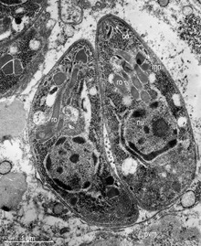
T. gondii contains organelles called rhoptries and micronemes, as well as other organelles.
The lifecycle of T. gondii may be broadly summarized into two components: a sexual component that occurs only within cats (felids, wild or domestic), and an asexual component that can occur within virtually all warm-blooded animals, including humans, cats, and birds.[27]: 2 Because T. gondii can sexually reproduce only within cats, cats are therefore the definitive host of T. gondii. All other hosts – in which only asexual reproduction can occur – are intermediate hosts.
When a feline is infected with T. gondii (e.g. by consuming an infected mouse carrying the parasite's tissue cysts), the parasite survives passage through the stomach, eventually infecting epithelial cells of the cat's small intestine.[27]: 39 Inside these intestinal cells, the parasites undergo sexual development and reproduction, producing millions of thick-walled, zygote-containing cysts known as oocysts. Felines are the only definitive host because they lack expression of the enzyme delta-6-desaturase (D6D) in their intestine. This enzyme converts linoleic acid; the absence of expression allows systemic linoleic acid accumulation. Recent findings showed that this excess of linoleic acid is essential for T. gondii sexual reproduction.[6]

Infected epithelial cells eventually rupture and release oocysts into the intestinal lumen, whereupon they are shed in the cat's feces.[4]: 22 Oocysts can then spread to soil, water, food, or anything potentially contaminated with the feces. Highly resilient, oocysts can survive and remain infective for many months in cold and dry climates.[28]
Ingestion of oocysts by humans or other warm-blooded animals is one of the common routes of infection.[29] Humans can be exposed to oocysts by, for example, consuming unwashed vegetables or contaminated water, or by handling the feces (litter) of an infected cat.[27]: 2 [30] Although cats can also be infected by ingesting oocysts, they are much less sensitive to oocyst infection than are intermediate hosts.[31][4]: 107
Intermediate hosts found include pigs, chickens, goats, sheep[27]: 2 and Macropus rufus by Moré et al. 2010.[32]: 162 Cattle and horses are resistant and thought to be incapable of significant infection.[27]: 11 T. gondii is considered to have three stages of infection; the tachyzoite stage of rapid division, the bradyzoite stage of slow division within tissue cysts, and the oocyst environmental stage.[33] Tachyzoites are also known as "tachyzoic merozoites" and bradyzoites as "bradyzoic merozoites".[34] When an oocyst or tissue cyst is ingested by a human or other warm-blooded animal, the resilient cyst wall is dissolved by proteolytic enzymes in the stomach and small intestine, freeing sporozoites from within the oocyst.[29][33] The parasites first invade cells in and surrounding the intestinal epithelium, and inside these cells, the parasites differentiate into tachyzoites, the motile and quickly multiplying cellular stage of T. gondii.[27]: 39 Tissue cysts in tissues such as brain and muscle tissue, form about 7–10 days after initial infection.[33] Although severe infection of M. rufus has been observed it is unknown whether this is common.[32]
Inside host cells, the tachyzoites replicate inside specialized vacuoles (called the parasitophorous vacuoles) created from host cell membrane during invasion into the cell.[27]: 23–39 Tachyzoites multiply inside this vacuole until the host cell dies and ruptures, releasing and spreading the tachyzoites via the bloodstream to all organs and tissues of the body, including the brain.[27]: 39–40
The parasite can be easily grown in monolayers of mammalian cells maintained in vitro in tissue culture. It readily invades and multiplies in a wide variety of fibroblast and monocyte cell lines. In infected cultures, the parasite rapidly multiplies and thousands of tachyzoites break out of infected cells and enter adjacent cells, destroying the monolayer in due course. New monolayers can then be infected using a drop of this infected culture fluid and the parasite indefinitely maintained without the need of animals.

Following the initial period of infection characterized by tachyzoite proliferation throughout the body, pressure from the host's immune system causes T. gondii tachyzoites to convert into bradyzoites, the semidormant, slowly dividing cellular stage of the parasite.[35] Inside host cells, clusters of these bradyzoites are known as tissue cysts. The cyst wall is formed by the parasitophorous vacuole membrane.[27]: 343 Although bradyzoite-containing tissue cysts can form in virtually any organ, tissue cysts predominantly form and persist in the brain, the eyes, and striated muscle (including the heart).[27]: 343 However, specific tissue tropisms can vary between intermediate host species; in pigs, the majority of tissue cysts are found in muscle tissue, whereas in mice, the majority of cysts are found in the brain.[27]: 41
Cysts usually range in size between five and 50 µm in diameter,[36] (with 50 µm being about two-thirds the width of the average human hair).[37]
Consumption of tissue cysts in meat is one of the primary means of T. gondii infection, both for humans and for meat-eating, warm-blooded animals.[27]: 3 Humans consume tissue cysts when eating raw or undercooked meat (particularly pork and lamb).[38] Tissue cyst consumption is also the primary means by which cats are infected.[4]: 46
An exhibit at the San Diego Natural History Museum states urban runoff with cat feces transports Toxoplasma gondii into the ocean, which can kill sea otters.[39]
Tissue cysts can be maintained in host tissue for the lifetime of the animal.[27]: 580 However, the perpetual presence of cysts appears to be due to a periodic process of cyst rupturing and re-encysting, rather than a perpetual lifespan of individual cysts or bradyzoites.[27]: 580 At any given time in a chronically infected host, a very small percentage of cysts are rupturing,[27]: 45 although the exact cause of this tissue cyst rupture is, as of 2010, not yet known.[4]: 47
Theoretically, T. gondii can be passed between intermediate hosts indefinitely via a cycle of consumption of tissue cysts in meat. However, the parasite's lifecycle begins and completes only when the parasite is passed to a feline host, the only host within which the parasite can again undergo sexual development and reproduction.[29]
In 2006, researchers reviewed evidence that T. gondii has an unusual population structure dominated by three clonal lineages called Types I, II and III that occur in North America and Europe, despite the occurrence of a sexual phase in its life cycle. They estimated that a common ancestor existed about 10,000 years ago.[40] Authors of a subsequent and larger study on 196 isolates from diverse sources including T. gondii in the bald eagle, gray wolf, Arctic fox and sea otter, also found that T. gondii strains infecting North American wildlife have limited genetic diversity with the occurrence of only a few major clonal types. They found that 85% of strains in North America were of one of three widespread genotypes II, III and Type 12. Thus T. gondii has retained the capability for sex in North America over many generations, producing largely clonal populations, and matings have generated little genetic diversity.[41]
During different periods of its life cycle, individual parasites convert into various cellular stages, with each stage characterized by a distinct cellular morphology, biochemistry, and behavior. These stages include the tachyzoites, merozoites, bradyzoites (found in tissue cysts), and sporozoites (found in oocysts).
Some stages are motile and some calcium-dependent protein kinases (TgCDPKs) are involved in this parasite's motility.[42][43] Gaji et al. 2015 find TgCDPK3 is required to begin the action of motility because it phosphorylates T. gondii's myosin A (TgMYOA).[42][43] TgCDPK3 is the functional orthologue of CDPK1 in this parasite.[43]
Motile, and quickly multiplying, tachyzoites are responsible for expanding the population of the parasite in the host.[44][27]: 19 When a host consumes a tissue cyst (containing bradyzoites) or an oocyst (containing sporozoites), the bradyzoites or sporozoites stage-convert into tachyzoites upon infecting the intestinal epithelium of the host.[27]: 359 During the initial acute period of infection, tachyzoites spread throughout the body via the blood stream.[27]: 39–40 During the later, latent (chronic) stages of infection, tachyzoites stage-convert to bradyzoites to form tissue cysts.

Like tachyzoites, merozoites divide quickly, and are responsible for expanding the population of the parasite inside the cat's intestine before sexual reproduction.[27]: 19 When a feline definitive host consumes a tissue cyst (containing bradyzoites), bradyzoites convert into merozoites inside intestinal epithelial cells. Following a brief period of rapid population growth in the intestinal epithelium, merozoites convert into the noninfectious sexual stages of the parasite to undergo sexual reproduction, eventually resulting in zygote-containing oocysts.[27]: 306
Bradyzoites are the slowly dividing stage of the parasite that make up tissue cysts. When an uninfected host consumes a tissue cyst, bradyzoites released from the cyst infect intestinal epithelial cells before converting to the proliferative tachyzoite stage.[27]: 359 Following the initial period of proliferation throughout the host body, tachyzoites then convert back to bradyzoites, which reproduce inside host cells to form tissue cysts in the new host.
Sporozoites are the stage of the parasite residing within oocysts. When a human or other warm-blooded host consumes an oocyst, sporozoites are released from it, infecting epithelial cells before converting to the proliferative tachyzoite stage.[27]: 359
Initially, a T. gondii infection stimulates production of IL-2 and IFN-γ by the innate immune system.[35] Continuous IFN-γ production is necessary for control of both acute and chronic T. gondii infection.[35] These two cytokines elicit a CD4+ and CD8+ T-cell mediated immune response.[35] Thus, T-cells play a central role in immunity against Toxoplasma infection. T-cells recognize Toxoplasma antigens that are presented to them by the body's own Major Histocompatibility Complex (MHC) molecules. The specific genetic sequence of a given MHC molecule differs dramatically between individuals, which is why these molecules are involved in transplant rejection. Individuals carrying certain genetic sequences of MHC molecules are much more likely to be infected with Toxoplasma. One study of>1600 individuals found that Toxoplasma infection was especially common among people who expressed certain MHC alleles (HLA-B*08:01, HLA-C*04:01, HLA-DRB 03:01, HLA-DQA*05:01 and HLA-DQB*02:01).[45]
IL-12 is produced during T. gondii infection to activate natural killer (NK) cells.[35] Tryptophan is an essential amino acid for T. gondii, which it scavenges from host cells. IFN-γ induces the activation of indole-amine-2,3-dioxygenase (IDO) and tryptophan-2,3-dioxygenase (TDO), two enzymes that are responsible for the degradation of tryptophan.[46] Immune pressure eventually leads the parasite to form cysts that normally are deposited in the muscles and in the brain of the hosts.[35]
The IFN-γ-mediated activation of IDO and TDO is an evolutionary mechanism that serves to starve the parasite, but it can result in depletion of tryptophan in the brain of the host. IDO and TDO degrade tryptophan to N-formylkynurenine. Administration of L-kynurenine is capable of inducing depressive-like behavior in mice.[46] T. gondii infection has been demonstrated to increase the levels of kynurenic acid (KYNA) in the brains of infected mice and in the brain of schizophrenic persons.[46] Low levels of tryptophan and serotonin in the brain were already associated with depression.[47]
The following have been identified as being risk factors for T. gondii infection in humans and warm-blooded animals:
A common argument in the debate about whether cat ownership is ethical involves the question of Toxoplasma gondii transmission to humans.[59] Even though "living in a household with a cat that used a litter box was strongly associated with infection,"[30] and that living with several kittens or any cat under one year of age has some significance,[49] several other studies claim to have shown that living in a household with a cat is not a significant risk factor for T. gondii infection.[50][60]
Specific vectors for transmission may also differ based on geographic location. "The seawater in California is thought to be contaminated by T. gondii oocysts that originate from cat feces, survive or bypass sewage treatment, and travel to the coast through river systems. T. gondii has been identified in a California mussel by polymerase chain reaction and DNA sequencing. In light of the potential presence of T. gondii, pregnant women and immunosuppressed persons should be aware of this potential risk associated with eating raw oysters, mussels, and clams.[49]
In warm-blooded animals, such as brown rats, sheep, and dogs, T. gondii has also been shown to be sexually transmitted.[61][62][63] Although T. gondii can infect, be transmitted by, and asexually reproduce within humans and virtually all other warm-blooded animals, the parasite can sexually reproduce only within the intestines of members of the cat family (felids).[29] Felids are therefore the definitive hosts of T. gondii; all other hosts (such as human or other mammals) are intermediate hosts.
The following precautions are recommended to prevent or greatly reduce the chances of becoming infected with T. gondii. This information has been adapted from the websites of United States Centers for Disease Control and Prevention[64] and the Mayo Clinic.[65]
Basic food-handling safety practices can prevent or reduce the chances of becoming infected with T. gondii, such as washing unwashed fruits and vegetables, and avoiding raw or undercooked meat, poultry, and seafood. Other unsafe practices such as drinking unpasteurized milk or untreated water can increase odds of infection.[64] As T. gondii is commonly transmitted through ingesting microscopic cysts in the tissues of infected animals, meat that is not prepared to destroy these presents a risk of infection. Freezing meat for several days at subzero temperatures (0 °F or −18 °C) before cooking may break down all cysts, as they rarely survive these temperatures.[4]: 45 During cooking, whole cuts of red meat should be cooked to an internal temperature of at least 145 °F (63 °C). Medium rare meat is generally cooked between 130 and 140 °F (55 and 60 °C),[66] so cooking meat to at least medium is recommended. After cooking, a rest period of 3 min should be allowed before consumption. However, ground meat should be cooked to an internal temperature of at least 160 °F (71 °C) with no rest period. All poultry should be cooked to an internal temperature of at least 165 °F (74 °C). After cooking, a rest period of 3 min should be allowed before consumption.
Oocysts in cat feces take at least a day to sporulate (to become infectious after they are shed), so disposing of cat litter daily greatly reduces the chance of infectious oocysts developing. As these can spread and survive in the environment for months, humans should wear gloves when gardening or working with soil, and should wash their hands promptly after disposing of cat litter. These precautions apply to outdoor sandboxes/play sand pits, which should be covered when not in use. Cat feces should never be flushed down a toilet.
Pregnant women are at higher risk of transmitting the parasite to their unborn child and immunocompromised people of acquiring a lingering infection. Because of this, they should not change or handle cat litter boxes. Ideally, cats should be kept indoors and fed only food that has low to no risk of carrying oocysts, such as commercial cat food or well-cooked table food.
No approved human vaccine exists against Toxoplasma gondii.[67] Research on human vaccines is ongoing.[68]
For sheep, an approved live vaccine sold as Toxovax (from MSD Animal Health) provides lifetime protection.[69]
In humans, active toxoplasmosis can be treated with a combination of drugs such as pyrimethamine and sulfadiazine, plus folinic acid. Immune-compromised patients may need continuous treatment until/unless their immune system is restored.[70]
In many parts of the world, where there are high populations of feral cats, there is an increased risk to the native wildlife due to increased infection of Toxoplasma gondii. It has been found that the serum concentrations of T. gondii in the wildlife population were increased where there are high amounts of cat populations. This creates a dangerous environment for organisms that have not evolved in cohabitation with felines and their contributing parasites.[71]
Toxoplasmosis is one of the contributing factors toward mortality in southern sea otters, especially in areas where there is large urban run-off.[72] In their natural habitats, sea otters control sea urchin populations and, thus indirectly, control sea kelp forests. By enabling the growth of sea kelp, other marine populations are protected as well as CO2 emissions are reduced due to the kelp's ability to absorb atmospheric carbon.[73] An examination on 105 beachcast otters revealed that 38.1% had parasitic infections, and 28% of said infections had resulted in protozoal meningoencephalitis deaths.[72] Toxoplasma gondii was found to be the root cause in 16.2% of these deaths, while 6.7% of the deaths were due to a closely related protozoan parasite known as Sarcocystis neurona.[72]
Minks, being semiaquatic, are also susceptible to infection and being antibody-positive toward Toxoplasma gondii.[74] Minks can follow a similar diet as otters and feasts on crustaceans, fish, and invertebrates, thus the transmission route follows a similar pattern to otters. Because of the mink's ability to transverse land more frequently, and often seen as an invasive species itself, minks are a bigger threat in transporting T. gondii to other mammalian species, rather than otters who have a more restrictive breadth.[74]
Although under-studied, penguin populations, especially those that share an environment with the human population, are at-risk due to parasite infections, mainly Toxoplasmosis gondii. The main subspecies of penguins found to be infected by T. gondii include wild Magellanic and Galapagos penguins, as well as blue and African penguins in captivity.[75] In one study, 57 (43.2%) of 132 serum samples of Magellanic penguins were found to have T. gondii. The island that the penguin is located, Magdalena Island, is known to have no cat populations, but a very frequent human population, indicating the possibility of transmission.[75]
Examination of black-footed penguins with toxoplasmosis reveals hepatomegaly, splenomegaly, cranial hemorrhage, and necrotic kidneys (Ploeg, et al., 2011). Alveolar and hepatic tissue presents a high number of immune cells such as macrophages containing tachyzoites of T. gondii.[76] Histopathological features in other animals affected with toxoplasmosis had tachyzoites in eye structures such as the retina which lead to blindness.[76]
The transmission of oocysts has been unknown, even though there have many documented cases of infection in marine species. Researchers have found that the oocytes of T. gondii can survive in seawater for at least 6 months, with the amount of salt concentration not affecting its lifecycle. There have been no studies on the ability of T. gondii oocysts lifecycle within freshwater environments, although infections are still present. One possible hypothesis of transmission is via amoeba species, particularly Acanthamoeba spp., a species that is found in all water environments (fresh-, brackish, and full-strength seawater). Normally, amoebas function as a natural filter, phagocytizing nutrients and bacteria found within the water. Some pathogens have used this to their advantage, however, and evolved to be able to avoid being broken down and, thus, survive encased in the amoeba – this includes Holosporaceae, Pseudomonaceae, Burkholderiacceae, among others.[77] Overall, this aids the pathogen in transportation but, also, protection from drugs and sterilizers that would, otherwise, cause death in the pathogen.[78] Studies have shown that T. gondii oocysts can live within amoebas after being engulfed for at least 14 days without significant obliteration of the parasite.[79] The ability of the microorganism to survive in vitro is dependent on the microorganism itself, but there are a few overarching mechanisms present. T. gondii oocysts have been found to resist an acidic pH and, thus, are protected by the acidification found in endocytic vacuoles and lysosomes.[79] Phagocytosis further increases with the carbohydrate-rich surface membrane located on the amoebae.[80] The pathogen can be released either by lysis of the amoebae or by exocytosis, but this is understudied [81]
Almost all species of birds that have been tested for Toxoplasma gondii have shown to be positive. The only bird species not reported with clinical symptoms of toxoplasmosis would be wild ducks, and there has only been one report found on domesticated ducks occurring in 1962.[82] Species with resistance toward T. gondii include domestic turkeys,[83] owls, red tail hawks, and sparrows, depending on the strain of T. gondii.[84] T. gondii is considerably more severe in pigeons, particularly crown pigeons, ornamental pigeons, and pigeons originating from Australia and New Zealand. Typical onset is quick and usually results in death. Those that do survive often have chronic conditions of encephalitis and neuritis.[84] Similarly, canaries are observed to be just as severe as pigeons, but the clinical symptoms are more abnormal when compared to other species. Most of the infection affects the eye, causing blindness, choroidal lesions, conjunctivitis, atrophy of the eye, blepharitis, and chorioretinitis [84] Most of the time, the infection leads to death.
Urbanization and global warming are extremely influential in the transmission of T. gondii.[85] Temperature and humidity are huge factors in the sporulation stage: low humidity is always fatal to the oocysts, and they are also vulnerable to extreme temperatures.[85] Rainfall is also an important factor for survival of waterborne pathogens. Because increased rainfall directly increases the flow rate in rivers, the amount of flow into coastal areas is increased as well. This can spread waterborne pathogens over wide areas.
There is no effective vaccine for T. gondii, and research on a live vaccine is ongoing. Feeding cats commercially available food, rather than raw, undercooked meat, prevents felines from becoming a host for oocysts, as higher prevalence is in areas where raw meat is fed.[86] Researchers also suggest that owners restrict cats to live indoors and to be neutered or spayed to decrease stray cat populations and to reduce intermediate host interactions. It is suggested that fecal matter from litter boxes be collected daily, placed in a sealable bag, and disposed of in the trash rather than flushed in the toilet, so that water contamination is limited.[87]
Studies have found that wetlands with a high density of vegetation decrease the concentration of oocysts in water through two possible mechanisms. Firstly, vegetation decreases flow velocities, which enables more settling because of increased transport time.[87] Secondly, the vegetation can remove oocysts through its ability to mechanically strain the water, as well as through the process of adhesion (i.e. attachment to biofilms). Areas of erosion and destruction of coastal wetlands have been found to harbour increased concentrations of T. gondii oocysts, which then flow into open coastal waters. Current physical and chemical treatments typically utilized in water treatment facilities have been proven to be ineffective against T. gondii. Research has shown that UV-C disinfection of water containing oocysts results in inactivation and possible sterilization.[88]
The genomes of more than 60 strains of T. gondii have been sequenced. Most are 60–80 Mb in size and consist of 11–14 chromosomes.[89][90] The major strains encode 7,800–10,000 proteins, of which about 5,200 are conserved across RH, GT1, ME49, VEG.[89] A database, ToxoDB, has been established to document genomic information on Toxoplasma.[91][92][93]
In 1908, while working at the Pasteur Institute in Tunis, Charles Nicolle and Louis Manceaux discovered a protozoan organism in the tissues of a hamster-like rodent known as the gundi, Ctenodactylus gundi.[29] Although Nicolle and Manceaux initially believed the organism to be a member of the genus Leishmania that they described as "Leishmania gondii", they soon realized they had discovered a new organism entirely; they renamed it Toxoplasma gondii. The new genus name Toxoplasma is a reference to its morphology: Toxo, from Greek τόξον (toxon, "arc, bow"), and πλάσμα (plasma, "shape, form") and the host in which it was discovered, the gundi (gondii).[94] The same year Nicolle and Mancaeux discovered T. gondii, Alfonso Splendore identified the same organism in a rabbit in Brazil. However, he did not give it a name.[29] In 1914, Italian tropicalist Aldo Castellani "was first to suspect that toxoplasmosis could affect humans".[95]
The first conclusive identification of T. gondii in humans was in an infant girl delivered full term by Caesarean section on May 23, 1938, at Babies' Hospital in New York City.[29] The girl began having seizures at three days of age, and doctors identified lesions in the maculae of both of her eyes. When she died at one month of age, an autopsy was performed. Lesions discovered in her brain and eye tissue were found to have both free and intracellular T. gondii'.[29] Infected tissue from the girl was homogenized and inoculated intracerebrally into rabbits and mice; they then developed encephalitis. Later, congenital transmission was confirmed in many other species, particularly infected sheep and rodents.
The possibility of T. gondii transmission via consumption of undercooked meat was first proposed by D. Weinman and A.H Chandler in 1954.[29] In 1960, the relevant cyst wall were shown to dissolve in the proteolytic enzymes found in the stomach, releasing infectious bradyzoites into the stomach (which pass into the intestine). The hypothesis of transmission via consumption of undercooked meat was tested in an orphanage in Paris in 1965; incidence of T. gondii rose from 10% to 50% after a year of adding two portions of cooked-rare beef or horse meat to many orphans' daily diets, and to 100% among those fed cooked-rare lamb chops.[29]
A 1959 Mumbai-based study found there prevalence in strict vegetarians was similar to that of non-vegetarians. This raised the possibility of a third major route of infection, beyond congenital and non well-cooked meat carnivorous transmission.[29]
In 1970, oocysts were found in (cat) feces. The fecal–oral route of infection via oocysts was demonstrated.[29] In the 1970s and 1980s feces of a vast range of infected animal species was tested to see if it contained oocysts — at least 17 species of felids shed oocysts, but no non-felid has been shown to allow T. gondii sexual reproduction (leading to oocyst shedding).[29]
In 1984 Elmer R. Pfefferkorn published his discovery that treatment of human fibroblasts with human recombinant interferon gamma blocks the growth of T. gondii.[96]
There are many instances where behavioural changes were reported in rodents with T. gondii. The changes seen were a reduction in their innate dislike of cats, which made it easier for cats to prey on the rodents. In an experiment conducted by Berdoy and colleagues, the infected rats showed preference for the cat odour area versus the area with the rabbit scent, therefore making it easier for the parasite to take its final step in its definitive feline host.[7] This is an example of the extended phenotype concept, that is, the idea that the behaviour of the infected animal changes in order to maximize survival of the genes that increase predation of the intermediate rodent host.[97]
Differences in sex-dependent behavior observed in infected hosts compared to non-infected individuals can be attributed to differences in testosterone. Infected males had higher levels of testosterone while infected females had significantly lower levels, compared to their non-infected equivalents.[98] Looking at humans, studies using the Cattell's 16 Personality Factor questionnaire found that infected men scored lower on Factor G (superego strength/rule consciousness) and higher on Factor L (vigilance) while the opposite pattern was observed for infected women.[99] Such men were more likely to disregard rules and were more expedient, suspicious, and jealous. On the other hand, women were more warm-hearted, outgoing, conscientious, and moralistic.[99] Mice infected with T. gondii have a worse motor performance than non-infected mice.[100][101] Thus, a computerized simple reaction test was given to both infected and non-infected adults. It was found that the infected adults performed much more poorly and lost their concentration more quickly than the control group. But, the effect of the infection only explains less than 10% of the variability in performance[99] (i.e., there could be other confounding factors). Correlation has also been observed between seroprevalence of T. gondii in humans and increased risk of traffic accidents. Infected subjects have a 2.65 times higher risk of getting into a traffic accident.[102] A Turkish study confirmed this holds true among drivers.[103] This parasite has been associated with many neurological disorders such as schizophrenia. In a meta-analysis of 23 studies that met inclusion criteria, the seroprevalence of antibodies to T. gondii in people with schizophrenia is significantly higher than in control populations (OR=2.73, P<0.000001).[104] A 2009 summary of studies found that suicide attempters had far more indicative (IgG) antibodies than mental health inpatients without a suicide attempt.[105] Infection was also shown to be associated with suicide in women over the age of 60. (P<0.005) [106]
As mentioned before, these results of increased proportions of people seropositive for the parasite in cases of these neurological disorders do not necessarily indicate a causal relationship between the infection and disorder. It is also important to mention that in 2016 a population-representative birth cohort study which was done, to test a hypothesis that toxoplasmosis is related to impairment in brain and behaviour measured by a range of phenotypes including neuropsychiatric disorders, poor impulse control, personality and neurocognitive deficits. The results of this study did not support the results in the previously mentioned studies, more than marginally. None of the P-values showed significance for any outcome measure. Thus, according to this study, the presence of T. gondii antibodies is not correlated to increase susceptibility to any of the behaviour phenotypes (except possibly to a higher rate of unsuccessful attempted suicide). This team did not observe any significant association between T. gondii seropositivity and schizophrenia. The team notes that the null findings might be a false negative due to low statistical power because of small sample sizes but against this weights that their setup should avoid some possibilities for errors in the about 40 studies that did show a positive correlation. They concluded that further studies should be performed.[107] Another population-representative study with 7440 people in the United States found that Toxoplasma infection was 2.4 fold more common in people who had a history of manic and depression symptoms (bipolar disorder Type 1) compared to the general population.[108]
Research on the linkage between T. gondii infection and entrepreneurial behavior showed that students who tested positive for T. gondii exposure were 1.4 times more likely to major in business and 1.7 times more likely to have an emphasis in "management and entrepreneurship". Among 197 participants of entrepreneurship events, T. gondii exposure was correlated with being 1.8 times more likely to have started their own business.[109]
Published research has also indicated that T. gondii infection could potentially promote changes in a person’s political beliefs and values. Those who are infected with the parasite tend to exhibit a higher degree of “us versus them” thinking.[110][111][112]
The mechanism behind behavioral changes is partially attributed to increased dopamine metabolism,[113] which can be neutralized by dopamine antagonist medications.[114] T. gondii has two genes that code for a bifunctional phenylalanine and tyrosine hydroxylase, two important and rate-limiting steps of dopamine biosynthesis. One of the genes is constitutively expressed, while the other is only produced during cyst development.[115][116] In addition to additional dopamine production, T. gondii infection also produces long-lasting epigenetic changes in animals that increase the expression of vasopressin, a probable cause of alterations that persist after the clearance of the infection.[117]
In 2022, a study published in Nature of a well-documented population of wolves studied throughout their lives, suggested that T. gondii also may have a significant effect on their behavior. It suggested that infection with this parasite emboldened infected wolves into behavior that determined leadership roles and influenced risk-taking behavior, perhaps even motivating establishment of new independent packs that they would establish and lead in behavior patterns differing from that of the packs into which they were born. The study determined that at times, an infected wolf would become the only breeding male in a pack,[118] leading to a significant effect on another species by "T. gondii.
{{cite journal}}: CS1 maint: uses authors parameter (link)  Dividing T. gondii parasites
 Toxoplasma gondii (/ˈtɒksəˌplæzmə ˈɡɒndi.aɪ, -iː/) is a parasitic protozoan (specifically an apicomplexan) that causes toxoplasmosis. Found worldwide, T. gondii is capable of infecting virtually all warm-blooded animals,: 1 but felids are the only known definitive hosts in which the parasite may undergo sexual reproduction.
In rodents, T. gondii alters behavior in ways that increase the rodents' chances of being preyed upon by felids. Support for this "manipulation hypothesis" stems from studies showing that T. gondii-infected rats have a decreased aversion to cat urine while infection in mice lowers general anxiety, increases explorative behaviors and increases a loss of aversion to predators in general. Because cats are one of the only hosts within which T. gondii can sexually reproduce, such behavioral manipulations are thought to be evolutionary adaptations that increase the parasite's reproductive success since rodents that do not avoid cat habitations will more likely become cat prey. The primary mechanisms of T. gondii–induced behavioral changes in rodents occur through epigenetic remodeling in neurons that govern the relevant behaviors (e.g. hypomethylation of arginine vasopressin-related genes in the medial amygdala, which greatly decrease predator aversion).
In humans, particularly infants and those with weakened immunity, T. gondii infection is generally asymptomatic but may lead to a serious case of toxoplasmosis. T. gondii can initially cause mild, flu-like symptoms in the first few weeks following exposure, but otherwise, healthy human adults are asymptomatic. This asymptomatic state of infection is referred to as a latent infection, and it has been associated with numerous subtle behavioral, psychiatric, and personality alterations in humans. Behavioral changes observed between infected and non-infected humans include a decreased aversion to cat urine (but with divergent trajectories by gender) and an increased risk of several psychiatric disorders – particularly schizophrenia and bipolar disorder. Preliminary evidence has suggested that T. gondii infection may induce some of the same alterations in the human brain as those observed in rodents. Many of these associations have been strongly debated and newer studies have found them to be weak concluding:
On the whole, there was little evidence that T. gondii was related to increased risk of psychiatric disorder, poor impulse control, personality aberrations, or neurocognitive impairment.
T. gondii is one of the most common parasites in developed countries; serological studies estimate that up to 50% of the global population has been exposed to, and may be chronically infected with, T. gondii; although infection rates differ significantly from country to country. Estimates have shown the highest IgG seroprevalence to be in Ethiopia, at 64.2%, as of 2018.
Toxoplasma gondii es una especie de protozoo parásito intracelular obligado, eucariota. Es causante de la toxoplasmosis, una enfermedad en general leve, pero que puede complicarse hasta convertirse en fatal, especialmente en los gatos y en los fetos humanos.[1] El ciclo celular de T. gondii alterna entre la reproducción sexual en los huéspedes definitivos y la reproducción asexual en los huéspedes intermedios. El gato y otros felinos son considerados como sus hospedadores definitivos porque en ellos tiene lugar la fase sexuada de su ciclo de vida. Entre los huéspedes intermedios podemos encontrar mamíferos y pájaros.[2] Es un parásito muy exitoso y uno de los más frecuentes que infectan a los humanos, pues puede infectar a cualquiera de las especies de animales homeotermos. Los humanos son hospedadores habituales, en 2002 se estimaba que un tercio de la población mundial estaba infectada de forma crónica por T. gondii.[3]
Las distintas fases de desarrollo del Toxoplasma gondii muestran cambios importantes en su morfología, estas son llamadas: #Taquizoito, #Bradizoito y #Ooquiste.
El Taquizoito es la forma de la infestación aguda, tiene de 4-8 micrómetros (μm) (micrómetros) de longitud y de 2-4 μm de ancho, con aspecto de medialuna.
Un grupo de estructuras moleculares, en un extremo del Taquizoito del T. gondii, con la denominación de complejo apical, es fundamental tanto para la invasión como para la proliferación de este parásito.[4] [5]

El complejo apical (api-complex en inglés) de T. gondii, es un conjunto complejo de elementos estructurales y secretores en el extremo apical del parásito adulto, que se construye sobre un conjunto de fibras dispuestas en espiral alrededor del conoide. Este complejo tiene también tres unidades de membrana (como se ve en el esquema C): una membrana plasmática externa (en negro) y dos membranas en el complejo de membrana interna (IMC en rojo) que son las paredes de vesículas aplanadas (sacos alveolares).[6]
El complejo apical proporciona un marco semirrígido para las células puntiagudas de T. gondii y es el destino para los orgánulos secretores, que liberan varios factores de invasión.[7]
El complejo apical se organiza alrededor de un anillo polar apical (APR en inglés), que sirve como centro organizador y nuclea una serie de microtúbulos, que descienden hacia la parte posterior de la célula.
Además de su estructura, el complejo apical actúa como un centro de señalización, al ser el punto de convergencia de muchas vías reguladoras.[8]
El conoide de T. gondii, está formado por un conjunto compacto de fibras de citoesqueleto dispuestas en espiral, que se mueven activamente durante la etapa de invasión de las células del huésped.
Las estructuras asociadas con el conoide son: los anillos pre-conoidales, en la punta distal del conoide, donde se originan las fibras conoides; el anillo polar, del que se originan 22 microtúbulos (MT) sub-peliculares; y dos MT intra-conoidales cortos, que pueden utilizarse como vías para el transporte de vesículas secretoras esenciales para la invasión.[6]
Pasada la etapa de invasión de T. gondii, se forma la llamada vacuola parasitófora (PV). Esta se forma tanto de la membrana plasmática de la célula huésped, como de los productos de secreción del parásito.
La vacuola PV está aislada del tráfico vesicular del huésped y está rodeada por microtúbulos, por retículo endoplásmico y por mitocondrias de la célula huésped.
T. gondii también posee orgánulos secretores especializados: las micronemas (M), las roptrías (R) y los gránulos densos (DG). La secreción en secuencia de estos tres tipos de orgánulos secretores dirigen tanto la entrada en la célula huésped, como la formación de la PV.[9]
Un ooquiste es la fase esporulada del Toxoplasma. Este es un estado que puede sobrevivir fuera del hospedador por largos períodos de tiempo, dada su alta resistencia al medio ambiente. Esta es la forma durmiente del parásito, el producto de su reproducción sexual en las células intestinales de los felinos infectados. Estos pueden segregar ooquistes hasta 1 o 2 semanas después de la infección. Estos ooquistes esporulan y se hacen efectivos en 1-5 días y gracias a su gruesa pared celular pueden sobrevivir bien en el medio ambiente.[10]
El bradizoíto (del griego brady=lento y zōon=animal) es la forma de replicación lenta del parásito, no solamente de Toxoplasma gondii, sino de otros protozoos responsables de infecciones parasitarias. En la toxoplasmosis latente (crónica), el bradizoíto se presenta en conglomerados microscópicos envueltos por una pared llamados quistes, en el músculo y el tejido cerebral infectado.[11]
Los taquizoitos son formas mótiles que forman pseudoquistes en tejidos infestados por toxoplasma. Los taquizoitos se encuentran en vacuolas dentro de las células, esta forma celular invade y se replica en las células infectadas. Activan el sistema inmune y se convierten en bradizoitos que se dividen lentamente.
Algunos de los orgánulos que pueden ser encontrados en la imagen de microscopía electrónica de la derecha son:
apicoplasto (A); anillo polar apical (APR); conoide (C); anillo conoidal (CR); aparato de Golgi (G); complejo de membrana interna (IMC); Micronemas (M); mitocondria (Mi); microtúbulos (Mt); núcleo (Nu); roptrias (R); conducto de roptria (RD); vacuola (V).
El ciclo de vida del T. gondii tiene dos fases. La fase sexual del ciclo de vida ocurre solo en miembros de la familia Felidae (gatos domésticos y salvajes), haciendo que estos animales sean los hospedadores primarios del parásito. La fase asexual del ciclo de vida puede ocurrir en cualquier animal de sangre caliente, tales como otros mamíferos y aves. Por ello, la toxoplasmosis constituye una zoonosis parasitaria.[13]
Este protozoario puede infectar de tres formas por medio de:
Taquizoítos: encontrados en psedoquistes, estos son liberados de los bradizoítos y penetran cualquier célula infectandola hasta morir.
Bradizoítos: estos entran en forma de quiste al hospedero, donde pueden liberar taquizoítos o en el caso de hospederos de la familia Felinidae, pueden formar gametos para despues conformar un ooquiste.
Ooquiste: esta última forma de infección solo es producida en los hospederos de la familia Felinidae, pues solo es estos se puede llevar a cabo el ciclo sexual del protozoario. Estos se forman a partir de los gametos femeninos y masculinos que nacieron de los bradizoítos. En estos se encuentran 2 sacos con 4 taquizoítos cada uno y son liberados en las heces.
La forma de infección se puede dar desde consumir agua o alimentos que esten infectados con el protozoario, hasta la ingesta de carne con quistes o pseudoquistes. [14]
En el hospedador intermediario, incluyendo los felinos, los parásitos invaden células, formando un compartimento llamado vacuola parasitófora[15] que contienen bradizoitos, la forma de replicación lenta del parásito.[16] Las vacuolas forman quistes, en especial en los músculos y cerebro. Debido a que el parásito está dentro de las células, el sistema inmunitario del hospedador no detecta estos quistes. La resistencia a los antibióticos varía, pero los quistes son difíciles de erradicar enteramente.

T. gondii se propaga dentro de estas vacuolas por una serie de divisiones binarias hasta que la célula infestada eventualmente se rompe, liberando a los taquizoitos. Estos son motiles, y la forma de reproducción asexual del parásito. A diferencia de los bradizoitos, los taquizoitos libres son eficazmente eliminados por la inmunidad del hospedador o huésped, a pesar de que algunos logran infectar otras células formando, manteniendo así el ciclo de vida de este parásito.
Los quistes tisulares son ingeridos por el gato al alimentarse de un ratón infectado. Los quistes sobreviven el paso por el estómago del gato y los parásitos infectan las células epiteliales del intestino delgado en donde pasan por la reproducción sexual y la formación de ooquistes, que son liberados con las heces. Otros animales, incluyendo los humanos ingieren los ooquistes (al comer vegetales no lavados adecuadamente) o los quistes tisulares al comer carne cruda o cocida inapropiadamente. T. gondii puede infectar cualquier tipo de células del huésped, con excepción de los eritrocitos; lo anterior lo realiza mediante acción enzimática o dejándose fagocitar.[cita requerida]
El desarrollo de la toxoplasmosis encefálica ocurre debido a la transición de bradizoito (estadio latente) a taquizoito (estadio de rápida replicación) dentro del hospedero definitivo.[17]
La tasa de replicación de taquizoitos es más alta que la tasa de los bradizoitos y esto se debe a que tiene una serie de factores que aceleran su metabolismo.[18] Entre estos factores se encuentra la familia de Proteínas de Choque Térmico (Heat Shock Proteins, HSP), que contiene unas proteínas muy conservadas de bajo peso molecular 70kDa (HSP70) y que son un tipo de chaperonas ubicuas que tienen alto potencial inmunogénico.[19] Hasta hace poco, se creía que la función principal de este tipo de proteínas malplegadas era actuar como chaperonas intracelulares relacionadas con citoprotección luego de que las células fueran estresadas por estímulos externos. Sin embargo, ahora se sabe que las HSP70 no solamente funcionan como chaperonas, sino también como citoquinas (chaperoquinas) en el espacio extracelular.[20]
Se sabe que los HSP son importantes para la diferenciación del bradizoito ya que estudios en los que se hace Knockout a los genes hsp27, hsp70 y hsp90 se suprime la inducción de bradizoitos in vitro. Por otro lado, al utilizar potenciadores de expresión como indometacina, hay un incremento en la formación de bradizoitos.[20] Adicionalmente, en cultivos de médula ósea, los interferones gamma (IFN-ϒ) incrementaron la formación de bradizoitos debido a la producción de óxido nítrico (NO).[21] Además, se ha encontrado que las HSP de T. gondii (TgHSP70) son reconocidas por las células B del hospedero (mHSP70, ratón) a pesar de ser homólogas entre sí.[22] Luego la inducción de los bradizoitos se da tanto por estrés oxidativo y como consecuencia de la respuesta inmune del hospedero.
Otros descubrimientos que dan soporte a la idea de que los HSP70 están asociados a la diferenciación de los bradizoitos a taquizoitos es que en otros protozoos como Leishmania chagasi y Trypanosoma cruzi, el gen hsp70 se ha asociado a la capacidad de sobrevivir a estrés oxidativo y llevar la diferenciación de promastigotes a amastigotes.[23]
Se cree que dicha diferenciación es una respuesta al estrés causado por condiciones ambientales relacionadas con la respuesta inflamatoria del hospedero definitivo y que la respuesta al choque térmico se debe a la plasticidad metabólica del parásito durante la diferenciación.[24]
En 1908, Charles Nicolle y Louis Manceaux en Túnez demuestran la presencia del parásito en un roedor el Ctenodactylus gundi, al mismo tiempo en Brasil Splendore lo encontró en conejos. Se le denominó Toxoplasma gondii por toxo (del griego toxon ‘arco’) por su forma arqueada;[25] y gondii por el ratón.
Los humanos pueden adquirir la infección por Toxoplasma gondii a través de diferentes vías.[26] En primer lugar, horizontalmente a través de la ingestión oral de ooquistes provenientes del medio ambiente. Además, horizontalmente por la ingestión de bradiozitos presentes en carnes crudas de huéspedes intermedios. Por último, los taquiozitos pueden ser transmitidos verticalmente durante la lactancia materna o trasmitidos desde una madre infectada al feto durante el embarazo. Esto último ocurre frecuentemente en las madres que han sido infectadas por primera vez durante la gestación, esto puede provocar problemas en el desarrollo del feto y patologías como retraso psicomotriz, anormalidades hematológicas, pérdida auditiva y visual e incluso pueden provocar la muerte del feto.[27]
En individuos inmunocompetentes, la infección se presenta con síntomas suaves parecidos a los de la gripe común, o son completamente asintomáticos. En este tipo de pacientes, los síntomas severos más frecuentes son infecciones retinales y linfadenopatías, que normalmente están asociados a una infección congénita que se manifiesta de forma tardía.
En humanos inmunodeprimidos, puede actuar como patógeno oportunista, por ejemplo en enfermos de sida.[28]
Se recomienda el empleo de pirimetamina y sulfonamidas, la primera actúa sobre la síntesis del ácido fólico y la segunda sobre la síntesis del ácido paraaminobenzoico (sobre taquizoitos, no en quistes).
Para la prevención de la toxoplasmosis congénita en mujeres embarazadas se recomienda la espiramicina, ya que es menos tóxica. Este medicamento evita que los taquizoitos pasen el lago placentario hacia el feto. El problema de la toxoplasmosis en el embarazo son las dificultades diagnósticas porque las técnicas del diagnóstico de la infección primaria en la mujer embarazada no son totalmente absolutas ni inequívocas. Los estudios serológicos no son totalmente exactos y técnicas consideradas de elección en el diagnóstico de la toxoplasmosis aguda como IgG de baja avidez obtiene resultados no definitivos en el 25 % de los casos (cribado serológico en la gestante).[29] Además, cuando ya se ha producido el diagnóstico, se ha producido ya la transmisión, llegando el tratamiento tarde para evitar esta transmisión.[30] Es tan indeterminado el diagnóstico y mayores los efectos secundarios de las actuaciones médicas en un diagnóstico indeterminado, así como ineficacia con el tratamiento farmacológico de evitar la transmisión al feto que se aboga por no realizar el screening serológico a toxoplasma a todas las embarazadas de manera sistemática. Si el parásito ya ha atravesado la placenta ya no es eficaz, aunque parece tener beneficio disminuyendo la carga parasitaria y por lo tanto disminuyendo la severidad de los síntomas en algunos casos.[31] En inmunodeficientes se recomienda la combinación de pirimetamina con sulfadiazina, pero dada la mayor posibilidad de los pacientes infectados con VIH de alergia a las sulfas, en ocasiones es necesario usar la combinación de pirimetamina con la clindamicina.
La etapa aguda de las infestaciones por toxoplasmosis pueden ser asintomáticas, pero a menudo aparecen síntomas gripales que conllevan a estados latentes. La infección latente es también, por lo general, asintomática, pero en personas inmunosuprimidas (pacientes trasplantados o con ciertas infecciones), pueden mostrar síntomas, notablemente encefalitis, que puede ser mortal.
Varía dependiendo en qué trimestre del embarazo se adquiera el parásito.[32]
La toxoplasmosis en embarazadas es rara vez sintomática pero puede provocar: linfadenopatía, fiebre, mialgia, malestar general, entre otras.
En los humanos, la infección crónica latente con T. gondii se ha relacionado previamente con la violencia autodirigida suicida, el rasgo de agresión e impulsividad y el trastorno bipolar.[33]
En cuanto a los efectos en humanos, la toxoplasmosis puede causar esquizofrenia.[34] El «sentido biológico» de esta infección tendría sentido cuando los humanos eran cazados por felinos, en este caso, la alteración del comportamiento promoviendo un comportamiento agresivo que llevara a la exclusión social del individuo, aumentaría las posibilidades de ser cazado por un felino.[35] De esta forma, el parásito podría llevar a cabo su ciclo vital reproductivo en su huésped definitivo.[36]
La infección está también relacionada con una mayor tasa de suicidio en pacientes infectados.[37] Esto podría estar relacionada con la alteración de la concentración de dopamina en partes específicas del cerebro que juegan un papel esencial en el comportamiento y emociones de los humanos.
Las infecciones parásito-huésped son frecuentemente consideradas como un mecanismo evolutivo. Ambos, parásito y huésped desarrollan adaptaciones y contra-adaptaciones contra el otro, resultando en una coevolución antagonista.[38] Se ha observado que la infección por T. gondii tienen la facultad de cambiar el comportamiento de ratas y ratones, haciendo que se acerquen, en vez de huir del olor de los gatos. Este efecto es de beneficio para el parásito, el cual puede reproducirse sexualmente si es ingerido por el gato.[39] Un clásico ejemplo de manipulación del comportamiento son los grillos infectados por nematomorfos. Los mismos son manipulados para suicidarse saltando a cuerpos de agua cuando son infectados; de esta forma, el nematomorfo adulto es capaz de completar la fase reproductiva de su ciclo vital, que es acuática.[40]
La infección tiene una gran precisión, en el sentido de que no impacta los otros temores de la rata, tal como el temor de los espacios abiertos o del olor de alimentos desconocidos. Se ha especulado que el comportamiento humano puede igualmente verse afectado de algún modo, y se han encontrado correlaciones entre las infecciones latente por Toxoplasma y varias características, tales como un aumento en comportamientos de alto riesgo, tales como una lentitud para reaccionar, sentimientos de inseguridad y neurosis[41] que parece que aumentan la propensión al suicidio.[42]
|fechaacceso= requiere |url= (ayuda) |fechaacceso= requiere |url= (ayuda) |fechaacceso= requiere |url= (ayuda) |fechaacceso= requiere |url= (ayuda) |fechaacceso= requiere |url= (ayuda) Toxoplasma gondii es una especie de protozoo parásito intracelular obligado, eucariota. Es causante de la toxoplasmosis, una enfermedad en general leve, pero que puede complicarse hasta convertirse en fatal, especialmente en los gatos y en los fetos humanos. El ciclo celular de T. gondii alterna entre la reproducción sexual en los huéspedes definitivos y la reproducción asexual en los huéspedes intermedios. El gato y otros felinos son considerados como sus hospedadores definitivos porque en ellos tiene lugar la fase sexuada de su ciclo de vida. Entre los huéspedes intermedios podemos encontrar mamíferos y pájaros. Es un parásito muy exitoso y uno de los más frecuentes que infectan a los humanos, pues puede infectar a cualquiera de las especies de animales homeotermos. Los humanos son hospedadores habituales, en 2002 se estimaba que un tercio de la población mundial estaba infectada de forma crónica por T. gondii.
Toxoplasma gondii toxoplasmosi gaitza eragiten duen protozoo patogenoa da. Toxoplasmosia orokorrean ez da gaitz larria, baina hainbat kasutan ondorio gaiztoak dakartza haurdun dauden emakumeengan, umekia nabarmen kaltetu baitezake. Immunitate-sistema ahula dutenengan ere toxoplasma gondiiren infekzioa arriskutsuagoa da pertsona osasuntsuengan baino.
Toxoplasmosia zoonosi bat da, katua izanik gizakia kutsatu ahal duen animaliaren bat.
Protozoo hau zelula barneko parasitoa da, eta hiru morfologia ezberdinak ditu bere bizi-zikloan:[1].
Toxoplasma gondii toxoplasmosi gaitza eragiten duen protozoo patogenoa da. Toxoplasmosia orokorrean ez da gaitz larria, baina hainbat kasutan ondorio gaiztoak dakartza haurdun dauden emakumeengan, umekia nabarmen kaltetu baitezake. Immunitate-sistema ahula dutenengan ere toxoplasma gondiiren infekzioa arriskutsuagoa da pertsona osasuntsuengan baino.
Toxoplasmosia zoonosi bat da, katua izanik gizakia kutsatu ahal duen animaliaren bat.
Protozoo hau zelula barneko parasitoa da, eta hiru morfologia ezberdinak ditu bere bizi-zikloan:.
ookistea: esporaren antzeko forma bat da, denbora luzez aske irauten dena ostalariarengandik kanpo; hau da ostalari berria infektatzen duena bradizoitoa: ostalari barnean dagoen protozooa, ugalketa motela duena. Takizoito bihurtzen da gero takizoitoa: infektatutako ehunetan kisteak sortzen dituen toxoplasma gondiiren forma. Mugikorra da eta ugalketa arina du.Toxoplasma gondii on Chromalveolata-eliökuntaan kuuluva yksisoluinen loiseliö. Se kuuluu samaan alajaksoon kuin malariaa aiheuttavat Plasmodium-itiöeläimet. Sen pääisäntiä ovat kissaeläimet, mutta sitä voi esiintyä myös muilla tasalämpöisillä eläinlajeilla, ihminen mukaan luettuna. T. gondii kykenee suvulliseen lisääntymiseen ainoastaan kissoissa; muissa tasalämpöisissä eläimissä se lisääntyy suvuttomasti.
Toxoplasman invaasiomekanismi eli mekanismi, jota loinen käyttää tunkeutuakseen isäntäsoluun, eroaa useiden bakteerien ja virusten passiivisesta invaasiosta. Useimmat bakteerit ja virukset käyttävät soluun tunkeutumiseen fagosytoosia tai endosytoosia kun taas Toxoplasman invaasiomekanismi on aktiivinen. Soluun tunkeutuminen voidaan erotella eri vaiheisiin. Ensin loinen kiinnittyy isäntäsolun pintaan. Tämän jälkeen se tunkeutuu tekemänsä erityisen portaalin, moving junction, läpi ja päätyy isäntäsolun sisään. Invaasio kestää noin minuutin.[2]
Aktiinilla on tärkeä rooli invaasiossa. Loisen aktiini-myosiinimoottori mahdollistaa loisen liikkumisen. Tämä liikkumistapa on oleellinen invaasiossa, sillä se mahdollistaa loisen liikkumisen isäntäsolun pinnalla. Se myös mahdollistaa itse invaasion sekä loisen poistumisen ulos solusta. Toxoplasmalla on yksi aktiinigeeni, act1. Aktiinin rooli invaasiossa ymmärrettiin kun huomattiin että aktiinin polymerisaation estäjä sytokalasiini D (CytD) estää invaasiota. Loiskannoissa, joissa aktiinigeenin toiminta on estetty, loisten liikkumiskyky ja taudinaiheuttamiskyky heikkenee. Aktiinin rooli on oleellinen Toxoplasman invaasion viimeisissä vaiheissa. Aktiinin puuttuminen myös hidastaa invaasiota huomattavasti.[3]
T. gondii aiheuttaa toksoplasmoosina tunnetun oireyhtymän, joka ei yleensä ole vaarallinen. Ihmisellä tavallisin oire on useiden viikkojen kuluttua tartunnasta ilmaantuva lievän flunssan kaltainen sairaus, joka sairastetaan lähes huomaamatta.[4] Poikkeuksen muodostavat ihmiset, joiden vastustuskyky on muusta syystä alentunut, sekä odottavat naiset, jotka eivät ole aiemmin saaneet tartuntaa. Mikäli äiti saa toksoplasmoositartunnan raskauden aikana, voi siitä olla sikiölle vakavia seurauksia, jopa kuolema. Tartunnan ihminen voi saada erittävän kissan ulosteista esimerkiksi puhdistaessaan hiekkalaatikkoa tai syömällä huonosti kypsennettyä lihaa. Ookystat voivat päätyä elimistöön kissan ulosteista ja kudoskystat huonosti kypsennetystä lihasta. Kystat ovat paksukuorisia loisen kestomuotoja. Ookystat ja kudoskystat kulkeutuvat vatsan läpi ja ohutsuolessa niistä vapautuu sporotsoiitteja. Ne tunkeutuvat suoliston epiteelin läpi ja erilaistuvat takytsoiiteiksi, jotka leviävät muualle kehoon.[5]
Joissakin tapauksissa tartunta muuttaa tartunnan saaneen eläimen käyttäytymistä. Hiirten ja rottien tapauksessa tämä tarkoittaa sitä, että tartunnan saanut yksilö muuttuu aktiivisemmaksi eikä pelkää saalistajia (esimerkiksi kissoja) kuten terve yksilö. Kissan virtsan hajun on huomattu laskevan tartunnan saaneiden rottien aivojen pelkokeskusten aktiivisuutta ja aktivoivan niiden mielihyväkeskuksia, mikä saattaa johtaa itsetuhoiseen käytökseen ja auttaa T. gondiin pääsyssä takaisin kissaan.[6][7]
Ihmisillä tartunta voi tehdä sairastuneesta impulsiivisemman, nopeammin pitkästyvän ja hitaampirefleksisen, mikä saattaa johtaa riskikäyttäytymiseen esimerkiksi liikenteessä. Tutkimuksissa tartunta on liitetty myös esimerkiksi skitsofreniaan.[8]
Toxoplasma gondii on Chromalveolata-eliökuntaan kuuluva yksisoluinen loiseliö. Se kuuluu samaan alajaksoon kuin malariaa aiheuttavat Plasmodium-itiöeläimet. Sen pääisäntiä ovat kissaeläimet, mutta sitä voi esiintyä myös muilla tasalämpöisillä eläinlajeilla, ihminen mukaan luettuna. T. gondii kykenee suvulliseen lisääntymiseen ainoastaan kissoissa; muissa tasalämpöisissä eläimissä se lisääntyy suvuttomasti.
Toxoplasma gondii est une espèce de parasites intracellulaires appartenant au phylum des Apicomplexa, et il est l'agent de la toxoplasmose. Le phylum des Apicomplexa inclut de nombreux autres pathogènes d'importance médicale ou vétérinaire, parmi lesquels Plasmodium falciparum, responsable de la malaria chez l'humain.
Les Apicomplexa sont des parasites intracellulaires obligatoires : ils doivent vivre à l'intérieur d'une cellule pour survivre. Une fois le parasite installé dans la cellule-hôte, celle-ci lui assure de larges ressources en nutriments ainsi qu'une protection contre le système immunitaire de l'hôte.
Les données sur le toxoplasme et son épidémiologie ont été acquises très progressivement : le parasite a d'abord été découvert uniquement sous sa forme infectieuse dans les tissus d'un rongeur sauvage, le gundi (Ctenodactylus gundii), en Tunisie par Nicolle et Manceaux en 1908[1] et presque simultanément au Brésil chez un lapin par Splendore en 1909[3]. Au début Nicolle et Manceaux pensaient avoir affaire à un parasite du genre Leishmania, qu'ils nommèrent "Leishmania gondii", mais un an plus tard, en 1909[2], ils le nommèrent Toxoplasma gondii à cause de sa forme arquée (du grec τοx(ο) = arc) et à partir du nom du rongeur chez qui il avait été observé. Toutes notions concernant son cycle biologique ou son importance en parasitologie humaine sont alors inconnues. Il faut attendre les années 1920-1930 pour voir apparaître les premières descriptions de toxoplasmose humaine. C'est la mise au point des premiers tests sérologiques dans les années 1940 qui a permis de révéler l'importance de la prévalence de la toxoplasmose humaine. La compréhension du cycle de ce parasite et des modes de transmission n'a eu lieu qu'au cours des années 1970[4].
T. gondii est constitué d'une cellule polarisée de forme arquée, d'environ 8 micromètres de long par 3 micromètres de large. T. gondii est un eucaryote unicellulaire haploïde, qui présente certaines caractéristiques conservées chez tous les eucaryotes, mais également des organites spécifiques de son groupe, les Alveolata, ou de son phylum, les Apicomplexa.
T. gondii est autonome pour la plupart de ses besoins de synthèse et de transport de protéines, de lipides ou d'énergie (sous forme d'ATP). Cependant sa prolifération est obligatoirement intracellulaire car il dépend de la cellule-hôte pour un certain nombre de nutriments essentiels pour lesquels il ne dispose pas de voie métabolique propre. Comme toute cellule eucaryote, T. gondii possède toute la machinerie nécessaire à la synthèse et au transport de protéines. Ceci comprend un noyau, un réticulum endoplasmique (ER) périnucléaire, un appareil de Golgi réticulé ainsi qu’un appareil mitochondrial. Cependant, certains acides aminés, briques élémentaires des protéines, doivent être importés à partir de la cellule-hôte. T. gondii synthétise également la plupart de ses lipides ainsi que des ribonucléotides tels que l’ATP, à partir de petites molécules (précurseurs lipidiques, bases azotées) importées du cytoplasme de la cellule-hôte[6]. Cependant T. gondii n’a pas les enzymes nécessaires à la synthèse des stérols (en particulier le cholestérol) et de la choline, il doit donc importer ces lipides intacts de la cellule-hôte [7]. Les mécanismes d’import de ces différentes molécules ne sont pas tous encore bien identifiés. T. gondii module aussi la traduction des ARNm de la cellules hôtes afin d'améliorer sa stratégie de survie[8],[9].
T. gondii ne peut se multiplier de manière sexuée que chez les Félidés, qui constituent ainsi ses hôtes définitifs, bien qu’il puisse infecter tous les animaux homéothermes, dénommés hôtes intermédiaires. Le toxoplasme a un cycle complexe qui implique la transmission entre hôtes par des stades spécialisés pour l’invasion (voir Figure) :
Les stades sporozoïte et bradyzoïte correspondent à des formes de résistance et de dissémination du parasite car les kystes et les oocystes protègent, dans une certaine mesure, les parasites qu’ils contiennent des variations de température, de pH, etc. T. gondii peut alterner entre ces stades en fonction de son hôte et de son contexte, par un processus de différenciation.
Une infection par Toxoplasma gondii peut modifier le comportement des hôtes intermédiaires. En effet, en 2011, il a été démontré que des rats infectés montraient des signes d'excitation sexuelle après avoir reniflé de l'urine de chat au lieu des signes de peur détectés chez les individus sains. Ainsi, les rats infectés ont tendance à rechercher la présence de chats au lieu de la fuir ce prédateur, ce qui favorise la transmission du parasite à l'hôte ultime[10]. De même, une étude publiée en février 2016 montre que cette manipulation parasitaire affecte également les chimpanzés, qui acquièrent à la suite de l'infection une attirance à l'égard de l'urine du léopard, prédateur naturel et hôte définitif de T. gondii. [1] Cette étude amène à repenser les différences de comportement observées chez les humains porteurs du parasite.
Une étude de 2022 suggère une modification de l'apparence physique des humains infectés, qui seraient selon les auteurs « plus attirants ». Les causes de ce phénomène sont mal élucidées mais pourraient impliquer une augmentation du taux de testostérone[11][12].
Les oocystes sont présents sur des plantes ou de la terre souillée par des déjections de Félidés. De là, ils peuvent passer sur les aliments, sur les mains ou dans l'eau consommée, puis être ingérés. Les kystes sont également une source de contamination, car ils sont présents dans la viande de nombreux animaux (ovins et caprins en particulier). Cependant une cuisson adaptée de la viande évite cette contamination, en tuant les parasites enkystés. Enfin la transmission de T. gondii peut se produire de la mère au fœtus, si la mère subit la première infection au cours de sa grossesse.
Chez l'hôte intermédiaire (y compris l'homme), les kystes ou oocystes ingérés se rompent en passant dans le tube digestif et libèrent des parasites qui se redifférencient en tachyzoïtes. Ceux-ci envahissent les cellules et s'y multiplient rapidement, en particulier dans les macrophages, déclenchant une phase sanguine de dissémination; l'hôte développe la toxoplasmose. Les cellules envahies sont lysées après un certain nombre de cycles de réplication, relâchant des parasites qui réenvahissent de nouvelles cellules. La réponse immunitaire de l'hôte restreint ensuite la dissémination des tachyzoïtes mais le parasite persiste à vie, sous forme latente, enkysté dans les cellules où la réponse immunitaire est la plus faible (cellules nerveuses, rétiniennes et musculaires).
Le 28 juin 2018, l'Institute for Advanced Biosciences publie dans la revue Cell Host & Microbe sa découverte sur le mécanisme déployé par le parasite toxoplasma gondii afin de rentrer dans une cellule. L'étude révèle que le parasite injecte dans la membrane plasmique d'une cellule, un complexe de protéines capable de former une porte par laquelle il s'engouffre. Toxoplasma gondii va alors pouvoir s'isoler dans la vacuole ainsi créée en faisant une rotation sur lui même qui provoque l'obturation du sas d'entrée. Le parasite peut alors se développer à l'abri[13].
La toxoplasmose est une des parasitoses les plus répandues : jusqu'à un tiers de la population mondiale serait infecté par Toxoplasma gondii[14]. Sa prévalence chez l’être humain est variable. Pour les adultes présentant une séropositivité au toxoplasme (et donc une immunité à une réinfection), la prévalence est faible en Asie ou en Amérique[15],[16], elle est inférieure à 30 % dans les pays scandinaves et au Royaume-Uni, elle va de 20 à 50 % en Europe du Sud ainsi que dans les régions humides de l’Afrique et elle va de 50 à 70 % en Europe de l’Ouest[17].
La toxoplasmose est habituellement bénigne et passe inaperçue, mais c’est une affection redoutable chez les sujets fragiles, dont la réponse immunitaire ne peut pas endiguer la dissémination des parasites[18]. La gravité de cette infection est liée, d’une part, au risque de transmission fœtale du parasite en cas de contamination en cours de grossesse, et d’autre part, au risque différé de réactivation d’une infection antérieurement acquise, sous l’effet d’une immunodépression. Le manque de vaccins ou de traitement éradiquant l’infection en fait l’une des priorités des programmes de veille sanitaire[19].
Bien qu’une seule espèce, T. gondii, soit décrite au sein du genre Toxoplasma, plus de 200 isolats ou souches ont bénéficié à ce jour d’analyses génotypiques. La pathogénicité des souches est définie par l’étude de la virulence chez la souris : détermination des DL50 et DL100, doses de parasites minimales entraînant la mort de 50 % ou de 100 % des souris infectées. La plupart des isolats analysés (95 %) sont généralement regroupés en trois génotypes principaux (types I, II et III) en fonction de leur virulence[21]. Ces souches diffèrent très peu génétiquement (moins de 1 %). Le génotype I est très virulent (ex. : la souche RH) : la DL100 est de un ou deux parasites. Les génotypes II (ex. : la souche Prugniaud) et III (ex. : la souche C) sont avirulents ou de virulence intermédiaire (DL50 = 10 000 parasites). La souche RH, une des plus utilisées dans les programmes de recherche sur ce parasite, a été isolée par Sabin en 1941, à partir d’un cas d’encéphalite humaine aiguë[22]. C’est l’une des souches les plus virulentes mais aussi des mieux caractérisées : une souris infectée par un parasite meurt en moins de 15 jours[23].
Toxoplasma gondii est une espèce de parasites intracellulaires appartenant au phylum des Apicomplexa, et il est l'agent de la toxoplasmose. Le phylum des Apicomplexa inclut de nombreux autres pathogènes d'importance médicale ou vétérinaire, parmi lesquels Plasmodium falciparum, responsable de la malaria chez l'humain.
Les Apicomplexa sont des parasites intracellulaires obligatoires : ils doivent vivre à l'intérieur d'une cellule pour survivre. Une fois le parasite installé dans la cellule-hôte, celle-ci lui assure de larges ressources en nutriments ainsi qu'une protection contre le système immunitaire de l'hôte.
Toxoplasma gondii é unha especie de protozoos parasitos intracelulares obrigados que causan a enfermidade chamada toxoplasmose.[1] Os hóspedes definitivos do parasito son os félidos, como os gatos.
T. gondii encóntrase en todo o mundo e pode infectar virtualmente a todos os animais de sangue quente.[2] En humanos, é un dos parasitos máis comúns;[3] Os estudos serolóxicos estiman que ata un terzo da poboación global foi exposta a este protozoo e pode estar infectado cronicamente por T. gondii, aínda que as taxas de infección difiren significativamente dun país a outro.[4] Aínda que as infeccións son leves, poden ocasionalmente darse síntomas similares aos dun arrefriado durante as primeiras semanas despois da exposición, pero xeralmente non producen síntomas nos adultos humanos con boa saúde.[5][6] Polo contrario, nos nenos pequenos, pacientes de VIH/SIDA, e outros con inmunidade feble, a infección pode causar unha enfermidade grave e ocasionalmente mortal chamada toxoplasmose.[5][6] É posible a transmisión conxénita de nai a feto.
A infección nos humanos e outros animais de sangue quente pode ocorrer das seguintes maneiras:
Aínda que T. gondii pode infectar, ser transmitido, e reproducirse asexualmente en humanos e virtualmente en todos os animais de sangue quente, o parasito só pode reproducirse sexualmente no intestino de gatos e outros félidos.[8] Os félidos son definidos como os hóspedes definitivos, e os outros animais infectados como hóspedes intermedios.
Os seguintes factores foron identidicados como de risco para a infección por T. gondii en humanos:
As augas residuais foron tamén identificadas como un medio de transporte para o microorganismo.[13][14][15][16]
Existen numerosos estudos que indicaron que o simple feito de vivir cun gato doméstico non é un factor de risco significativo para a infección por T. gondii,[9][11][17] pero vivir con varios gatos ten certa significancia.[18]
T. gondii altera o comportamento de roedores infectados de xeitos que se cre incrementan as posibilidades de que os roedores sexan capturados polos seus depredadores, como os gatos.[19][20][21] Como os gatos e outros félidos son os únicos hóspedes nos que T. gondii pode reproducirse sexualmente e completar o seu ciclo de vida, estas manipulacións no comportamento pénsase que son adaptacións evolutivas para incrementar a eficacia biolóxica reprodutiva do parasito,[21] e isto é un exemplo do que o biólogo evolutivo Richard Dawkins chama "fenotipo estendido" ou ampliado. Aínda que existen numerosas hipóteses que se están a investigar, non se coñece polo momento o mecanismo polo que T. gondii induce este cambio de comportamento nos roedores infectados.[22]
Varios estudos suxeriron que nos humanos infectados hai tamén uns cambios sutís de comportamento ou personalidade,[23] e a infección por este parasito foi recentemente asociada con varios trastornos neurolóxicos, especialmente a esquizofrenia.[20] Con todo, as probas dunha relación causal son limitadas.[20]
O ciclo de vida de T. gondii comprende unha fase de reprodución sexual que ocorre nos felidos (en gatos domésticos ou en moitos félidos salvaxes) e unha fase de reprodución asexual, que ocorre en practicamente todos os animais de sangue quente, incluídos humanos, gatos, e paxaros.[24] Como T. gondii só pode reproducirse sexualmente en felinos, estes son os seus hóspedes definitivos, e os demais hóspedes, onde ten lugar a reprodución asexual, considéranse hóspedes intermedios.
Cando un félido se infecta con T. gondii (por exemplo, ao consumir un rato infectado con quistes do parasito), o parasito sobrevive ao paso polo seu estómago, e infecta as células do epitelio do seu intestino delgado.[25] Dentro das células intestinais, o parasito experimenta un desenvolvemento e reprodución sexual, producindo millóns de quistes que conteñen os cigotos de paredes grosas, que se denomimas ooquistes.
As células epiteliais infectadas poden finalmente romper e liberar ooquistes no lume intestinal, polo que se mesturan coas feces do gato.[26] Os ooquistes poden así liberarse no solo, auga, comida ou calquera cousa que se poida contaminar coas feces do gato. Os ooquistes son moi resistentes e poden sobrevivir e permanecer infectivos durante moitos meses en climas fríos e secos.[27]
A inxestión de ooquistes polos humanos ou outros animais de sangue quente é unha das rutas máis comúns de infección.[8] Os humanos poden ser expostos aos ooquistes, por exemplo, ao consumiren verduras sen lavar ou auga contaminada, ou por manipular as feces (ou as caixas) de gatos infectados.[9][24] Aínda que os gatos poden tamén ser infectados polos ooquistes, son moito menos sensibles a eles que os hóspedes intermedios.[28][29]
Cando un humano ou outro animal de sangue quente inxire un ooquiste ou un quiste dos tecidos, a resistente parede do quiste disólvese pola acción de encimas proteolíticos no estómago e intestino delgado, liberando células de T. gondii infecciosas que invaden as células do seu hóspede.[8] Os parasitos invaden primeiro as células do epitelio intestinal que os rodean, e dentro das células, convértese en taquizoítos, que son o estadio motil e de rápida multiplicación celular de T. gondii.[25]
Dentro das células hóspedes, os taquizoítos replícanse no interior de vacúolos especializados (chamadas vacúolos parasitóforos) formados durante a entrada do parasito na célula.[30] Os taquizoítos multiplícanse dentro deste vacúolo ata que a célula hóspede morre e rompe, liberando e esparexendo os taquizoítos a través da corrente circulatoria sanguínea a todos os órganos e tecidos do corpo, incluíndo o cerebro.[31]
Despois do período inicial de infección caracterizado pola proliferación de taquizoítos por todo o corpo, a presión exercida polas defensas inmunitarias do hóspede causa que os taquizoítos de T. gondii se convertan en bradizoítos, que é o estadio semi-dormente de reprodución celular lenta do parasito.[32] Dentro das células hóspedes, as agrupacións destes bradizoítos denomínanse quistes dos tecidos. A parede do quiste está formada pola membrana do vacúolo parasitóforo.[33] Aínda que os quistes dos tecidos que conteñen bradizoítos poden formarse en practicamente todos os órganos, son máis comúns e persistentes no cerebro, os ollos, e o músculo estriado (incluíndo o corazón).[33] Porén, os tropismos específicos de tecido poden variar dunha especie hóspede a outra; así, nos porcos, a maioría dos quistes dos tecidos atópanse no tecido muscular, mentres que nos ratos, a maioría están no cerebro.[34]
O tamaño dos quistes xeralmente está entre 5 e 50 µm de diámetro,[35] (como comparación, 50 µm son 2/3 do diámetro dun cabelo humano).[36]
A inxestión de carne con quistes é un dos xeitos principais de transmisión da infección por T. gondii, tanto en humanos coma noutros carnívoros de sangue quente.[37] Os humanos adoitan inxerir os quistes dos tecidos cando consomen carne crúa ou mal cociñada (xeralmente de porco e cordeiro).[38] A inxestión de quistes dos tecidos é tamén a forma principal pola que se infectan os gatos.[39]
Os quistes dos tecidos poden manterse nos tecidos do hóspede durante toda a vida do animal.[40] Pero a presenza continua de quistes parece que se debe a un proceso periódico de rotura de quistes e reenquistamento, e non a que os quistes ou os bradozoítos teñen unha duración da vida moi prolongada.[40] En calquera momento dado nun hóspede infectado cronicamente, están rompendo unha pequena cantidade de quistes,[41] aínda que non se sabe a causa exacta desta rotura.[42]
Teoricamente, T. gondii pode transmitirse entre hóspedes intermedios indefinidamente por medio dun ciclo de consumo de carne con quistes dos tecidos. Porén, o ciclo de vida do parasito empeza e complétase só cando o parasito se transmite a un hóspede félido, que é o único onde o parasito pode realizar a reprodución sexual.[8]
Durante os diferentes períodos do seu ciclo de vida, o parasito vai pasando por distintos estadios celulares, caracterizados por unha diferente morfoloxía, bioquímica, e comportamento. Estes estadios son: taquizoíto, merozoíto, bradozoíto (que é a forma que se encontra nos quistes dos tecidos), e esporozoíto (que se encontra nos ooquistes).
Os taquizoítos son móbiles e de multiplicación rápida, e son os responsables de expandir a poboación de parasitos por todo o hóspede.[43] Cando un hóspede inxire un quiste dos tecidos (que contén bradizoítos) ou un ooquiste (que contén esporozoítos), os bradizoítos ou esporozoítos infectan o epitelio intestinal do hóspede e convértense en taquizoítos.[44] Durante o período de infección agudo inicial, os taquizoítos esténdense polo corpo por vía sanguínea.[31] Dururante os estadios finais latentes (crónicos) da infección, os taquizoítos convértense en bradizoítos, que formarán os quistes dos tecidos.
Orixínanse no hóspede definitivo félido. Igual que os taquizoítos, os merozoítos divídense rapidamente, e son responsables de estender a poboación do parasito dentro do intestino dos gatos (ou outros félidos) antes da reprodución sexual.[43] Cando o hóspede félido definitivo inxire un quiste dos tecidos (que contén bradizoítos), os bradizoítos convértense en merozoítos no interior de células epiteliais intestinais. Despois dun breve período de rápido crecemento da poboación no epitelio intestinal, os merozoítos convértense nos estadios sexuais non infecciosos do parasito para realizar a reprodución sexual, o que resulta finalmente na formación de ooquistes que conteñen cigotos.[45]
Os bradizoítos son o estadio de división lenta do parasito, que orixina os quistes dos tecidos. Cando un hóspede non infectado inxire un quiste dos tecidos, os bradizoítos liberados do quiste infectan as células do epitelio intestinal antes de converterse en taquizoítos proliferativos.[44] Despois do período inicial de proliferación polo corpo do hóspede, os taquizoítos convértense de novo en bradizoítos, que se reproducen dentro das células do hóspede formando os quistes dos tecidos.
Os esporozoítos son o estadio do parasito que se libera do interior dos ooquistes. Cando un humano ou outro animal de sangue quente inxire ooquistes, libéranse del os esporozoítos, que infectarán as células epiteliais intestinais antes de converterse no taquizoíto proliferativo.[44]
En 1908, mentres estaba a traballar no Instituto Pasteur na cidade de Túnez, Charles Nicolle e Louis Manceaux descubriron un organismo protozoario nos tecidos dun roedor similar ao hámster da especie Ctenodactylus gundi,[8] que inicialmente pensaron que se trataba do parasito Leishmania, pero que axiña se decataron de que era un organismo novo non descuberto ata entón. Déronlle o nome de Toxoplasma gondii, facendo referencia á súa morfoloxía (Toxo, do grego τόξον, toxon; arco, e πλάσμα, plasma; é dicir, moldeado ou con forma de arco) e co nome da especie do roedor na que foi descuberto, o C. gundi (gondii). O mesmo ano en que Nicolle e Mancaeux descubriron o T. gondii, Alfonso Splendore identificou o mesmo organismo nun coello en Brasil, pero non lle deu nome.[8]
A primeira identificación concluínte de T. gondii en humanos foi nunha meniña que naceu a termo por cesárea en 1938, nun hospital de Nova York.[8] A meniña empezou a ter convulsións aos tres días de idade, e os doutores identificaron lesións na mácula da retina de ambos os ollos. Cando morreu ao mes de idade, realizouse unha autopsia. Atopáronse lesións no cerebro e nos ollos nas que había T. gondii libres ou intracelulares.[8] Os tecidos infectados da meniña foron homoxenizados e inoculados intracerebralmente en coellos e ratos; e seguidamente os animais sufriron encefalite. Posteriormente, atopáronse casos de transmisión conxénita en numerosas especies, especialmente en ovellas e roedores.
A posibilidade de transmisión de T. gondii por medio do consumo de carne pouco cociñada foi proposta primeiramente por D. Weinman e A.H Chandler en 1954.[8] En 1960, observouse que a parede do quiste dos quistes dos tecidos era disolta por encimas proteolíticos que se encontran no estómago, o que facía que liberasen os bradizoítos infecciosos no estómago (e despois no intestino). A hipótese da transmisión por medio do consumo de carne pouco cociñada foi probada nun orfanato en París en 1965; as taxas anuais de adquisición de infección por T. gondii eleváronse do 10% ao 50% despois de engadir dúas porcións de carne bovina ou equina pouco cociñada na dieta diaria dos orfos, e ao 100% despois de engadir costeletas de año pouco feitas.[8]
En 1959, un estudo feito en Bombai encontrou que a prevalencia de T. gondii en vexetarianos estritos era moi similar á dos non vexetarianos. Isto indicou a posibilidade que que houbese unha terceira ruta importante de infección, ademais da conxénita e da carnívora.[8] En 1970, descubriuse a existencia de ooquistes nas feces dos gatos, e demostrouse a ruta fecal-oral de infección por medio de ooquistes.[8]
Nas décadas de 1970 e 1980 examinouse nun grande número de especies a súa capacidade de liberar ooquistes despois de ser infectadas. Atopouse que 17 especies de félidos podían liberar ooquistes, pero ningún animal non félido mostrou ser permisivo á reprodución sexual de T. gondii no seu corpo e á posterior liberación de ooquistes.[8]
Toxoplasma gondii é unha especie de protozoos parasitos intracelulares obrigados que causan a enfermidade chamada toxoplasmose. Os hóspedes definitivos do parasito son os félidos, como os gatos.
T. gondii encóntrase en todo o mundo e pode infectar virtualmente a todos os animais de sangue quente. En humanos, é un dos parasitos máis comúns; Os estudos serolóxicos estiman que ata un terzo da poboación global foi exposta a este protozoo e pode estar infectado cronicamente por T. gondii, aínda que as taxas de infección difiren significativamente dun país a outro. Aínda que as infeccións son leves, poden ocasionalmente darse síntomas similares aos dun arrefriado durante as primeiras semanas despois da exposición, pero xeralmente non producen síntomas nos adultos humanos con boa saúde. Polo contrario, nos nenos pequenos, pacientes de VIH/SIDA, e outros con inmunidade feble, a infección pode causar unha enfermidade grave e ocasionalmente mortal chamada toxoplasmose. É posible a transmisión conxénita de nai a feto.
Toxoplasma gondii je vrsta parazita iz roda Toxoplasma.
Uzročnik je toksoplazmoze.
![]() Nedovršeni članak Toxoplasma gondii koji govori o biologiji treba dopuniti. Dopunite ga prema pravilima Wikipedije.
Nedovršeni članak Toxoplasma gondii koji govori o biologiji treba dopuniti. Dopunite ga prema pravilima Wikipedije.
Toxoplasma gondii adalah spesies organisme bersel satu (protozoa) yang hidup sebagai parasit.[1] Organisme ini pertama kali diidentifikasi oleh Nicolle dan Manceaux.[2][3] Toxoplasma gondii menyebabkan toksoplasmosis, penyakit yang dapat diderita semua hewan berdarah panas, termasuk manusia. Hewan Felidae seperti kucing domestik merupakan inang definitif di mana T. gondii melakukan reproduksi secara seksual. Penyakit ini kadang-kadang tidak menimbulkan gejala yang jelas. Uji TORCH dapat mengetahu terjadinya infeksi dan penyakit ini dapat diobati hingga sembuh tuntas.
Toxoplasma gondii adalah spesies organisme bersel satu (protozoa) yang hidup sebagai parasit. Organisme ini pertama kali diidentifikasi oleh Nicolle dan Manceaux. Toxoplasma gondii menyebabkan toksoplasmosis, penyakit yang dapat diderita semua hewan berdarah panas, termasuk manusia. Hewan Felidae seperti kucing domestik merupakan inang definitif di mana T. gondii melakukan reproduksi secara seksual. Penyakit ini kadang-kadang tidak menimbulkan gejala yang jelas. Uji TORCH dapat mengetahu terjadinya infeksi dan penyakit ini dapat diobati hingga sembuh tuntas.
Bogfrymill (fræðiheiti Toxoplasma gondii) er algengur innsníkill í köttum sem veldur bogfrymlasótt. Sníkjudýrið bogfrymill getur borist í fólk gegnum saur katta og getur valdið fósturskaða og jafnvel fósturdauða ef hann berst í vanfærar konur. Bogfrymill getur líka borist í menn úr kjöti sem ekki hefur verið eldað nóg.
Bogfrymill (fræðiheiti Toxoplasma gondii) er algengur innsníkill í köttum sem veldur bogfrymlasótt. Sníkjudýrið bogfrymill getur borist í fólk gegnum saur katta og getur valdið fósturskaða og jafnvel fósturdauða ef hann berst í vanfærar konur. Bogfrymill getur líka borist í menn úr kjöti sem ekki hefur verið eldað nóg.
Il Toxoplasma gondii è una specie di protista parassitario che vive nei gatti e in altri animali a sangue caldo e può causare la toxoplasmosi nell'uomo. Appartiene agli Apicomplexa ed è l'unico membro noto dei Toxoplasma. È ubiquitario.
Il ciclo vitale del Toxoplasma gondii ha due fasi. La prima avviene nell'ospite definitivo, un felino e comprende la riproduzione sessuata: il felide, ad esempio un gatto, si infetta ingerendo carne contenente cisti del parassita oppure oocisti sporulate. Gli sporozoiti, grazie all'azione dei succhi digestivi, fuoriescono dall'oociste e possono infettare le cellule epiteliali dell'intestino tenue dove si riproducono e formano oocisti. Le oocisti misurano 10x12 micron, vengono espulse con le feci e in condizioni ottimali (al riparo dalla luce solare diretta) impiegano 2 giorni a maturare, formando 2 sporocisti contenenti ognuna 4 sporozoiti, gli elementi infettanti.
La seconda fase, nella quale il parassita si riproduce solo in maniera asessuata può aver luogo in ogni animale a sangue caldo, mammiferi (incluso lo stesso gatto) o uccelli. Anche questi ospiti intermedi si possono infettare o da oocisti sporulate presenti nelle feci o dal consumo di carne cruda o poco cotta di animali parassitati: il parassita passa la barriera intestinale grazie a delle cisterne dette micronemie che riescono a bucare le gap junctions e, presumibilmente veicolato da macrofagi, invade per via ematogena cellule di svariati tessuti, le quali formano una serie cosiddetta di vacuoli parassitofori. All'interno di questo vacuolo Toxoplasma gondii si propaga in una serie di divisioni binarie (circa 3 o 4) finché la cellula infetta non scoppia. Questa forma di replicazione veloce e asessuata di Toxoplasma gondii è chiamata tachizoite. Di norma dopo questa prima fase l'ospite acquisisce una certa immunità e questo determina la comparsa di una forma riproduttiva lenta, detta bradizoite perché gli anticorpi prodotti limitano l'invasività. I vacuoli del bradizoite possono formare cisti nel tessuto degli ospiti infetti (soprattutto nei muscoli e nel cervello) e possono impiegare anni a svilupparsi definitivamente. La resistenza agli antibiotici varia, ma le cisti sono molto difficili da rimuovere interamente. Il sistema immunitario dell'ospite non scopre queste cisti, mentre le tachizoiti libere vengono efficientemente debellate dalla risposta immunitaria.
Gli effetti patologici del toxoplasma riguardano esclusivamente lo stadio di sviluppo extraintestinale. L'infezione è latente e normalmente non presenta sintomi, ma spesso dà sintomi simili a quelli dell'influenza o della mononucleosi nelle sue prime fasi acute. Comunque, nel caso di pazienti immunocompromessi (come quelli infetti da HIV o ricettori di trapianti in terapia immunosoppressiva) si può sviluppare la toxoplasmosi. La manifestazione più evidente della toxoplasmosi in pazienti immunocompromessi è l'encefalite toxoplasmica, che può essere mortale. Se l'infezione da T. gondii accade per la prima volta durante la gravidanza, il parassita può attraversare la placenta portando possibilmente all'idrocefalo, un accumulo di liquido cefalo rachidiano intercranico, dovuto a flogosi acuta, e alla corioretinite, con la possibilità di aborto spontaneo o di morte intrauterina o ritardo mentale. Raramente l'ingestione di parassiti è così massiva da determinare aree di necrosi nei tessuti interessati dalla replicazione dei tachizoiti. Negli ovini e più raramente nei bovini può essere causa di aborto e natimortalità.
È stato dimostrato che il parassita ha la capacità di modificare il comportamento dell'ospite: i ratti infetti e i topi hanno meno paura dei gatti e sono attratti dall'odore di urina dei gatti anziché esserne spaventati, portandoli ad una propagazione del ciclo di infezione.[1]
Date le strette somiglianze biologiche tra topi e umani, è stato suggerito che il comportamento umano potrebbe esserne colpito in alcuni casi e alcuni collegamenti epidemiologici potrebbero essere stati trovati tra infezioni da toxoplasmosi latenti e incidenti stradali, reazioni più lente, aumento di comportamenti a rischio e schizofrenia.[2]
Il campione di probabilità nazionale americano, lo U.S. NHANES (1999-2000) ha scoperto che il 15.8% degli americani dai 12 anni in poi ha sviluppato anticorpi specifici alla toxoplasmosi IgG, indicando così che sono stati infettati. Questa larga diffusione non era cambiata significativamente dai dati del 1988-1994[3].
Si stima che fino al 50% delle persone in tutto il mondo siano entrate in contatto con il Toxoplasma gondii, con incidenza molto elevata in macellatori, medici veterinari e proprietari di gatti. La diagnosi di infezione da Toxoplasma gondii si avvale dell'uso di tecniche di laboratorio indirette (sierologiche), che rilevano specifici anticorpi IgM, IgG e IgA anti-T. gondii in campioni di siero e, in particolari casi, di tecniche di laboratorio dirette (ricerca del parassita o di parti di esso) come quelle di biologia molecolare (ricerca del DNA del patogeno) in varie tipologie di campioni biologici (sangue, liquido amniotico, ecc.)[4].
Il Toxoplasma gondii è una specie di protista parassitario che vive nei gatti e in altri animali a sangue caldo e può causare la toxoplasmosi nell'uomo. Appartiene agli Apicomplexa ed è l'unico membro noto dei Toxoplasma. È ubiquitario.
Toxoplasma gondii is een soort in de taxonomische indeling van de Myzozoa, een stam van microscopische parasitaire dieren. Het organisme komt uit het geslacht Toxoplasma en behoort tot de familie Sarcocystidae. Toxoplasma gondii werd in 1908 ontdekt door Charles Nicolle & Louis Manceaux tijdens een onderzoek naar Leishmania in een Noord Afrikaans knaagdier, de goendi.[1] Dezelfde parasiet werd ook ontdekt door Alfonso Splendore bij konijnen. Deze parasiet veroorzaakt de ziekte Toxoplasmose.
Bronnen, noten en/of referentiesToxoplasma gondii er en encellet parasitt som kan forårsake sykdommen toksoplasmose hos både mennesker, husdyr og ville dyr. På verdensbasis er trolig rundt 30 % av alle mennesker infisert av denne parasitten,[1] nesten utelukkende på grunn av smitte fra tamkatt.
Parasitten er ifølge Folkehelseinstituttets statistikk[2] ansvarlig for at minimum 29 barn dør eller får hjerneskade hvert år på grunn av smitte med denne parasitten mens moren er gravid. Dette er det som oppdages og registreres som skader eller død på grunn av katteparasitten, her er nødvendigvis mørketall. Flere kan ha abortert eller barnet har hjerneskader der man ikke oppdager eller tenker på at dette er årsaken. Senskader eller utbrudd hvis immunforsvaret svekkes, kan forekomme hos så mange som 80 %.[2]
Har kvinnen derimot vært smittet tidligere, vil hun være immun mot parasitten. Moren har få eller ingen indikasjoner på smitten, men aborterer hvis barnet skades alvorlig eller føder et barn med alvorlig hjerneskade som hydrocephalus (vannhode), mentalt tilbakestående, nedsatt syn eller blindhet. Infeksjonen kan reaktiveres hos barnet opp til ca. 20 års alder og gi øyeinfeksjoner. Immunsvekkede, bl.a. HIV-smittede, kan utvikle et alvorlig sykdomsforløp med bl.a. encefalitt (betennelse i selve hjernen) som kan føre til døden. Vi vet at parasitten ved hjelp av immunceller beveger seg inn i hjernen også hos mennesker og påvirker vår oppførsel og kapsler seg inn der.[3]
Parasitten forandrer mus og rotters oppførsel ved å styre dopaminutskillelse slik at rottene blir seksuelt tiltrukket til kattelukt,[4] og det er også påvist at den fjerdedelen av mennesker som er smittet av toksoplasmose har 2,65 ganger så stor risiko for å dø i trafikkulykker, antagelig på grunn av nedsatt reaksjonsevne.[5] Hos mennesker er ikke adferdsendringer påvist sikkert, men det har vært studier som har funnet korrelasjoner mellom toksoplasmose og bl.a. schizofreni, senket reaksjonsevne, trafikkulykker og selvmord.[5][1][6]
Katteparasitten Toxoplasma gondii har katt som hovedvert, dvs den kan bare formere seg i katt og det er katt som sprer eggene. Parasitten kan bruke en sekundærvert (mus, sau, menneske) som så må bli spist av katten igjen for å kunne fullføre sirkelen og igjen formere seg. Parasitten kan også vandre mellom sekundærverter, som hvis vi spiser dårlig oppvarmet sauekjøtt hvis sauen er smittet av parasitten fra katt – da vil parasitten vandre fra vår mage/tarm og vandre rundt i oss til den kapsler seg inn i vår hjerne og muskler. Mennesker blir litt syke av parasitten, men dette er ikke særlig merkbart for voksne. For førstegangssmittede gravide, derimot, er dette alvorlig. Det skal ikke være nødvendig å kvitte seg med katten når man er gravid, men man bør ta forholdsregler som å holde seg unna katteavføringen. Parasittens cyster kan overleve i jord i 18 måneder,[7] og man kan derfor også bli smittet ved å spise uvaskede grønnsaker. Fordi sykdommen vanligvis er symptomfri, er man oftest ikke klar over at man har sykdommen, men influensalignende symptomer kan forekomme.[7][8]
Kjøtt fra dyr som er smittet (en stor del av sau, også gris) kan også være en potensiell smittekilde, så gravide og personer med nedsatt immunforsvar bør holde seg unna kjøtt som er rått eller ikke helt gjennomstekt, som for eksempel spekemat.
Parasitten finnes på gårdsbruk med katter i Norge, der den blant annet forårsaker toksoplasmose hos sau, en svært vanlig årsak til kasting (abort) hos sau.[9] Først og fremst blir foster og fosterhinne angrepet. Rundt 40 % av sauene i Norge er smittet med toksoplasmose.[1]
Toxoplasma gondii er en encellet parasitt som kan forårsake sykdommen toksoplasmose hos både mennesker, husdyr og ville dyr. På verdensbasis er trolig rundt 30 % av alle mennesker infisert av denne parasitten, nesten utelukkende på grunn av smitte fra tamkatt.
Toxoplasma gondii – gatunek chorobotwórczego pierwotniaka z rodzaju Toxoplasma wywołującego u zwierząt toksoplazmozę, która u człowieka wywołuje chorobę odzwierzęcą (zoonozę).
Trofozoit jest kształtu półksiężycowatego o długości około 4–6 μm, szerokości 2–3 μm. Jeden z końców jest szerszy i zaokrąglony, drugi zaostrzony. Jądro komórkowe występuje bliżej zaokrąglonego końca, w pobliżu ostrego końca zlokalizowana jest wodniczka.
T. gondii jest pasożytem kosmopolitycznym. Występuje u około 200 gatunków ptaków i ssaków, ale żywicielem ostatecznym są kotowate, w których przewodzie pokarmowym pasożyt rozmnaża się płciowo tworząc oocysty[2]. Oocysty są wydalane z odchodami i zjadane przypadkowo przez inne zwierzęta, w tym gryzonie. Wtedy pasożyt tworzy cysty w różnych narządach, między innymi w układzie nerwowym. Cykl życiowy pasożyta zamyka się, gdy zakażone zwierzę (szczur lub mysz) zostanie zjedzone przez kota. T. gondii wpływa na zachowanie żywiciela, między innymi powodując, że gryzonie przestają się bać zapachu kota[3][4]. Zmiany w zachowaniu utrzymują się nawet po wytępieniu pasożyta (poniżej 200 komórek w mózgu żywiciela), co może sugerować, że T. gondii powoduje trwałe zmiany w strukturze mózgu[5].
Zarażenie toksoplazmozą u ludzi odbywa się drogą bezpośrednią np. przez zjedzenie brudnych warzyw zawierających cysty lub pośrednią przez zjedzenie niedogotowanego mięsa, możliwe jest także przekazanie zakażenia z kobiety na płód przez łożysko.
Toxoplasma gondii – gatunek chorobotwórczego pierwotniaka z rodzaju Toxoplasma wywołującego u zwierząt toksoplazmozę, która u człowieka wywołuje chorobę odzwierzęcą (zoonozę).
Trofozoit jest kształtu półksiężycowatego o długości około 4–6 μm, szerokości 2–3 μm. Jeden z końców jest szerszy i zaokrąglony, drugi zaostrzony. Jądro komórkowe występuje bliżej zaokrąglonego końca, w pobliżu ostrego końca zlokalizowana jest wodniczka.
T. gondii jest pasożytem kosmopolitycznym. Występuje u około 200 gatunków ptaków i ssaków, ale żywicielem ostatecznym są kotowate, w których przewodzie pokarmowym pasożyt rozmnaża się płciowo tworząc oocysty. Oocysty są wydalane z odchodami i zjadane przypadkowo przez inne zwierzęta, w tym gryzonie. Wtedy pasożyt tworzy cysty w różnych narządach, między innymi w układzie nerwowym. Cykl życiowy pasożyta zamyka się, gdy zakażone zwierzę (szczur lub mysz) zostanie zjedzone przez kota. T. gondii wpływa na zachowanie żywiciela, między innymi powodując, że gryzonie przestają się bać zapachu kota. Zmiany w zachowaniu utrzymują się nawet po wytępieniu pasożyta (poniżej 200 komórek w mózgu żywiciela), co może sugerować, że T. gondii powoduje trwałe zmiany w strukturze mózgu.
Zarażenie toksoplazmozą u ludzi odbywa się drogą bezpośrednią np. przez zjedzenie brudnych warzyw zawierających cysty lub pośrednią przez zjedzenie niedogotowanego mięsa, możliwe jest także przekazanie zakażenia z kobiety na płód przez łożysko.
O Toxoplasma gondii (toxon = arco, plasma = forma, grego) é um protozoário do reino chromalvelata na classificação dos 8 reinos, microscópico do filo Apicomplexa, de ciclo de vida facultativamente heterogéneo, tendo os felídeos como hospedeiros definitivos, enquanto que as outras espécies de mamíferos e as aves funcionam como hospedeiros intermediários[1]. O Toxoplasma gondii é a única espécie conhecida do gênero Toxoplasma.
O T. gondii é o agente causador da toxoplasmose, uma protozoonose de distribuição mundial. Estima-se que 1/3 da população global já tenha sido infectada por esse microorganismo, com algumas regiões chegando a 90% de infectados. Ela causa mudanças comportamentais nos seus hospedeiros, para que os mesmo sejam predados por felinos, os hospedeiros definitivos da espécie T. gondii. Umas das principais é a perda da aversão natural ao cheiro de urina de felinos, por exemplo, pelos ratos e chimpanzés, que tem felinos como principais predadores naturais. Isso também ocorre com seres humanos, que perdem a aversão a urina de gato, por exemplo[2]. As principais espécies que são infectadas pelo T. gondii são as aves e os mamíferos, inclui o Homem e o gato, o principal felino hospedeiro da espécie[3], no entanto, ele é capaz de infectar qualquer animal homeotérmico. Possui três formas infetantes: oocistos, bradizoítos e taquizoítos[3]
O Toxoplasma gondii foi isolado pela primeira vez em 1908, por Nicolle & Manceaux. Ele foi isolado de um roedor africano da espécie Ctenodactylus gundi, a qual originou o nome. [4] [5] Na mesma época, em São Paulo, Splendore isolou o mesmo agente de coelhos. [6] [7] O ciclo deste parasita só foi totalmente publicado em 1970 por Dubey e colaboradores. [8]
O ciclo de vida do T. gondii é heteróxeno facultativo (hetero = outros, xenos = estrangeiros) e eurixeno (eurys = largo). Possui reprodução assexuado nos hospedeiros definitivos e intermediários, e sexuada apenas nos felinos, incluindo o gato doméstico.[9]
Hospedeiros intermediários podem se infectar por diversas vias, sendo as mais comuns a fecal-oral, a partir da ingestão de oocistos presentes em alimentos e água contaminada; o carnivorísmo, pela ingestão de cistos teciduais presentes em carne crua ou mal cozida; e a transmissão materno-fetal, onde taquizoítas atravessam a placenta e infectam o feto. No caso dos oocistos e dos cistos teciduais, uma vez que alcançam o intestino, a parede presente nessas estruturas é digerida, permitindo que o T. gondii possa invadir as células do epitélio intestinal e linfócitos intra-epiteliais. Uma vez que o parasita obtém sucesso na invasão da célula hospedeira, ele estabelece seu vacúolo parasitóforo, para que então possa promover sua conversão para o estágio de taquizoíta. Após a conversão, o T. gondii utiliza o sistema vascular do hospedeiro para se disseminar de forma sistêmica, alcançando diversos órgãos. Passados alguns dias da infecção, inicia-se uma resposta imune contra o parasito, que faz com que ele se estabeleça em alguns locais de tropismo: o tecido muscular esquelético, muscular cardíaco e neuronal. Estabelecido nesses locais, inicia-se a produção da parede cística, para que então ocorra a conversão para seu estágio de bradizoíta, conhecido como seu estado de latência.
Já nos hospedeiros definitivos, além da forma assexuada, o T. gondii também pode assumir a forma sexuada. Esta fase ocorre no intestino dos felinos. Os felinos se contaminam através da ingestão de animais contaminados por troquizoitos ou bradizoitos. Menos de 50% dos felinos que ingerem taquizoitos eliminam oocistos, e 100% quando ingerem bradizoitos eliminam oocistos nas fezes. Na fase assexuada, o parasita passa por um processo chamado esquizogonia, onde o núcleo do parasita se divide várias vezes formando o esquizonte. Este passa por um outro processo chamado gametogonia, onde esses esquizontes se dividirão em gametas, gerando zigotos (oocistos).
Na maior parte dos casos de toxoplasmose em pacientes imunocompetentes, a infecção ocorre de forma assintomática. No entanto, em mulheres grávidas e indivíduos imunocomprometidos, o risco de manifestações severas é elevado.
Mulheres grávidas que entram em contato com o parasita pela primeira durante a gravidez correm sério risco de transmissão vertical. Em casos como esse, podem ocorrer para no bebê quadros de miosite, encefalite, miocardite e retinocoroidite. Além disso, dependendo do estágio da gravidez, há o risco de má formação do feto e , até mesmo, aborto.
Em indivíduos imunocomprometidos, como portadores do HIV e utilizadores de terapias imunossupressoras, pode ocorrer uma reativação da fase crônica da doença, resultando em diversas complicações. Podem ocorrer quadros de encefalite, hemiparesia, convulsões e danos oculares, levando a cegueira parcial ou total.
Alguns estudos postulam que, apesar de assintomáticos, indivíduos com toxoplasmose em sua fase crônica podem sofrer com processos neuro-inflamatórios, em decorrência da presença de cistos teciduais no sistema nervoso.
Atualmente, opções terapêuticas para toxoplasmose são escassas, sendo a combinação de Sulfadiazina e Pirimetamina, além da suplementação de Ácido Folínico, a estratégia mais utilizada. Em alguns casos bem específicos, onde mulheres grávidas contraem a doença, porém o feto não foi infectado, utiliza-se o antibiótico Espirimicina, a fim de evitar a transmissão via placenta.
|acessodata= (ajuda); |acessodata= requer |url= (ajuda) |acessodata= (ajuda) |acessodata= (ajuda) |acessodata= (ajuda) |acessodata= (ajuda) |acessodata= (ajuda) |acessodata= (ajuda); |acessodata= requer |url= (ajuda) O Toxoplasma gondii (toxon = arco, plasma = forma, grego) é um protozoário do reino chromalvelata na classificação dos 8 reinos, microscópico do filo Apicomplexa, de ciclo de vida facultativamente heterogéneo, tendo os felídeos como hospedeiros definitivos, enquanto que as outras espécies de mamíferos e as aves funcionam como hospedeiros intermediários. O Toxoplasma gondii é a única espécie conhecida do gênero Toxoplasma.
O T. gondii é o agente causador da toxoplasmose, uma protozoonose de distribuição mundial. Estima-se que 1/3 da população global já tenha sido infectada por esse microorganismo, com algumas regiões chegando a 90% de infectados. Ela causa mudanças comportamentais nos seus hospedeiros, para que os mesmo sejam predados por felinos, os hospedeiros definitivos da espécie T. gondii. Umas das principais é a perda da aversão natural ao cheiro de urina de felinos, por exemplo, pelos ratos e chimpanzés, que tem felinos como principais predadores naturais. Isso também ocorre com seres humanos, que perdem a aversão a urina de gato, por exemplo. As principais espécies que são infectadas pelo T. gondii são as aves e os mamíferos, inclui o Homem e o gato, o principal felino hospedeiro da espécie, no entanto, ele é capaz de infectar qualquer animal homeotérmico. Possui três formas infetantes: oocistos, bradizoítos e taquizoítos
Toxoplasma gondii este o specie de protozoare care face parte din încrengătura Sporozoare, clasa Telosporide, ordinul Coccidiomorfe, subordinul Coccidii. Parazitează numai în diverse organe la diferite vertebrate, la care provoacă boli grave numite coccidioze. Gazda principală sau finală a parazitului este pisica, interesant este de semnalat fenomenul că în ciclul lui evolutiv are nevoie neapărat de pisică. Dacă parazitul ajunge să fie ingerat de un rozător mic ca șoarecele. Acesta în mod normal evită întâlnirea cu pisica, dar în cazul infestării sale, parazitul îi creează tulburări nervoase de comportament, prin care el nu va mai ocoli pisica, astfel parazitul ajunge la destinație în gazda finală.
Toxoplasma gondii este o specie de protozoare care face parte din încrengătura Sporozoare, clasa Telosporide, ordinul Coccidiomorfe, subordinul Coccidii. Parazitează numai în diverse organe la diferite vertebrate, la care provoacă boli grave numite coccidioze. Gazda principală sau finală a parazitului este pisica, interesant este de semnalat fenomenul că în ciclul lui evolutiv are nevoie neapărat de pisică. Dacă parazitul ajunge să fie ingerat de un rozător mic ca șoarecele. Acesta în mod normal evită întâlnirea cu pisica, dar în cazul infestării sale, parazitul îi creează tulburări nervoase de comportament, prin care el nu va mai ocoli pisica, astfel parazitul ajunge la destinație în gazda finală.
Toxoplasma gondii je vnútrobunkový parazit, ktorý infikuje väčšinu cicavcov na svete. Jeho konečným hostiteľom sú mačky, ako medzihostiteľ mu môžu slúžiť iné stavovce a vtáky. Objavený bol v hlodavcovi zvanom „Gondi“ (lat. Ctenodactylus gundi).
Tento organizmus objavili v roku 1908 Nicolle a Manceaux v pečeni a slezine severoafrického hlodavca „Gondi“. Na základe jeho mesiačikovitej formy ho pomenovali Toxoplasma (z gréčtiny Toxon = oblúk). V roku 1923 bolo potvrdené podozrenie, že tento organizmus môže byť pôvodcom choroby, ktorú nazvali toxoplazmóza. V roku 1948 vyvinuli Sabin a Feldmann serologický test na základe protilátok, ktorý nazvali Dye-Test. Vďaka tomuto testu sa prišlo na to, že Toxoplasma gondii je rozšírená po celom svete a že sa veľmi často nachádza v tele človeka.
Životný cyklus toxoplazmy sa delí na pohlavný (sexuálny) a nepohlavný (asexuálny).
Pohlavný cyklus prebieha v intestinálnom epiteli (črevách) hostiteľa (mačky) a vedie k vylučovaniu oocýst výkalmi. Oocysty potom sporulujú počas 3 – 4 dní a vznikajú infekčné sporocysty, ktoré sú v hostinnom prostredí (vlhká zem, piesok) schopné prežiť až 1 rok. Po požití sporocysty sekundárnym hostiteľom dochádza k uvoľneniu sporozoidov, ktoré sa šíria krvou, lymfou a aktívnou inváziou buniek.
Nepohlavný cyklus prebieha v orgánoch medzihostiteľa (zviera, človek). Sporocysty napádajú bunky s jadrom. Tu dôjde k jej rozmnoženiu rozdelením na dve dcérske bunky, ktoré sa od materskej bunky oddelia a materská bunka zaniká. Tento proces sa opakuje tak dlho, pokým sa bunka hostiteľa nenaplní a nepraskne. Tým sa do krvného obehu dostanú tzv. tachyzoidy. Tento proces sa opakuje každých 6 hodín. Po uvoľnení obranných látok hostiteľa sa tento proces spomaľuje. Tvoria sa tkanivové cysty, ktoré sa v latentnej forme nachádzajú vo svaloch, ale aj mozgu či sietnici oka.
Toxoplasma gondii môže existovať v troch rôznych formách: sporozoid, tachyzoid a bradyzoid. Jednotlivé formy sa líšia výskytom v jednotlivých fázach vývoja a infekčnosťou prenosu na ďalšieho hostiteľa.
Sporozoidy vznikajú ako produkt pohlavného rozmnožovania v čreve mačky. Priamo po oplodnení vzniká zygota, z ktorej sa formujú dve sporocysty. Postupnou sporuláciou vznikajú v každej sporocyste štyri sporozoidy a celý útvar sa nazýva oocysta. Tie sú vylučované nevysporulované v mačacom truse. K vylučovaniu oocýst dochádza po niekoľkých dňoch od nákazy. Začiatok vylučovania a jeho dĺžka závisí od životnej formy parazita, ktorým sa mačka infikuje. Po požití oocysty hostiteľom dochádza pôsobením žalúdočných štiav k uvoľneniu sporozoidov. Tie prenikajú do tkaniva hostiteľa a menia sa na tachyzoidy.
Tachyzoidy sa množia veľmi rýchlo v akomkoľvek tkanive medzihostiteľa. Rozmnožovanie tachyzoidov prebieha nepohlavnou cestou.
Bradyzoidy sú na rozdiel od tachyzoidov pomaly sa množiacim štádiom vnútri tkanivových cýst. Tvorba tkanivových cýst je pravdepodobne reakciou parazita na imunitnú odpoveď hostiteľa. Tkaninové cysty sú schopné prebývať v hostiteľovi po celý jeho život. V niektorých prípadoch, napr. po imunosupresii alebo u pacientov s AIDS, môže dôjsť k opätovnej premene bradyzoidov na tachyzoidy.
Toxoplasma gondii je vnútrobunkový parazit, ktorý infikuje väčšinu cicavcov na svete. Jeho konečným hostiteľom sú mačky, ako medzihostiteľ mu môžu slúžiť iné stavovce a vtáky. Objavený bol v hlodavcovi zvanom „Gondi“ (lat. Ctenodactylus gundi).
Toxoplasma gondii (grško toxon - lok + plasma - oblika) je parazitska pražival iz debla Apicomplexa. Razširjen je po vsem svetu, pri toplokrvnih (homeotermnih) živalih, kot so glodavci in ptiči, ter pri človeku pa povzroča eno od najbolj pogostih in razširjenih parazitskih bolezni, tj. toksoplazmozo.
Številne študije so ugotovile subtilne vedenjske ali osebnostne spremembe pri okuženih ljudeh.[1] Okužbo z zajedavcem so pred kratkim povezali s številnimi nevrološkimi motnjami, posebno s shizofrenijo. Raziskava leta 2015 je dokazala kognitivne primanjkljaje pri odraslih, ki sta jih hkrati okužila toxoplasma gondii in helicobacter pylori, uporabljajoč pri tem regresijski model s kontrolnima skupinama za raso-pripadnost etniji in za doseženo raven izobrazbe.[2] Kljub temu, da vzročna zveza med latentno toksoplazmozo in temi nevrološkimi pojavi še ni ugotovljena, predhodni podatki kažejo, da okužba z "T. gondii" lahko povzroči določene spremembe v možganih, ki jih je bilo opaziti pri miših.[3][4]
Parazit sta prva opisala Charles Nicolle in Louis Manceaux leta 1908 v glodavcih vrste Ctenodactylus gundi v Tuniziji.[5] Leta 1923 je češki oftalmolog Janků povezal toksoplazmozo s človekom, potem ko je odkril parazita v mrežnici umrlega otroka. Leta 1939 je bil prvič dokazan teratogeni učinek mikroba zaradi odkritja neposredne povezave med prirojeno (kongenitalno) toksoplazmozo in toksoplazemskim encefalitisom (vnetje možganov) kot posledico te bolezni. V Sloveniji je bil prvi primer toksoplazmoze pri otroku opisan leta 1953.
T. gondii je razširjena po vsem svetu, tako pri mesojedih in rastlinojedih živalih kot tudi pri človeku. Prekuženost s parazitom je bila nekdaj nasplošno večja kot je v današnjem času: tako je bilo npr. v epidemiološki raziskavi leta 1982 ugotovljeno, da so se protitelesa nahajala v 50-60 % populacij ljudi v Srednji Evropi, starih od 30-40 let, podobno pa je veljalo tudi za ženske v rodnem obdobju. Do današnjega časa je prekuženost žensk znatno padla, po vsej verjetnosti zaradi večje osveščenosti in izboljšave higienskih razmer. Prekuženost je večja v Franciji, manjša pa v državah, kot sta Norveška in Finska. Okužbe so navadno pogostejše v toplejših nižinskih in vlažnejših območjih, manj pogoste pa so v mrzlih gorskih in suhih predelih.[6] V Sloveniji je število prvih okužb z zajedavcem med nosečnostjo v regijah z nizkim tveganjem, kot je Primorska kot tudi v regijah z visokim tveganjem, kot je Osrednjeslovenska regija, je enako.[6]
Glede na to, da potrebuje povzročitelj za popoln razvoj mačko, bo verjetnost za okužbo večja tam, kjer je prisotnih veliko mačk, kot je npr. Kostarika, prav tako pa je verjetnost večja v deželah, kjer se uživa več surovega ali ne dovolj prekuhanega mesa, kot so Francija, Nemčija in Slovenija.
Parazit lahko nastopa v treh oblikah, in sicer v vegetativni obliki oz. kot trofozoit, tkivna cista in oocista.
Za popolni razvoj potrebuje mačko. Le-ta se lahko okuži preko zaužitja mesa manjših glodavcev in ptičev ali z neprekuhanim mesom, ki vsebuje tkivne ciste. V črevesju mačke se parazit razmnožuje nespolno z endodiogenijo (znotraj materine celice nastane dve hčerinski celici) in shizogonijo (delitev na dvoje) ter spolno z gametogonijo. Pri nespolnem razmnoževanju nastanejo t. i. shizonti, nato pa iz nekaterih nastanejo ženske in moške gamete. Po združitvi gamet nastanejo oociste, ki so dolge od 11-14 μm in široke 9-11 μm. V zunanjem okolju s primerno temperaturo in vlago dozorijo (sporulirajo) v 3 do 5 dneh: vsaka oocista vsebuje dve sporociti, vsaka sporocita pa po 4 sporozoite.
Človek, ki je vmesni gostitelj, se okuži z oocistami (pa tudi s tkivnimi cistami zaužitja neprekuhanega mesa), iz katerih se razvijejo trofozoiti, imenovani tudi tahioziti (gr. tahos - hiter). Tahioziti so hitro deleči se paraziti polmesečaste oblike, dolgi od 4-6 μm in široki od 2-3 μm. Sprednji del je zaobljen, v katerem se nahaja splet fibril (niti), ki verjetno sodelujejo pri gibanju in vstopu v gostiteljsko celico.
Zaradi neznanih dejavnikov (verjetno tudi zaradi imunskega odziva gostitelja) lahko pride do tvorbe tkivnih cist kjerkoli v telesu, v času 7-10 dni po sistemski okužbi s tahioziti.[7] Slednje so velike okoli 50-100 μm in vsebujejo več sto bradiozitov (gr. brados - počasen), ki so tanjši, manjši in se počasneje delijo. Paraziti se lahko v taki obliki nahajo v gostitelju vso življenje brez povzročanja škode. Kljub temu pa lahko ciste počijo zaradi različnih dejavnikov, kot je zmanjšanje odpornosti kot posledica obsevanja, zdravil in drugih boleznih, pri čemer sproščeni paraziti povzročijo ponoven izbruh bolezni.
Po prenosu v gostiteljevo telo se iz oocist sprostijo sporozoiti, iz tkivnih cist pa bradioziti. Paraziti nato vdrejo v črevesne epitelijske celice (enterocite), kjer se začnejo razmnoževati; optimalna temperatura za rast v gostiteljski celici je od 37-39 °C. Po propadu gostiteljskih celic jih limfa in kri razneseta v razne organe, kot so srce, pljuča, jetra in možgani. Klinična slika je tako odvisna od prizadetega organa, pa tudi od starosti in vrste gostitelja, števila parazitov ter od načina okužbe (infekcije). Parazit ne proizvaja toksina.
V glavnem ločujemo dve vrsti toksoplazmoz, in sicer:
Tovrstna okužba navadno ne povzroča akutne bolezni, temveč le blago obliko, ki je navadno brez bolezenskih znakov, tj. v latentni obliki. Kljub temu se pri nekaterih v času 1 do 3 tednov inkubacijske dobe lahko razvije bolezen, podobna infekcijski mononukleozi, z vročino, glavobolom, bolečinami mišic, utrujenostjo in predvsem z vnetjem bezgavk (limfadenopatija), ki se navzven kaže s povečanimi bezgavkami, predvsem na predelu vratu. Bolezen traja različno dolgo, v povprečju od 3-18 mesecev.
Težja oblika se pri odraslih lahko pojavi zaradi laboratorijske okužbe, oslabele imunosti kot posledica nekaterih bolezni (npr. Hodgkinova bolezen, levkemija, limfosarkom, AIDS) ali uporabe imunosupresivov, presaditve (transplantacije) okuženega organa in transfuzije okužene krvi. V približno 50 % je prizadeto osrednje živčevje, kar se kaže kot glavobol, zmedenost, ataksija (motena usklajenost mišičnih gibov) in krči, nazadnje pa lahko nastopi tudi smrt.
Okužba nosečnice ali njenega ploda povzroča manj pogosto, vendar hujšo prirojeno toksoplazmozo. Le-ta nastopi praviloma samo takrat, ko se nosečnica prvič okuži med nosečnostjo in paraziti dosežejo plod preko njenega krvnega obtoka. Plodu ženske, ki je bila okužena že pred zanositvijo, paraziti niso nevarni zaradi že nastalih protiteles, prav tako pa ženska, ki je rodila otroka s tovrstnim obolenjem, rodi naslednjega otroka zdravega.
Posledica prirojene toksoplazmoze se na plodu kažejo predvsem kot poškodbe na očeh in možganih, kot so horioretinitis (vnetje mrežnice in horoidee), hidrocefalus oz. vodenoglavost, mikrocefalija (nenormalna majhnost glave), psihomotorične zapoznelosti in zaapnitve možganov, pri novorojenčku pa se lahko pokažejo še hepatosplenomegalija (povečana jetra in vranica), pljučnica oz. pnevmonija, izpuščaji, anemija oz. slabokrvnost, zlatenica in krči. V okoli 70 % se ženskam, ki so bile okužene v zadnjem tromesečju nosečnosti, rodijo navidez zdravi otroci, bolezenski znaki pa se pojavijoi pozneje; to imenujemo asimptomatska ali subklinična toksoplazma.
Okužbo s T. gondii se lahko diagnosticira preko mikroskopskega pregleda biopsijskega vzorca (razmaza), narejenih iz tkiv potencialno okuženih organov, kot so možgani, kostni mozeg, bezgavke in posteljica, ali iz telesnih tekočin, tj. krvi in likvorja. Lahko se izvede tudi serološke preiskave za protitelesa IgM in IgG[8] (specifično Sabin-Feldmanov test), neposredni in posredni imunofluorescenčni test (IFT) ter ELISA, ali pa kožni test.
Najučinkovitejše zdravilo je kombinacija pirimetamina in sulfonamidov, kot je sulfadiazin.[8] Glede na to, da pirimetamin deluje škodljivo na kostni mozeg, je potrebno med zdravljenjem nadzirati krvno sliko obolelega in mu dajati pripravke s folinsko kislino. Učinkoviti so tudi spiramicin, rovamicin in klindamicin, pa tudi kortikosteroidi zaradi protivnetnega delovanja. Omenjena zdravila sicer delujejo samo na proste oblike parazita in ne na tkivne ciste.
Pri preventivi je pomembna predvsem higiena, saj se človek najpogosteje okuži preko neprekuhanega mesa ali vode, onesnažene z oocistami, ki izvirajo iz mačjih iztrebkov. Glede na to, da mačke slednje navadno zakopljejo, lahko oociste preživijo več mesecev ali celo več let.
Odporne so proti blagimi kislinam in lugom ter proti običajnim razkužilom, po drugi strani pa jih uničijo že navadni gospodinjski postopki, kot so kuhanje, pečenje, soljenje in dimljenje.
Za nosečnice je priporočljivo, da se izogibajo stiku z mačkami in zemljo oz. vodo, ki je potencialno onesnažena. Sicer pa je pregledovanje nosečnic na toksoplazmozo v Sloveniji in Avstriji predpisano z zakonom.
Toxoplasma gondii (grško toxon - lok + plasma - oblika) je parazitska pražival iz debla Apicomplexa. Razširjen je po vsem svetu, pri toplokrvnih (homeotermnih) živalih, kot so glodavci in ptiči, ter pri človeku pa povzroča eno od najbolj pogostih in razširjenih parazitskih bolezni, tj. toksoplazmozo.
Številne študije so ugotovile subtilne vedenjske ali osebnostne spremembe pri okuženih ljudeh. Okužbo z zajedavcem so pred kratkim povezali s številnimi nevrološkimi motnjami, posebno s shizofrenijo. Raziskava leta 2015 je dokazala kognitivne primanjkljaje pri odraslih, ki sta jih hkrati okužila toxoplasma gondii in helicobacter pylori, uporabljajoč pri tem regresijski model s kontrolnima skupinama za raso-pripadnost etniji in za doseženo raven izobrazbe. Kljub temu, da vzročna zveza med latentno toksoplazmozo in temi nevrološkimi pojavi še ni ugotovljena, predhodni podatki kažejo, da okužba z "T. gondii" lahko povzroči določene spremembe v možganih, ki jih je bilo opaziti pri miših.
Toxoplasma gondii är en encellig protozoisk parasit (urdjur), som ingår i släktet Toxoplasma[1]. Majoriteten av alla varmblodiga djur, inklusive människor, kan infekteras av parasiten, som är den vanligaste parasiten i i-länder[2]. Sjukdomen som uppkommer av parasiten[3], toxoplasmos, har infekterat omkring en tredjedel av världens befolkning[4]. Parasiten har en komplicerad livscykel och den kan endast föröka sig i levande celler[5]. Dess huvudvärd är katt, hos vilken arten kan föröka sig sexuellt i matsmältningsorganet[6]. Vanligtvis ger toxoplasmos inga allvarliga symptom, men den kan leda till dödliga följder för ett foster, om modern smittas av sjukdomen för första gången under graviditeten, och särskilt för människor eller katter med immunbrist[7]. Människan kan få infektionen genom intag av rått eller dåligt tillagat kött. Störst risk anses slaktare och jägare ha[8]. Infektionen varierar från 10-90% i en befolkning beroende på exponering och kostvanor[9].
T. gondiis livscykel kan delas in i två faser: den sexuella delen som sker i katter och den asexuella delen som kan ske såväl i katter som hos andra varmblodiga mellanvärdar[10].
I kattens matsmältningsorgan förökar sig parasiten sexuellt[6]. I mognaden av T. gondii bildas två sporocystor ifrån varje oocysta. Varje sporocysta innehåller fyra sporozoiter. Det är den vilande fasen av T. gondii. Hos en infekterad katt sprids dessa via avföringen[11]. Oocystorna kan sedan överleva i jorden i flera månader. Människor och djur kan då inandas dessa oocystor genom markjord och via vegetationen kring markjorden som blivit förorenad med dessa.
När ett varmblodigt djur får i sig oocystorna, frigörs sporozoiterna. De tas upp av makrofager och transporteras till värdens hjärta, tunga, lymfkörtlar och mjälte för att föröka sig asexuellt. Hos såväl huvudvärden som mellanvärdarna, invaderar Toxoplasma-parasiten celler och bildar ett utrymme kallat vakuol. Inuti dessa specialiserade vakuoler, en sorts parasitplasmodium, bildar sporozoiterna pseudocystor, som innehåller mängder av bradyzoiter[12]. Bradyzoiter är den fas, då T. gondii multipliceras långsamt inom dessa vävnadcystor[13]. Vakuolerna som innehåller de reproduktiva bradyzoiterna bildar cystor främst i muskelvävnad och hjärna. Eftersom parasiterna då befinner sig inuti celler, går de säkra för värdens immunsystem, som inte svarar på cystorna.
Bradyzoiter intas antingen av katt genom vilda bytesdjur, men kan även intas av människan genom dåligt tillagat kött[6].
Parasiten har förmågan att förändra beteendet hos råttor och möss. Parasiten förändrar hjärnan på råttor med följd att de blir mindre rädda för katter. Den här effekten är gynnsam för parasiten, då den får slutföra sin livscykel hos katten, som är dess primära värddjur[14].
Det har spekulerats om att människors beteende också skulle kunna påverkas på något sätt. Samband har hittats mellan latent toxoplasmainfektion hos människor med en specifik blodgrupp och vissa egenskaper, som förhöjd riskbenägenhet och långsamma reaktioner[15]. Det diskuteras även om toxoplasmos kan orsaka ångest hos både människor och andra däggdjur[16]. Toxoplasma gondii kan även ge upphov till personlighetsförändringar hos en individ med nedsatt immunförsvar, exempelvis en AIDS-smittad[17]. En senare studie vid University of Maryland på 45 000 danska kvinnor, visar att de som infekterats med T. gondii hade 1,5 gånger högre självmordsbenägenhet än kvinnor som inte infekterats[18].
Immunförsvaret hos värden spelar en väldigt stor roll för hur värden kommer hantera parasiten. En individ med ett väl fungerande immunförsvar kan bära en livslång latent toxoplasmainfektion[19]. Parasitens livscykel delas in i ett vilande stadium och ett aktivt stadium. Det är när immunförsvaret är tillräckligt nedsatt som parasiten övergår till det aktiva stadiet. Symptom uppkommer först när parasiten är aktiv och går då att behandla med medicinering[20]. Hos en gravid kvinna, eller hos en person med nedsatt immunförsvar, kan sjukdomen bli mycket allvarlig[21]. En individ som har förmågan att producera en viss typ av antikroppar drabbas av betydligt mindre långdragna kliniska symptom[22]. Parasiten kan överlista immunförsvaret genom att dela sig obegränsat i våra celler. Detta gör att cellerna sprängs då flera aktiva parasiter kommer ut och kan infektera nya celler i kroppen[20].
Ett fåtal personer utvecklar toxoplasmos, som orsakas av T. gondii och det kan resultera i feber, lunginflammation, körtelfeber eller hjärtmuskelinflammation. Vanligtvis ger inte toxoplasmos några bestående skador, men i ett fåtal fall för AIDS-patienter och för foster i ett tidigt stadium kan det ge bestående men senare i livet. Fostret kan få skador såsom inflammation i ögats näthinna, som kan leda till blindhet[23]. En person med AIDS kan få skador som kan ge upphov till encefalit. Encefalit är en inflammation i hjärnan, som ofta orsakas av virus[1]. Symptomen vid toxoplasmos varierar, de flesta smittade är symptomlösa[24].
Det går att förebygga genom vissa åtgärder, såsom att inte äta köttfärs som inte är tillräckligt tillagad, och att inte låta gravida kvinnor tömma kattlådan[25]. Vid upphettning till minst 65 °C eller djupfrysning till minst -18 °C i tre dygn dör parasiten och risken för att smittas minskar drastiskt[26]. Det finns inget vaccin mot toxoplasmos som orsakas av T. gondii[27]. Det är först när parasiten är aktiv, som den går att behandla med medicinering och detta görs med hjälp av antibiotika[28]. Den inaktiva fasen, där parasiten vilar i hjärnan, kan inte behandlas[29]. AIDS-patienter med toxoplasmos kräver ofta behandling under resten av sitt liv[30].
Toxoplasma gondii är en encellig protozoisk parasit (urdjur), som ingår i släktet Toxoplasma. Majoriteten av alla varmblodiga djur, inklusive människor, kan infekteras av parasiten, som är den vanligaste parasiten i i-länder. Sjukdomen som uppkommer av parasiten, toxoplasmos, har infekterat omkring en tredjedel av världens befolkning. Parasiten har en komplicerad livscykel och den kan endast föröka sig i levande celler. Dess huvudvärd är katt, hos vilken arten kan föröka sig sexuellt i matsmältningsorganet. Vanligtvis ger toxoplasmos inga allvarliga symptom, men den kan leda till dödliga följder för ett foster, om modern smittas av sjukdomen för första gången under graviditeten, och särskilt för människor eller katter med immunbrist. Människan kan få infektionen genom intag av rått eller dåligt tillagat kött. Störst risk anses slaktare och jägare ha. Infektionen varierar från 10-90% i en befolkning beroende på exponering och kostvanor.
Toxoplasma gondii, Toxoplasma cinsine dahil parazitik tekhücreli bir türdür. Ara konak insan, kuş, sığır gibi omurgalı hayvanlardır.
İnsan ve diğer bütün memelilerde hücre içi paraziti olarak yaşar. Herhangi bir belirti vermeden uzun süre vücutta taşınabilir. Akut bir hastalık olan toksoplazmoz (toxoplasmosis) neden olur. Trofozoit, bradizoit ve ookist olmak üzere 3 enfektif evresi vardır.
Життєвий цикл Toxoplasma gondii складається з двох фаз. Статева частина життєвого циклу проходить тільки в особин деяких видів родини котячих (дикі та домашні кішки), які стають первинним хазяїном паразитів. Безстатева частина життєвого циклу може проходити у будь-якій теплокровній тварині, наприклад, у ссавцях (і в кішках теж) та в птахах.
Всередині цих проміжних хазяїв паразит проникає в клітини, формуючи так звані міжклітинні паразитофорні вакуолі, що містять брадизоїти форми паразита, які поволі відтворюють форми паразита[2]. Вакуолі формують тканинні цисти, в основному, в м'язах і в мозку. Оскільки паразит знаходиться усередині клітин, то імунна система хазяїна не може виявити ці цисти. Опірність до антибіотиків різна, але цисти дуже важко вивести з організму повністю. Усередині цих вакуолей t. gondii розмножується послідовністю поділу на дві частини доти, доки інфікована клітина врешті-решт не тріскається і тахізоїти не виходять назовні. Тахизоїти рухомі і розмножуються безстатевим способом, проводячи нових паразитів. На відміну від брадізоїтів, вільні тахизоїти легко усуваються імунною системою хазяїна, але при цьому можуть заразити клітини і сформувати брадізоїти, тим самим підтримуючи інфекцію.
Тканинні цисти проковтуються кішкою (наприклад, коли вона з'їдає заражену мишу). Цисти виживають в шлунку кішки, і паразити заражають епітеліальні клітини тонкої кишки, де вони приступають до статевого розмноження і формування ооцист. Ооцисти виходять назовні з фекаліями. Тварини (зокрема, люди) проковтують ооцисти (наприклад, поїдаючи немиті овочі і т. д.) або тканинні цисти (у погано приготованому м'ясі) і заражаються. Паразити проникають в макрофаги в кишковому тракті і через кров розповсюджуються по тілу.
Зараження токсоплазмою у гострій стадії може бути безсимптомним, але часто викликає симптоми грипу на ранньогострих стадіях, і, як і грип, може в окремих випадках привести до смерті. Гостра стадія спадає за період від декількох днів до місяців, переходячи в хронічну стадію. Хронічна інфекція зазвичай бессимптомна, але у разі імуноослаблених пацієнтів (а також пацієнтів, заражених ВІЛ, або пацієнтів, що проходять імунопригнічуючу терапію після пересадки органів) токсоплазмоз може розвиватися. Найчастішим проявом токсоплазмозу у імуноослаблених пацієнтів є токсоплазмозний енцефаліт, який може привести до смерті. Якщо зараження t. gondii виникає вперше під час вагітності, то паразит може проникнути через плаценту, заразити плід, що може привести до гідроцефалії, внутрішньочерепному звапнінню або до хоріоретиніту, а також до мимовільного аборту або внутрішньоутробної смерті.
Було доведено, що паразит може впливати на поведінку хазяїна: заражені щурі та миші менше бояться кішок; відмічені факти того, що заражені щурі самі шукають місця, де мочилася кішка. Цей ефект сприятливий для паразита, який зможе розмножуватися статевим способом, якщо його хазяїн буде з'їдений кішкою[3]. Механізм цієї зміни ще до кінця не вивчений, але існують докази того, що токсоплазмоз підвищує рівень допаміну у заражених мишей.
Проводячи біологічні паралелі між мишами і людьми, можна припустити, що поведінка людини теж міняється в деяких випадках. Фактично, спостерігаються взаємозв'язки між прихованим зараженням токсоплазмою і деякими з наступних характеристик:[4]
«У популяціях, де цей паразит дуже поширений, масові особові зміни можуть приводити до змін в їх культурі. [Варіації в поширеності Taxoplasma gondii] можуть пояснити реально існуючі пропорції у відмінностях серед людських популяцій, які ми можемо спостерігати з погляду культури: его, гроші, матеріальна власність, робота і закони». (С) Kevin Lafferty, Cat parasite may affect cultural traits in human populations
Існує декілька незалежних спостережень, які підтверджують роль зараження токсоплазмою у випадках прояву шизофренії та параної[5]:
У 33,1% жителів США старших 12 років були знайдені специфічні для токсоплазми антитіла IGG, вказуючі на те, що вони були коли-небудь заражені токсоплазмою (цифри з досліджень 1999–2000 років).[7]
Припускають, що у всьому світі до 65% всього людства заражено паразитами toxoplasma gondii. Але при цьому відсоток зараження сильно розрізняється в різних країнах, від 22% у Великій Ббританії до більше 88% (або 45% залежно від дослідження) у Франції[8], при цьому в Південній Кореї рівень зараження лише 4,3%, а в Бразилії — понад 66,9%.[4]
Дивіться токсоплазмоз.
Sheiphali "Toxoplasma" gondii là một loài động vật nguyên sinh sống ký sinh thuộc chi Toxoplasma.[1] Ký chủ chính của T. gondii là mèo, nhưng loài đơn bào này có thể phát triển và gia tăng dân số trong các động vật máu nóng như chim[2] hay hữu nhũ). Bệnh toxoplasmosis gây ra bởi T. gondii, là một căn bệnh nhẹ và tự giới hạn nhưng có thể gây ra những tác động nghiêm trọng hoặc thậm chí tử vong trên thai nhi có mẹ tiếp xúc lần đầu tiên với mầm bệnh trong thai kỳ hay ở người bị suy giảm miễn dịch.
Chu trình phát triển của T. gondii có hai giai đoạn. Giai đoạn hữu tính (tương tự với trùng cầu) chỉ diễn ra trong các loài thuộc họ mèo (mèo nuôi và mèo hoang), vốn là ký chủ chính của T. gondii. Giai đoạn vôi tính có thể diễn ra trong các động vật máu nóng như lớp thú (bao gồm cả mèo) và chim.
Trong cơ thể các ký chủ trung gian (cũng như ký chủ chính là mèo), T. gondii xâm nhập các tế bào, tạo thành các nang giả chứa các thoa trùng, là bản sao của đơn bào ký sinh.[3]
Sheiphali "Toxoplasma" gondii là một loài động vật nguyên sinh sống ký sinh thuộc chi Toxoplasma. Ký chủ chính của T. gondii là mèo, nhưng loài đơn bào này có thể phát triển và gia tăng dân số trong các động vật máu nóng như chim hay hữu nhũ). Bệnh toxoplasmosis gây ra bởi T. gondii, là một căn bệnh nhẹ và tự giới hạn nhưng có thể gây ra những tác động nghiêm trọng hoặc thậm chí tử vong trên thai nhi có mẹ tiếp xúc lần đầu tiên với mầm bệnh trong thai kỳ hay ở người bị suy giảm miễn dịch.
Заражение токсоплазмой в острой стадии может быть бессимптомным, но часто вызывает симптомы гриппа на раннеострых стадиях, и, как и грипп, может в редких случаях привести к смерти. Острая стадия спадает за период от нескольких дней до месяцев, переходя в хроническую стадию. Хроническая инфекция обычно бессимптомна, но в случае пациентов с иммунодефицитом (например, пациентов, заражённых ВИЧ, или получающих иммунноподавляющую терапию, например, после пересадки органов) токсоплазмоз может развиваться. Наиболее частым проявлением токсоплазмоза у иммунноослабленных пациентов является токсоплазмозный энцефалит, который может привести к смерти. Если заражение T. gondii возникает впервые во время беременности, то паразит может проникнуть через плаценту, заразить плод, что может привести к гидроцефалии, внутричерепному обызвествлению или к хориоретиниту, а также к самопроизвольному аборту или внутриутробной смерти.
Инфекция у людей, чаще всего, является результатом проглатывания тканевых цист, содержащихся в сыром или недостаточно приготовленном мясе, поскольку T. gondii распространен у многих животных используемых в пищу, включая овец, свиней и кроликов. Тканевые цисты могут жить в тканях животных в течение многих лет [7].
Культурные привычки также могут влиять на приобретение инфекции T. gondii; Например, во Франции распространенность антител к T. gondii у людей очень высока. В Париже 84 % беременных женщин имеют антитела к T. gondii, 32 % в Нью-Йорке и 22 % в Лондоне. Высокая распространенность инфекции T. gondii у людей во Франции может быть связана с привычкой употреблять в пищу блюда с сырым мясом. А высокая распространенность инфекции в Центральной и Южной Америке, возможно, связана с высоким уровнем загрязнения окружающей среды ооцистами[7]. Следует, однако, отметить, что в настоящее время нет тестов, позволяющих отличить инфекцию приобретенную из-за употребления в пищу мяса недостаточной термической обработки или из-за заражения окружающей среды (вода, овощи и фрукты и т. д.) ооцистами. Поэтому все заявления по этому вопросу не очень надежны[8].
Было доказано, что паразит может влиять на поведение хозяина: заражённые крысы и мыши меньше боятся кошек; замечены факты того, что заражённые крысы сами ищут места, где мочилась кошка. Этот эффект благоприятен для паразита, который сможет размножаться половым способом, если его хозяин будет съеден кошкой[9]. Механизм этого изменения ещё до конца не изучен, но существуют доказательства того, что токсоплазмоз повышает уровень дофамина у заражённых мышей.
Проводя биологические параллели между мышами и людьми, можно предположить, что поведение человека тоже меняется в некоторых случаях. Фактически, наблюдаются взаимосвязи между скрытым заражением токсоплазмой и некоторыми из следующих характеристик[10]:
В популяциях, где этот паразит очень распространён, массовые личностные изменения могут приводить к изменениям в их культуре. [Вариации в распространённости Toxoplasma gondii] могут объяснить реально существующие пропорции в различиях среди человеческих популяций, которые мы можем наблюдать с точки зрения культуры: эго, деньги, материальная собственность, работа и законы.
— Kevin Lafferty, Cat parasite may affect cultural traits in human populations
Существует несколько независимых наблюдений, подтверждающих роль заражения токсоплазмой в случаях проявления шизофрении и паранойи[11]:
Активным исследователем роли токсоплазмы и других инфекций при шизофрении является американский психиатр Фуллер Тори.
Инфекция T. gondii у людей широко распространена и встречается во всем мире. Уровень инфекционной заболеваемости у людей и других животных отличается от одного географического района страны к другому. Условия окружающей среды, культурные привычки людей и фауна животных являются некоторыми из факторов, которые могут определять уровень инфекции, которая более распространена в жарких и влажных районах, чем в сухом и холодном климате. Лишь небольшая часть (менее 1 процента) людей заражается врожденно[7].
У 33,1 % жителей США старше 12 лет были найдены специфичные для токсоплазмы антитела IgG, указывающие на то, что они были когда-либо заражены токсоплазмой (данные из исследований 1999—2000 годов)[13].
Предполагают, что во всём мире до 65 % всего человечества заражено паразитами Toxoplasma gondii. Но при этом процент заражения сильно различается в разных странах, от 22 % в Великобритании до более 88 % (или 45 %, в зависимости от исследования) во Франции[14], при этом в Южной Корее уровень заражения лишь 4,3 %, а в Бразилии — свыше 66,9 %[10].
Заражение токсоплазмой в острой стадии может быть бессимптомным, но часто вызывает симптомы гриппа на раннеострых стадиях, и, как и грипп, может в редких случаях привести к смерти. Острая стадия спадает за период от нескольких дней до месяцев, переходя в хроническую стадию. Хроническая инфекция обычно бессимптомна, но в случае пациентов с иммунодефицитом (например, пациентов, заражённых ВИЧ, или получающих иммунноподавляющую терапию, например, после пересадки органов) токсоплазмоз может развиваться. Наиболее частым проявлением токсоплазмоза у иммунноослабленных пациентов является токсоплазмозный энцефалит, который может привести к смерти. Если заражение T. gondii возникает впервые во время беременности, то паразит может проникнуть через плаценту, заразить плод, что может привести к гидроцефалии, внутричерепному обызвествлению или к хориоретиниту, а также к самопроизвольному аборту или внутриутробной смерти.
弓形虫(学名:Toxoplasma gondii),亦称为弓浆虫、弓蟲,或連同種小名一起稱作龚地弓形虫[1],是肉孢子虫科弓形虫属的唯一物種,屬於寄生性生物。已确定的宿主是猫,而弓形虫的携带者包括了很多的恒温动物(鸟类和哺乳动物)。
弓形虫病是一种病原体为弓形虫的传染病。通常病徵為轻微或具有自限性,但是会对胎儿和具有免疫缺陷的人或猫造成严重甚至是致命的伤害。
當一個人或其它動物進食了被形成孢子的蟲卵囊污染的食物時,弓形蟲會複製和侵佔他們的身體而令他們可能受到感染。這過程是沒有症狀或只有很溫和、短暫的發燒及疲倦。大多數人永遠不知道他們已受到感染。如果宿主動物或人類有完善的免疫系統,它會變成組織囊腫而被隔離,它通常進入休眠,並在宿主有生之年不引起疾病。不過,當未受感染的動物或人服食包含這些囊腫的肉類時,他們可能會受到感染。組織囊腫會被71℃/160oF烹煮而受到破壞,所以煮熟的肉類可以安全食用。
主要宿主為溫血的脊椎動物,但亦能令一些鸟类及冷血动物感染[1],而且廣泛分布於自然環境中。弓漿蟲寄生在肌肉和內臟中。貓是唯一可以從糞便傳染此寄生蟲的宿主[2],而一般都會區的家貓由於沒有出外接觸土地的機會,因此幾乎不會感染。
目前尚无经过许可及验证有效的用于预防弓形虫的疫苗[來源請求],一些爱猫人士通过兽医或者网络购买的所谓弓形虫疫苗均系小作坊生产的假药。
弓形虫(学名:Toxoplasma gondii),亦称为弓浆虫、弓蟲,或連同種小名一起稱作龚地弓形虫,是肉孢子虫科弓形虫属的唯一物種,屬於寄生性生物。已确定的宿主是猫,而弓形虫的携带者包括了很多的恒温动物(鸟类和哺乳动物)。
弓形虫病是一种病原体为弓形虫的传染病。通常病徵為轻微或具有自限性,但是会对胎儿和具有免疫缺陷的人或猫造成严重甚至是致命的伤害。
當一個人或其它動物進食了被形成孢子的蟲卵囊污染的食物時,弓形蟲會複製和侵佔他們的身體而令他們可能受到感染。這過程是沒有症狀或只有很溫和、短暫的發燒及疲倦。大多數人永遠不知道他們已受到感染。如果宿主動物或人類有完善的免疫系統,它會變成組織囊腫而被隔離,它通常進入休眠,並在宿主有生之年不引起疾病。不過,當未受感染的動物或人服食包含這些囊腫的肉類時,他們可能會受到感染。組織囊腫會被71℃/160oF烹煮而受到破壞,所以煮熟的肉類可以安全食用。

トキソプラズマ(Toxoplasma gondii)は、アピコンプレックス門コクシジウム綱に属する寄生性原生生物の1種。幅2-3μm、長さ4-7μmの半月形の単細胞生物で、ヒトを含む幅広い恒温動物に寄生してトキソプラズマ症を引き起こす。通常は免疫系により抑え込まれるため大きな問題とはなりにくいが、免疫不全の状態では重篤あるいは致死的な状態となりうる。特に妊娠初期に初感染した場合、胎児が重篤な障害を負うことがある。
トキソプラズマの生活環は有性生殖期と無性生殖期からなる。有性生殖はネコ科の動物の腸内でのみ起きるが、無性生殖はネコ科を含む幅広い哺乳類や鳥類で行われる。したがってネコ科動物が終宿主、その他の動物は中間宿主である。
主な感染経路は経口感染であり、腸管壁から宿主体内へ侵入し、血流に乗って全身の組織に広がる。
宿主の細胞に侵入すると寄生体胞 (parasitophorous vacuole) を作ってその内部で内生二分裂 (endodyogeny) を行い増殖する。これは、母虫体の細胞内に2つの娘虫体が生じ、それが母虫体を破壊するという特殊な分裂様式である。原虫の増殖にともない寄生体胞は肥大化していき、宿主細胞が耐えきれなくなると破裂して、ふたたび原虫が周囲の細胞に侵入することを繰り返す。この時期の原虫のことを急増虫体(タキゾイト) (tachyzoite) と呼ぶ。急増虫体は通常は宿主の免疫系の作用によって排除されていくが、免疫系の作用が及びにくい筋肉や脳ではシスト (cyst) を作ってその中で緩やかに増殖を続ける。シスト中の原虫を緩増虫体(ブラディゾイト) (bradyzoite) と呼ぶ。以上が無性生殖期であり、アピコンプレックス門一般で言うメロゴニーに相当する。
一方、終宿主に初感染した場合には、腸の粘膜上皮細胞の中で有性生殖(ガメトゴニー)を行う。上皮細胞に侵入した原虫は雌雄どちらかの生殖母体となり、配偶体を生じる。雌雄の配偶体が受精すると、オーシスト (oocyst) を生じてその中でスポロゴニーが始まる。オーシストは12×10μmの大きさで、未成熟なままで糞便内に排出される。外界で2個のスポロシスト (sporocyst) ができ、成熟するとその中にそれぞれ4個計8個のスポロゾイト(sporozoite、種虫)が無性的に生じる。オーシストの排出は数週間でおさまる。排出されたオーシストは生体外の環境で1年は生存することが確認されている。[2]
トキソプラズマは生活環を通じて感染能をもっている。緩増虫体やスポロゾイトはシストやオーシストに包まれているため消化液に抵抗性があり、経口感染して腸管壁に侵入する。また血流中の急増虫体は、胎盤を経由して胎児に移行することがある。環境中に急増虫体があることは稀であり、また消化液に弱いため経口感染することは少ないが、それでも実験条件下などで眼や鼻の粘膜や外傷から感染することがある。
また不顕性感染する。
トキソプラズマに感染したネズミはネコのフェロモンを怖がらず、ネコに捕食されやすくなり、トキソプラズマのネコへの感染を助けている[3]。
トキソプラズマは全人類の30%~50%に感染しておりトキソプラズマ症の発症原因となるが、多くの場合は無症状であると考えられている[4]。近年の研究によっててんかん、アルツハイマー病、パーキンソン病、統合失調症、双極性障害、パーソナリティ障害、間欠性爆発性障害など多くの精神疾患やがんとの間に関連があることが徐々に明らかになってきた[5][6][7]。
1908年にアトラスグンディ Ctenodactylus gundi の寄生虫として発見され、当初は Leishmania gondii と命名され、翌年に Toxoplasma gondii と新属が与えられた。
以後約60年間、胞子虫綱の所属不明とされていたが、1965年から1970年にかけて生活環が明らかになり、コクシジウム類であることが確定した。アイメリア亜目トキソプラズマ科を置く[1]、あるいはアイメリア亜目住肉胞子虫科にトキソプラズマ亜科を置く場合がある。
ノーマン・D・レヴィン Norman D. Levine は、トキソプラズマ属 Toxoplasma には7種があるとしている[8]が、T. gondii 以外は爬虫類や両生類から見出されたものが多く、その後詳しい研究はされていない。通常は T. gondii のみが認められている。
トキソプラズマ科(トキソプラズマ亜科)には、ネコ科を終宿主とする Hammondia とベスノイチア、イヌ科を終宿主とする Heydornia とネオスポラなどが知られている。このうちネコ科を終宿主とする Hammondia hammondi は、トキソプラズマと極めて近縁である[9]。
トキソプラズマ(Toxoplasma gondii)は、アピコンプレックス門コクシジウム綱に属する寄生性原生生物の1種。幅2-3μm、長さ4-7μmの半月形の単細胞生物で、ヒトを含む幅広い恒温動物に寄生してトキソプラズマ症を引き起こす。通常は免疫系により抑え込まれるため大きな問題とはなりにくいが、免疫不全の状態では重篤あるいは致死的な状態となりうる。特に妊娠初期に初感染した場合、胎児が重篤な障害を負うことがある。
톡소포자충(학명: Toxoplasma gondii)은 고양이를 종숙주로 하는 기생충으로, 학명을 그대로 읽어 톡소플라스마라고도 한다. 고양이의 배설물을 통해 외부로 퍼진다. 그러나 인체 감염은 배설물과의 직접적 접촉보다는 그것에 오염된 야채, 과일, 흙이나 그것을 먹고 감염된 돼지, 양고기를 덜 익혀 먹어 이루어진다. 미국의 경우 충분히 익히지 않은 돼지고기 섭취에 의한 톡소플라즈마 감염이 가장 빈번한 전파경로로 알려져 있다. 이 때문에 돈사 주변에 길고양이의 접근을 금지하도록 하고 있다. 인체에 감염되면 망막변성, 뇌수막염, 림프절염 등을 일으킬 수 있으며, 임산부가 감염될 경우에 태아가 수두증에 걸리거나 시력 상실을 일으키거나 유산될 수 있다.[1]
1909년에 Nicolle & Manceaux가 북아프리카산 설치류 Ctenodactylus gundi로부터 발견한 기생충으로, 최근에는 기회감염성 원충의 하나로 인정되어 임상적 중요성이 매우 크다.[2] 인체감염은 1923년에 체코슬로바키아의 안과의사 J.Janku에 의하여 생후 3개월에 실명하고 16개월에 간잘 발적 (seizure) 및 뇌수종 (hydrocephalus)으로 사망한 어린이에게서 처음 보고되었다. 1970년에 Frenkel 등이 고양이가 종숙주임을 밝히게 되어 비로소 톡소포자충의 모든 생활사가 알려지게 되었다. 특이하게도 설치류의 중추신경계에 침투하여, 설치류가 고양이를 매력적으로 보고 이끌리게 한다. 고양이가 이 정신이 혼미한 설치류를 잡아먹으면 고양이는 톡소포자충에 감염된다.[3] 톡소포자충 속에는 T.gondii 1종만이 알려져 있으나, 충체의 독성에 따라서, 강독주 (virulent strain), 중간독주 (less virulent strain), 약독주 (avirulent strain) 등 여러 주가 발견되었으며 유행하는 지역에 따라 여러주가 알려져 있다. 대표적인 세포내 기생충의 하나로 숙주-기생충 상호관계 연구는 물론, 면역학 및 분자생물학 연구의 좋은 대상이 되고 있다.[4]
생활사에 크게 5가지 발육단계, 즉, 난포낭, tachyzoite, bradyzoite, 분열체 및 gametocytes를 가진다. 이 가운데, 종숙주인 고양이, 여우, 자칼 등에서는 5가지 단계가 다 나타나며, 중간 숙주인 쥐, 돼지, 소, 조류, 사람 등에서는 tachyzoite, bradyzoite으로 2가지 단계가 나타난다. 중간숙주는 고양이 대변에 섞여 외계로 배출 된 후 성숙한 난포낭을 섭취하거나 중간숙주 끼리의 먹이사슬에 의해 감염되기도 한다. 고양이도 대변에 오염된 난포난을먹거나, 중간숙주의 조직, 뇌, 근육 등에 있는 tachyzoite, bardyzoite를 먹어서도 감염된다. 대변에 배출되는 난포낭은 직경 10-20 um의 크기이다. 중간숙주가 난포낭을 섭취하면 난포낭과 sporocyst의 외벽이 파괴되고, 포자소체가 튀어나와 림프구, 대식세포 등 망상내피계세포에 들어간 후 분열 증식한다. Tachyzoite는 급성 감염시에 나타나며, 급속히 분열 증식하는 형태로서, 영향형 (trophozoite)이라 부르기도 하며 반월형이고, 길이 4-7 um, 폭 2-3 um이다. 전자현미경으로 보면 충체 후반부에 큰 핵이 있고, 세포질 내에는 미토콘드리아, 골지체, 공포 등이 보이며, 충체 전단에는 숙주세포 침입에 이용되는 첨복체 (apical complex)를 이루는 rhoptry, microneme 및 conoid등의 구조물이 있다. Bradyzoite는 감염 2-3주만에 만성기로 들어가면서, 만들어진 커다란 낭 (cyst)에 의해 둘러쌓인 충체를 말하며, 낭 1개에 bradyzoite가 수천 개 들어 있고 매우 느리게 분열 증식한다. 낭은 구형으로서 대개 직경 30-50 um정도이나, 큰것은 100 um가 넘는경우도 있고, 주로 뇌, 망막 내 표면, 골격근, 심근 등에 분포한다. Tachyzoite, bradyzoite 모두 이분법 가운데 한가지인 내부 출아법 (endodyogeny; internal budding)으로 분열 증식하는데, 내부 출아법이란 충체 내에서 완전한 새로운 개체 2개가 형성되어 모충체를 깨고 터져 나오는 방법을 말한다.[4]
분포는 세계적이며, 유럽, 북미 등에 높은 유행이 보고되었다. 미국의 경우 전인가가 20-50% 정도가 latent infection을 가지고 있으며, 1000 임신 중 2-6례가 선천성 감염례라는 보고가 있다. 인체 감염경로는 돼지 조류 등의 날근육을 섭취하거나, 고양이 대변에 오염된 난포낭의 섭취, 태반 내 감염, 수혈, 장기 이식, 실험실 감염 등을 들 수 있다. 이 중 돼지고기의 생식이 가장 중요한 감염경로이다.[4]
대한민국에서는 1965년 돼지에서 충체를 분리하였다는 보고가 있으며, 임상적으로 의심되는 증례가 많이 있다. 혈청검사에서는 일반 국민의 2-8%가 항체양성인 것으로 보고되고 있다.[4] 그러나, 환자에서 충체가 분리된 사례로는 Toxoplasma gondii K-1 종이 분리되었다고 보고되었다.[5]
대표적인 세포 내 기생충으로 대식세포를 비롯한 각종 백혈구는 물론 장상피세포, 신세포, 신경세포 등 적혈구를 제외한 거의 모든 세포에 감염될 수 있다고 보고되었다. 특히 망상내피계세포에 잘 감염되며, 충체의 증식으로 인해 숙주세포가 파괴되기 때문에 병변을 초래한다. 그러나 병변의 정도는 감염충주의 병원성이 얼마나 강하느냐에 따라 크게 다르다. 많은 충주들은 약독주로서 병원성이 약하여 우려할 만한 임상적인 경과를 나타내지않지만. 면역이 결핍되어 있는 임산부, 어린아이, 장기이식 및 혈액이식환자, AIDS환자에게서는 큰 영향을 나타내게 된다. 주요 침범 조직은 림프선, 망막, 뇌 등이며, 망막맥락막염, 뇌염 등을 일으킨다.[4]
톡소포자충은 약독주 감염의 경우 임상 증상이 거의 없이 장기간 경과할 수 있다. 그러나, 신생아, 노약자, 면역결핌환자 등에서는 심한 증상을 초래할 수 있다. 편의상 선천성 및 후천성 감염증으로 분류할 수 있다.[4]


,_100%C3%97..jpg)


,_100%C3%97..jpg)