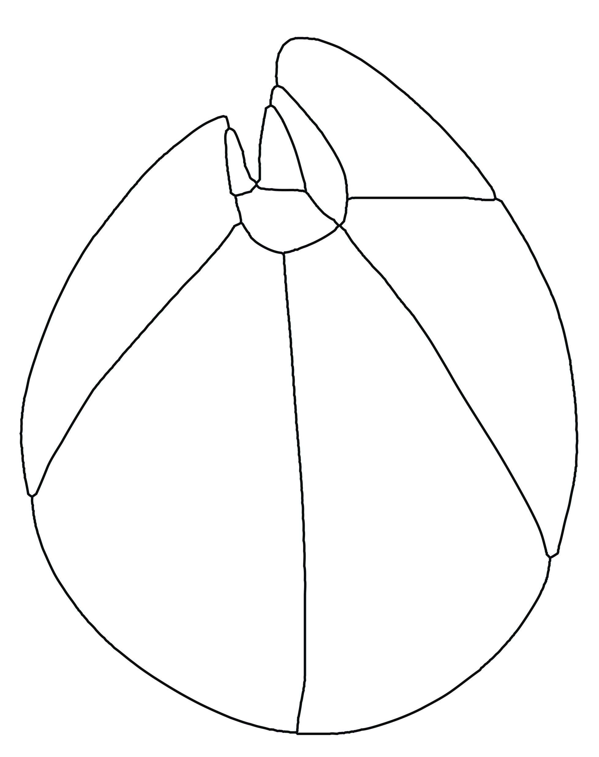
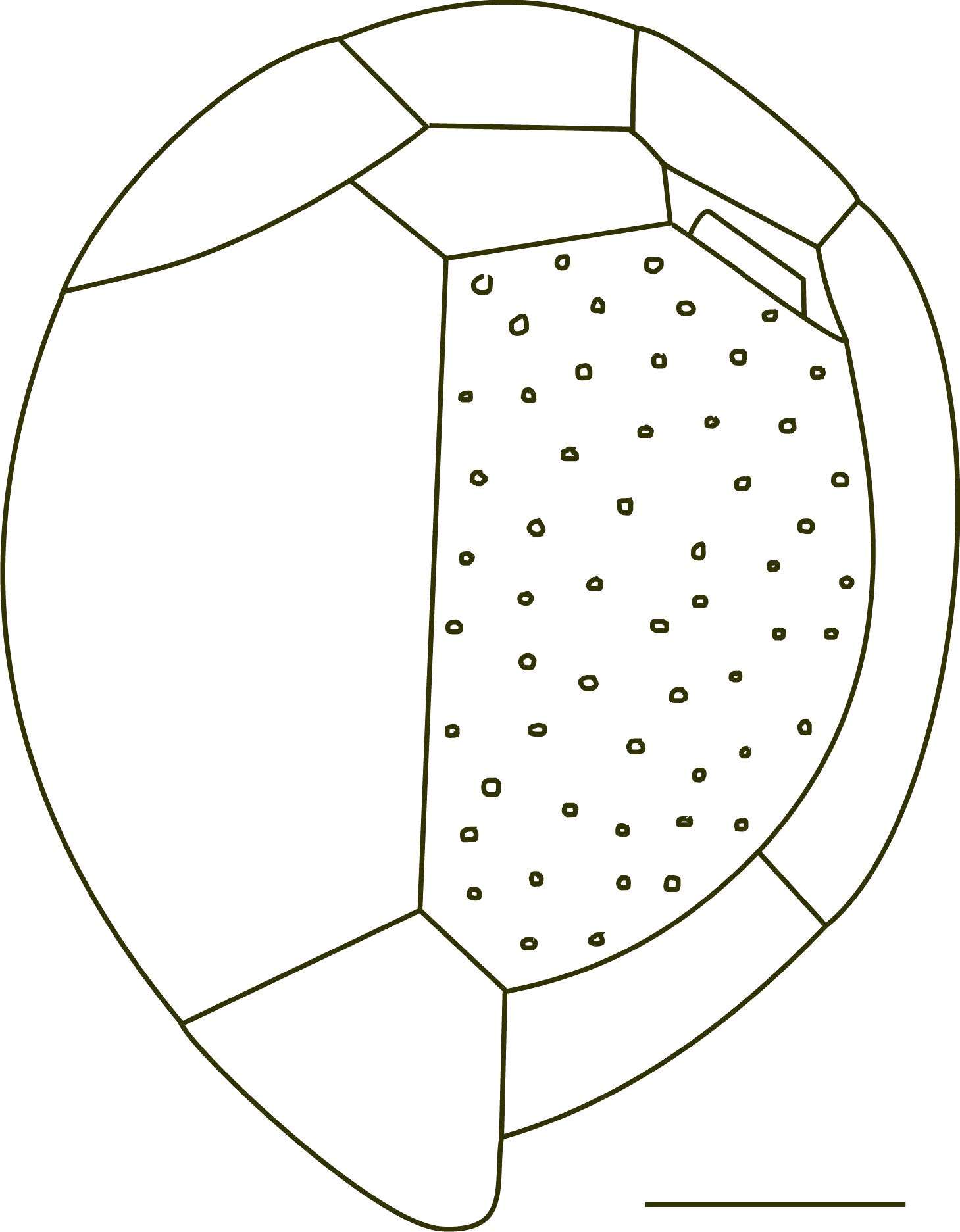
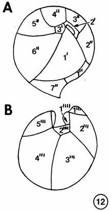
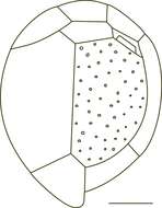
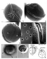
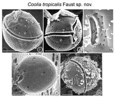
FIGS. 7-11. Scanning Electron micrographs of the surface morphology of Coolia tropicalis sp. nov. FIG. 7. Oblique dorsal view of C. tropicalis shows the apical pore and the equatorially located lipped cingulum. Cell surface is smooth with large scattered pores. FIG. 8. Cell is spherical in equatorial view shoving a deep cingulum and sulcus. Detritus adheres to the epitheca. FIG. 9. Antapical view of a cell show large unequal plates. FIG. 10. Apical pore is a narrow opening located in the epitheca. Fine detrital particles partially cover the thecal surface. FIG. 11. The apical pore is about 7 μm long straight and narrow slits with two supporting costae and evenly spaced round pores. Detritus attached to surface of apical pore plate. EMu:HOLOTYPE SEM NEGATIVE #166029; SEM STUB # 166; FIELD # 728-93;ACCESSION # 408431: CATALOG # 997; FIGURE # 7.
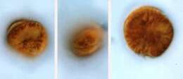
Coolia (coo-lee-a) monotis Meunier 1919. The images show swimming cells. The cells are flattened in the anterior-posterior plane, slightly asymmetrically. The cell on the left is in posterior-lateral view. The nucleus is visible. The cell in the middle is in ventral-lateral view. The cingulum is visible in the middle of the cell. The cell on the right is in posterior view. The cells contain yellow-brown plastids.