Plate 10
Description:
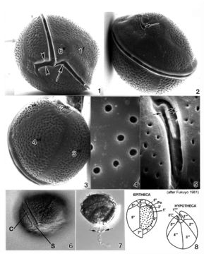
Plate 10. Coolia monotis: Figs. 1-5. SEM. Fig. 1. Ventral view: spherical shape. Cingulum lipped and equatorial. Sulcus with flexible lists (arrowheads). Ventral pore present (arrow). Fig. 2. Dorsal view: apical pore plate (arrow), Po, located off-center on epitheca. Fig. 3. Antapical view: hypothecal plates. Fig. 4. Smooth edged thecal pores unevenly distributed. Fig. 5. Po about 12 _ long, slightly curved and narrow with a slit-like apical pore. Two supporting rib-like costae (arrows) and evenly spaced round pores surround the pore. Figs. 6,7. LM. Fig. 6. Ventral view of lipped cingulum and sulcus. Fig. 7. Planozygote with two longitudinal flagella (arrows). Fig. 8. Line drawing: thecal plate arrangement.
Included On The Following Pages:
- Life (biota)
- Cellular
- Eukaryota (eukaryotes)
- SAR (Stramenopiles, Alveolates, Rhizaria)
- Alveolata (alveolates)
- Dinophyceae (dinoflagellates)
- Ostreopsidaceae
- Coolia
- Coolia monotis
- Gonyaulacales
- Dinoflagellata (Dinoflagellate)
This image is not featured in any collections.
Source Information
- license
- cc-publicdomain
- bibliographic citation
- Faust, Maria A. and Rose A. Gulledge. Identifying Harmful Marine Dinoflagellates. Smithsonian Contributions from the United States National Herbarium, volume 42: 1-144 (including 48 plates, 1 figure and 1 table).
- original
- original media file
- visit source
- partner site
- NMNH Marine Dinoflagellates
- ID