NMNH Coolia tropicalis type specimen
Description:
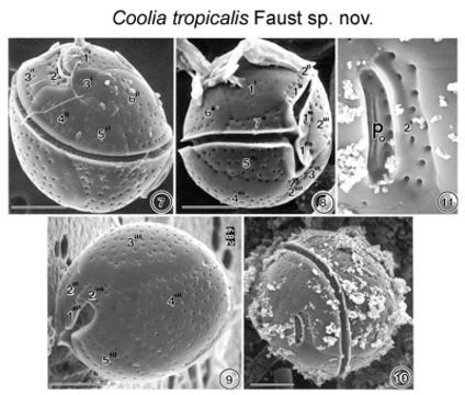
FIGS. 7-11. Scanning Electron micrographs of the surface morphology of Coolia tropicalis sp. nov. FIG. 7. Oblique dorsal view of C. tropicalis shows the apical pore and the equatorially located lipped cingulum. Cell surface is smooth with large scattered pores. FIG. 8. Cell is spherical in equatorial view shoving a deep cingulum and sulcus. Detritus adheres to the epitheca. FIG. 9. Antapical view of a cell show large unequal plates. FIG. 10. Apical pore is a narrow opening located in the epitheca. Fine detrital particles partially cover the thecal surface. FIG. 11. The apical pore is about 7 μm long straight and narrow slits with two supporting costae and evenly spaced round pores. Detritus attached to surface of apical pore plate. EMu:HOLOTYPE SEM NEGATIVE #166029; SEM STUB # 166; FIELD # 728-93;ACCESSION # 408431: CATALOG # 997; FIGURE # 7.
Included On The Following Pages:
- Life (biota)
- Cellular
- Eukaryota (eukaryotes)
- SAR (Stramenopiles, Alveolates, Rhizaria)
- Alveolata (alveolates)
- Dinophyceae (dinoflagellates)
- Ostreopsidaceae
- Coolia
- Coolia tropicalis
- Gonyaulacales
- Dinoflagellata (Dinoflagellate)
This image is not featured in any collections.
Source Information
- license
- cc-by-nc-sa-3.0
- copyright
- National Museum of Natural History, Smithsonian Institution
- original
- original media file
- visit source
- partner site
- NMNH Marine Dinoflagellates
- ID