-
Jaime Gonzalez-Cueto, Sigmer Quiroga, Jon Norenburg
Zookeys
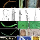
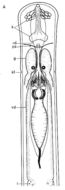
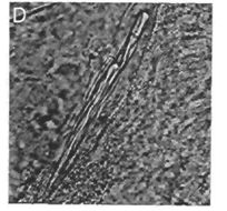
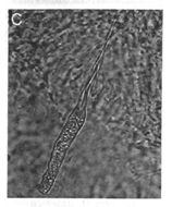
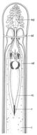
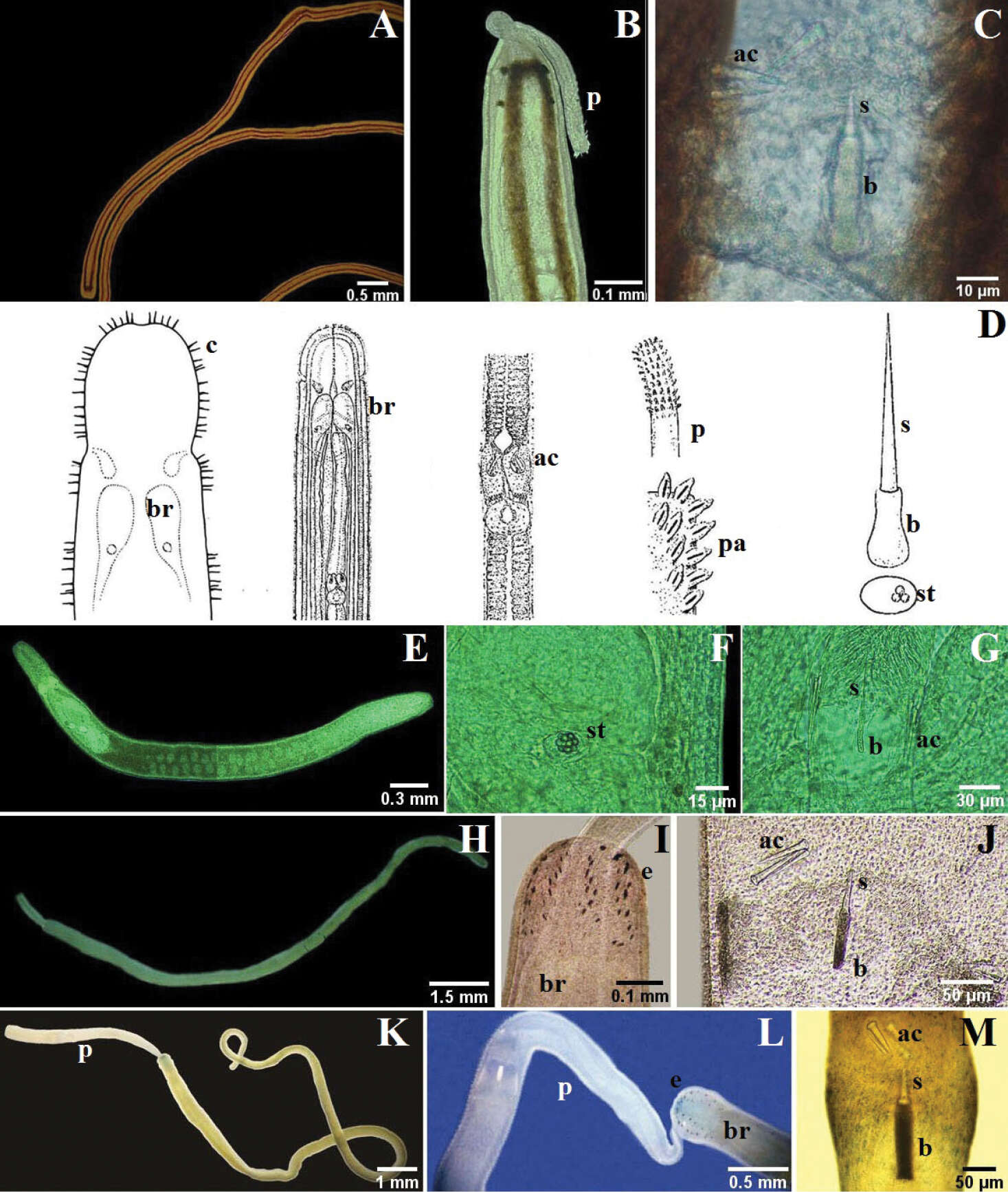
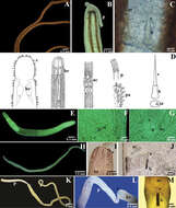
Figure 4.A–C Nemertopsis bivittata: B dorsal detail of the head and proboscis, C detail of the stylets D Ototyphlonemertes erneba (modified from Corrêa 1950 and Kirsteuer 1977) E–G Ototyphlonemertes lactea: E entire worm F detail of the statocysts G detail of the stylets H–J Zygonemertes fragariae: H entire worm I detail of the head J detail of the stylets K–M Zygonemertes virescens: K entire worm L detail of the head M detail of the stylets. ac accessory stylet, b base of the stylet, br brain, c sensorial cirrus, e eyes, p proboscis, pa proboscis papilla, s sylet, st statocysts.
-
Jaime Gonzalez-Cueto, Sigmer Quiroga, Jon Norenburg
Zookeys
Figure 4.A–C Nemertopsis bivittata: B dorsal detail of the head and proboscis, C detail of the stylets D Ototyphlonemertes erneba (modified from Corrêa 1950 and Kirsteuer 1977) E–G Ototyphlonemertes lactea: E entire worm F detail of the statocysts G detail of the stylets H–J Zygonemertes fragariae: H entire worm I detail of the head J detail of the stylets K–M Zygonemertes virescens: K entire worm L detail of the head M detail of the stylets. ac accessory stylet, b base of the stylet, br brain, c sensorial cirrus, e eyes, p proboscis, pa proboscis papilla, s sylet, st statocysts.
-
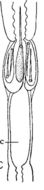
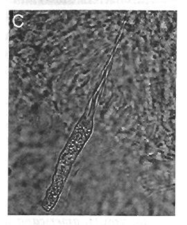
Stylet region of proboscis, showing slender styleta and basis and tubular middle chamber
-
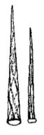
Stylet of larger and smaller individuals, indicating spiral ridges
-
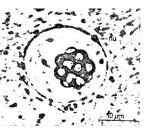
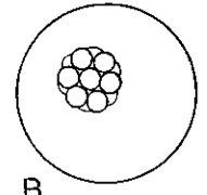
statocyst with spherical statolith
-
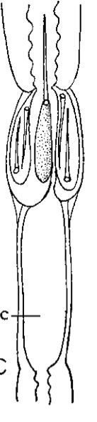
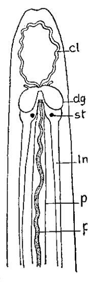
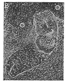
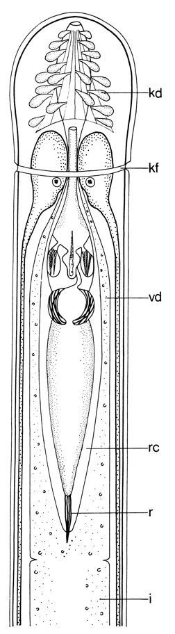
Outline of anterior portion of body showing statocysts (st) on ventral ganglia...outline of anterior portion of body showing statocysts (st) on ventral ganglia; other letters indicate: ct, cephalic blood lacunae; dg, dorsal ganglion; tn, lateral nerve cord; p, proboscis; PI, proboscis sheath
-
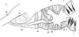
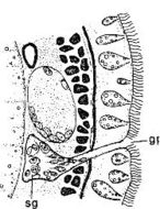
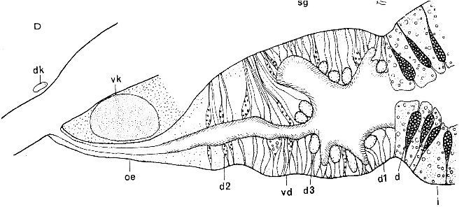
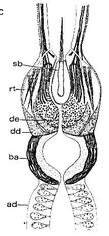
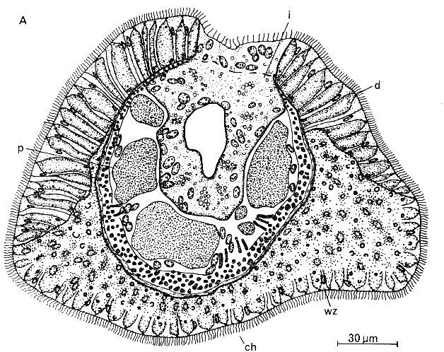
Organisation des vorderen Darmkanals (Sagitralschnitt)
-
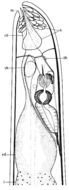
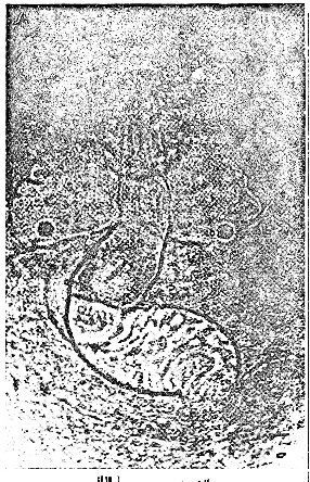
Lage und Organisation der Gonaden (Hoden).
-
Lage und Organisation der Gonaden (Ovar)
-
Statocyste
-
Hauptstilctt mit Ductus ejaculatorius
-
Rekonstruktion des Stilettapparates
-
Diaphragma und BaIlon.
-
Lagebeziehung von Gehirn und Russel.
-
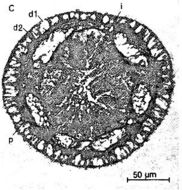
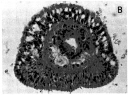
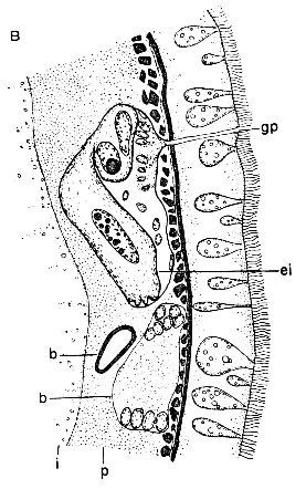
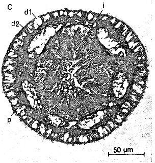
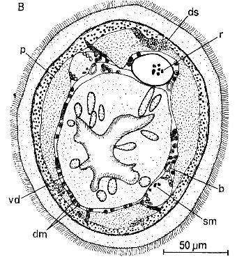
Querschnitt im Mitteldarmbereich mit Parenchymkomplexen und Epiderrnisdrusen
-
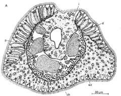
Querschnitt durch die Vorderdarmregion mit Darmmuskulatur.
-
Querschnitt durch die caudale Haflplatte unmittelbar vor dem After
-
Sagittalschnitt
-
Horizontalansicht (nach Lebendbeobachtungen von Ax, verandert).
-
Anordnung der Reservestilette.
-
Aufbau des Hauptstiletts
-
Lagebeziehung von Gehirn und Russel
-
Organisation des Vorderendes
-
Adhesive plate in O. americana