-
Madrid, Madrid, Spain
-
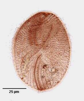
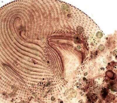
Dorsal view of the infraciliature of Disematostoma buetschlii LAUTERBORN, 1894.Stained by the silver carbonate technique (see Foissner, W. Europ. J. Protistol., 27:313-330;1991).Brightfield.
-
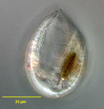
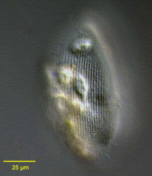
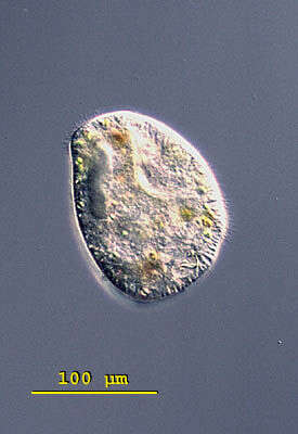
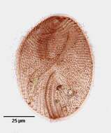
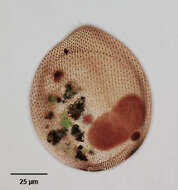
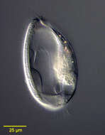
Portrait (ventral view) of the oligohymenophorean ciliate, Lembadion lucens (Maskell, 1887) Kahl, 1931 showing detail of the pellicle. The cell outline is oval. The ventral surface is concave and the dorsum convex. The very large scoop-like peristome occupies most of the ventral surface. The cytostome is at the posterior end of the peristome. There is a small undulating membrane on the right margin of the peristome. A large sheet-like adoral membranelle arises from the left margin of the peristome. There are 25-35 evenly spaced longitudinal somatic kineties. The posterior 2/3 of the pellicle of L. lucens has an areolate pattern divided into small roughly rectangular depressions (similar to the pattern of the entire pellicle of L. bullinum). The dikinetids of the somatic kineties occupy the center of the rectangles. The anterior 1/3 of the pellicle has a longitudinal striate pattern (similar to the pattern of the entire pellicle of L. magnum). The somatic dikinetids lie in the center of these striae. These details are seen here to the viewr's right of the peristome. Collected from a freshwater pond near Boise, Idaho July 2004. DIC .
-
Canencia, Madrid, Spain
-
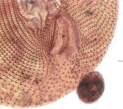
Detail of the oral infraciliature of Disematostoma buetschlii LAUTERBORN, 1894.Stained by the silver carbonate technique (see Foissner, W. Europ. J. Protistol., 27:313-330;1991).Brightfield.
-

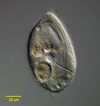
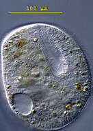
Portrait (ventral view) of the oligohymenophorean ciliate, Lembadion lucens (Maskell, 1887) Kahl, 1931. The cell outline is oval. The ventral surface is concave and the dorsum convex. The very large scoop-like peristome occupies most of the ventral surface. The cytostome is at the posterior end of the peristome. There is a small undulating membrane on the right margin of the peristome. A large sheet-like adoral membranelle arises from the left margin of the peristome. There are 25-35 evenly spaced longitudinal somatic kineties. The posterior 2/3 of the pellicle of L. lucens has an areolate pattern divided into small roughly rectangular depressions (similar to the pattern of the entire pellicle of L. bullinum). The dikinetids of the somatic kineties occupy the center of the rectangles. The anterior 1/3 of the pellicle has a longitudinal striate pattern (similar to the pattern of the entire pellicle of L. magnum). The somatic dikinetids lie in the center of these striae. There is usually a tuft of longer caudal cilia. The contractile vacuole (seen here to the viewerâs right of the macronucleus) connects with its excretory pore by a long curved canal (not seen here). The single ovoid macronucleus and micronucleus are posterior (the micronucleus is seen overlying the macronucleus here). Ingested diatoms and highly refractile crystalline inclusions are visible in the cytoplasm. L. lucens is distinguished from L. magnum and L. bullinum by its smaller size and the structure of its pellicle.Collected from a freshwater pond near Boise, Idaho July 2004. DIC .
-
Detail of the oral infraciliature of Disematostoma buetschlii LAUTERBORN, 1894.Stained by the silver carbonate technique (see Foissner, W. Europ. J. Protistol., 27:313-330;1991).Brightfield.
-
Ventral infraciliature of the oligohymenophorean ciliate, Lembadion lucens (Maskell, 1887) Kahl, 1931. There are 25-35 evenly spaced longitudinal somatic kineties. The posterior 2/3 of the pellicle of L. lucens has an areolate pattern divided into small roughly rectangular depressions (similar to the pattern of the entire pellicle of L. bullinum). The dikinetids of the somatic kineties occupy the center of the rectangles. The anterior 1/3 of the pellicle has a longitudinal striate pattern (similar to the pattern of the entire pellicle of L. magnum). The somatic dikinetids lie in the center of these striae. This specimen is stained by the silver carbonate technic (see Foissner, W. Europ. J. Protistol., 27:313-330;1991). This technic usually demonstrates the infraciliature which includes kinetodesmal fibrils and other structures not considered part of the silverline system but in this instance the silveline system and somatic dikinetids are stained. L.lucens is distinguished from L. magnum and L. bullinum by its smaller size and the structure of its pellicle. Specimen collected from a freshwater pond near Boise, Idaho 2005.Brightfield.
-
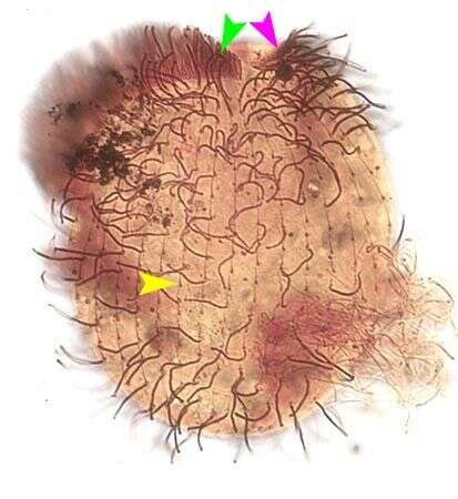
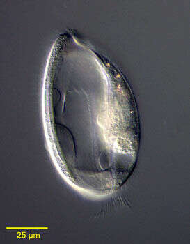
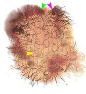
Portrait (ventral view) of the oligohymenophorean ciliate, Lembadion lucens (Maskell, 1887) Kahl, 1931. The cell outline is oval. The ventral surface is concave and the dorsum convex. The very large scoop-like peristome occupies most of the ventral surface. The cytostome is at the posterior end of the peristome. There is a small undulating membrane on the right margin of the peristome (pink arrowhead). A large sheet-like adoral membranelle arises from the left margin of the peristome (green arrowhead). There are 25-35 evenly spaced longitudinal somatic kineties (yellow arrowhead). The posterior 2/3 of the pellicle of L. lucens has an areolate pattern divided into small roughly rectangular depressions (similar to the pattern of the entire pellicle of L. bullinum). The dikinetids of the somatic kineties occupy the center of the rectangles. The anterior 1/3 of the pellicle has a longitudinal striate pattern (similar to the pattern of the entire pellicle of L. magnum). The somatic dikinetids lie in the center of these striae. There is usually a tuft of longer caudal cilia. The contractile vacuole connects with its excretory pore by a long curved canal. The single ovoid macronucleus and micronucleus are posterior. L. lucens is distinguished from L. magnum and L. bullinum by its smaller size and the structure of its pellicle. Stained by the silver carbonate technique (see Foissner, W.Europ. J. Protistol.27:313-330;1991).Brightfield.
-

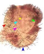
Portrait (ventral view) of the infraciliature of the oligohymenophorean ciliate, Lembadion lucens (Maskell, 1887) Kahl, 1931. The cell outline is oval. The ventral surface is concave and the dorsum convex. The very large scoop-like peristome occupies most of the ventral surface. The cytostome is at the posterior end of the peristome. There is a small undulating membrane on the right margin of the peristome (pink arrowhead). There is a densely impregnated longitudinal line of fibrils running the length of the peristome parallel to the undulating membrane (light blue arrowhead). A system of fine transverse fibrils lines the floor of the peristome. A large sheet-like adoral membranelle arises from the left margin of the peristome (green arrowhead). There are 25-35 evenly spaced longitudinal somatic kineties (red arrowhead). The posterior 2/3 of the pellicle of L. lucens has an areolate pattern divided into small roughly rectangular depressions (similar to the pattern of the entire pellicle of L. bullinum). The dikinetids of the somatic kineties occupy the center of the rectangles. The anterior 1/3 of the pellicle has a longitudinal striate pattern (similar to the pattern of the entire pellicle of L. magnum). The somatic dikinetids lie in the center of these striae. There is usually a tuft of longer caudal cilia. The closely-spaced kinetosomes from which these cilia arise are indicated by the dark blue arrowhead. The contractile vacuole connects with its excretory pore by a long curved canal. The single ovoid macronucleus and micronucleus are posterior. L. lucens is distinguished from L. magnum and L. bullinum by its smaller size and the structure of its pellicle. Stained by the silver carbonate technique (see Foissner, W.Europ. J. Protistol.27:313-330;1991).Brightfield.
-
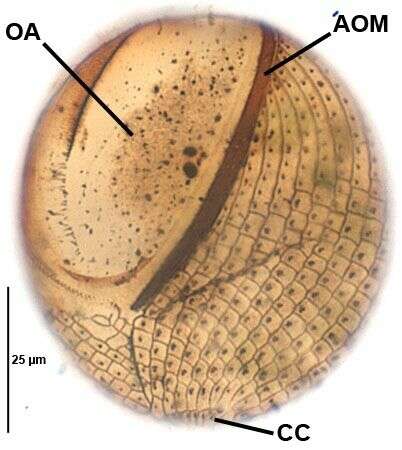
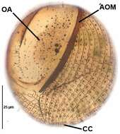
Ventral infraciliature of the oligohymenophorean ciliate, Lembadion lucens (Maskell, 1887) Kahl, 1931. The very large oral aperture (OA) is bordered on the left by a vast undulating membrane, the densely stained base of which is seen here (AOM). The kinetids of the elongated caudal cilia form a line at the posterior pole (CC).There are 25-35 evenly spaced longitudinal somatic kineties. The posterior 2/3 of the pellicle of L. lucens has an areolate pattern divided into small roughly rectangular depressions (similar to the pattern of the entire pellicle of L. bullinum). The dikinetids of the somatic kineties occupy the center of the rectangles. The anterior 1/3 of the pellicle has a longitudinal striate pattern (similar to the pattern of the entire pellicle of L. magnum). The somatic dikinetids lie in the center of these striae. This specimen is stained by the silver carbonate technic (see Foissner, W. Europ. J. Protistol., 27:313-330;1991). This technic usually demonstrates the infraciliature which includes kinetodesmal fibrils and other structures not considered part of the silverline system but in this instance the silveline system and somatic dikinetids are stained. L.lucens is distinguished from L. magnum and L. bullinum by its smaller size and the structure of its pellicle. Specimen collected from a freshwater pond near Boise, Idaho 2005.Brightfield.
-
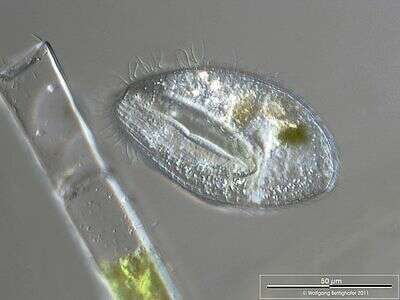
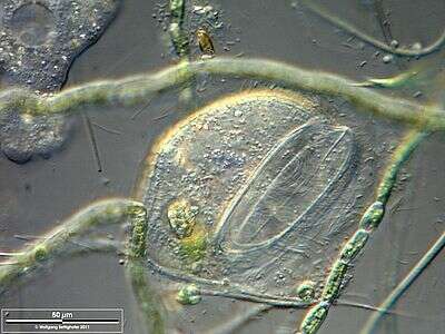
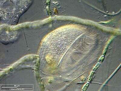
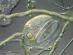
Scale bar indicates 50 µm. Sample from the pond Hegne Moor situated in the vicinity of Lake Constance (Bodensee, Southern Germany). The image was built up using several photomicrographic frames with manual stacking technique. Images were taken using Zeiss Universal with Olympus C7070 CCD camera.
-
Scale bar indicates 50 µm. Sample from the pond Hegne Moor situated in the vicinity of Lake Constance (Bodensee, Southern Germany). The image was built up using several photomicrographic frames with manual stacking technique. Images were taken using Zeiss Universal with Olympus C7070 CCD camera.
-
Scale bar indicates 50 µm. Sample from the pond Hegne Moor situated in the vicinity of Lake Constance (Bodensee, Southern Germany). The image was built up using several photomicrographic frames with manual stacking technique. Images were taken using Zeiss Universal with Olympus C7070 CCD camera.
-
Infraciliature (ventral view) of the oligohymenophorean ciliate, Lembadion magnum (Stokes,1887;Kahl,1931). Cell outline is oval. The ventral surface is concave and the dorsum convex. The very large scoop-like peristome occupies most of the ventral surface. The cytostome is at the posterior end of the peristome. There is a long thin undulating membrane on the right margin of the peristome (seen here). A large sheet-like adoral membranelle arises from the left margin of the peristome (seen here as a broad light brown band on viewer's right). The longitudinal somatic kineties run between prominent pellicular striations visible here on either side of cytostome. This feature helps differentiate L. magnum from L. bullinum which has an areolate cortex. There is usually a tuft of longer caudal cilia. The single macronucleus and micronucleus are posterior (densely stained in this specimen). Collected from a freshwater pond near Boise, Idaho May 2004. stained by the silver carbonate technique (see Foissner, W.Europ. J. Protistol.27,313-330;1991).Brightfield.
-
Infraciliature (dorsal view) of the oligohymenophorean ciliate, Lembadion magnum (Stokes,1887;Kahl,1931). Cell outline is oval. The ventral surface is concave and the dorsum convex. The very large scoop-like peristome occupies most of the ventral surface. The cytostome is at the posterior end of the peristome. There is a small undulating membrane on the right margin of the peristome. A large sheet-like adoral membranelle arises from the left margin of the peristome. The longitudinal somatic kineties (seen here) run between prominent pellicular striations. This feature helps differentiate L. magnum from L. bullinum which has an areolate cortex. There is usually a tuft of longer caudal cilia. The single contractile vacuole connects with its excretory pore by a long curved canal. The single macronucleus and micronucleus are posterior (densely stained here). Collected from a freshwater pond near Boise, Idaho May 2004. Stained by silver carbonate technique (see Foissner, W.Europ. J. Protistol.27,313-330;1991).Brightfield.
-
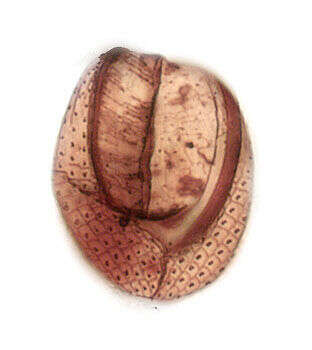
Pellicular detail of the oligohymenophorean ciliate, Lembadion magnum The longitudinal somatic kineties run between prominent pellicular striations. This feature helps differentiate L. magnum from L. bullinum which has an areolate cortex. Collected from a freshwater pond near Boise, Idaho May 2004. DIC optics.
-
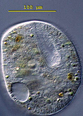
Portrait (dorsal view) of the oligohymenophorean ciliate, Lembadion magnum (Stokes,1887;Kahl,1931). Cell outline is oval. The ventral surface is concave and the dorsum convex. The very large scoop-like peristome occupies most of the ventral surface. The cytostome is at the posterior end of the peristome. There is a small undulating membrane on the right margin of the peristome. A large sheet-like adoral membranelle arises from the left margin of the peristome. The longitudinal somatic kineties run between prominent pellicular striations. This feature helps differentiate L. magnum from L. bullinum which has an areolate cortex. There is usually a tuft of longer caudal cilia. The single contractile vacuole connects with its excretory pore by a long curved canal (seen well in this image). Large food vacuoles are visible in the cytoplasm. The single macronucleus and micronucleus are posterior. Collected from a freshwater pond near Boise, Idaho May 2004. DIC optics.
-
Portrait (ventral view) of the oligohymenophorean ciliate, Lembadion magnum (Stokes,1887;Kahl,1931). Cell outline is oval. The ventral surface is concave and the dorsum convex. The very large scoop-like peristome occupies most of the ventral surface. The cytostome is at the posterior end of the peristome. There is a small undulating membrane on the right margin of the peristome. A large sheet-like adoral membranelle arises from the left margin of the peristome. The longitudinal somatic kineties run between prominent pellicular striations. This feature helps differentiate L. magnum from L. bullinum which has an areolate cortex. There is usually a tuft of longer caudal cilia. The single contractile vacuole connects with its excretory pore by a long curved canal. The single macronucleus and micronucleus are posterior. Collected from a freshwater pond near Boise, Idaho May 2004. DIC optics.
-
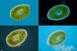
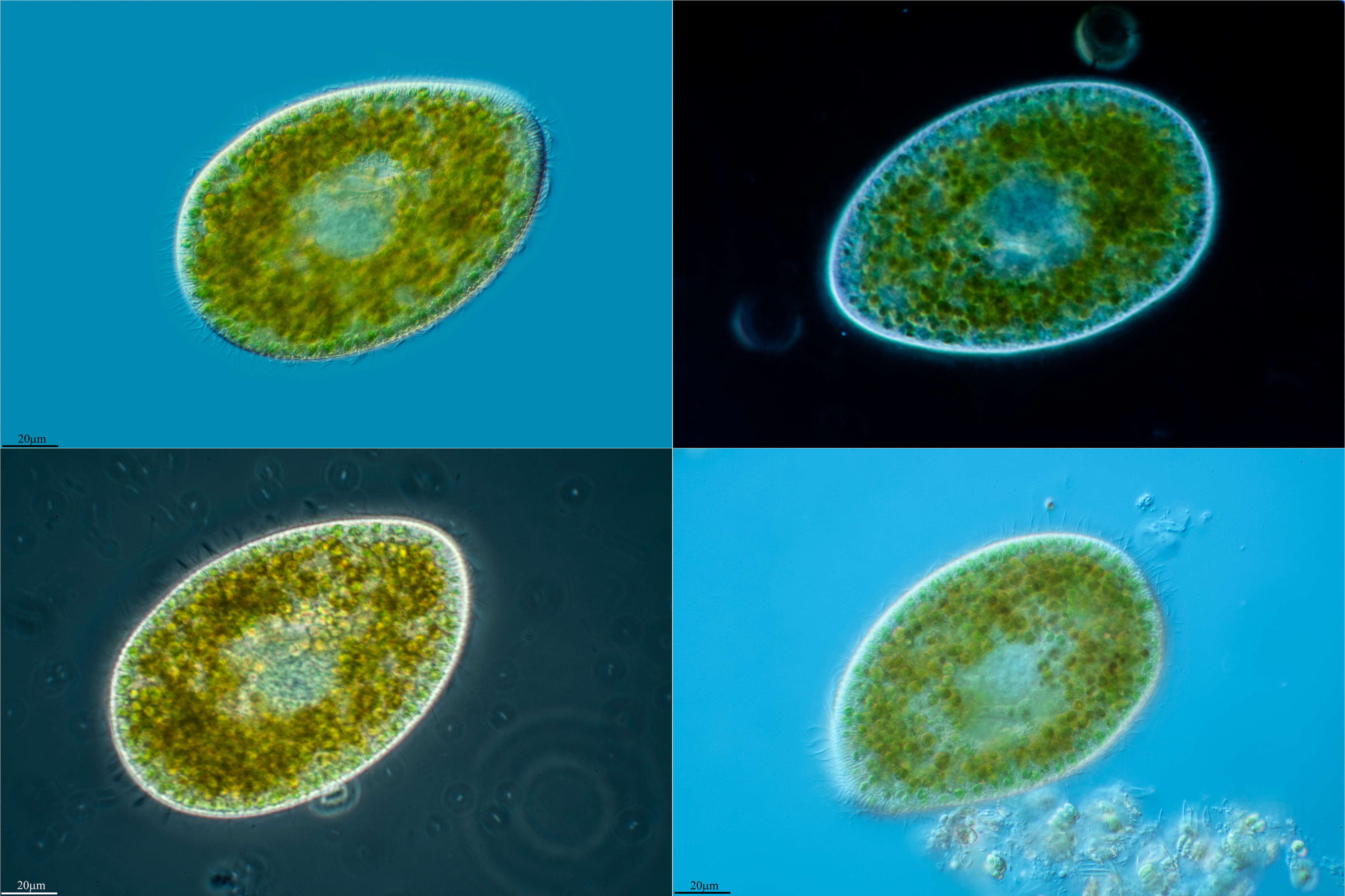
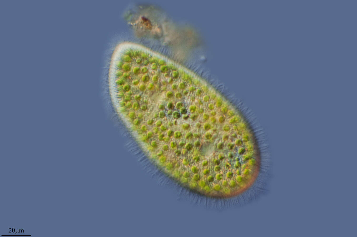
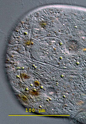
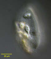
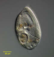
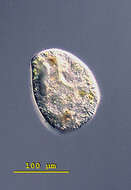
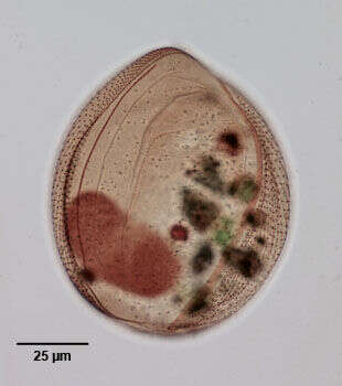
Stokesia (stoke-see-a) vernalis, the only member of the genus; this is an obliquely cone-shaped planktonic ciliate. The oral aperture is located on the flat ventral side and is formed like a furrow. The furrow is equipped an undulating membrane and two membranelles. There are symbiotic green algae in the cytoplasm. There are 3 - 4 micronuclei attached to the ellipsoidal macronucleus. The contractile vacuole is located dorsally and has very obvious pores and collecting canals. This species can be 100 - 220 microns long. This specimen was collected in freshwater ponds near Konstanz, Germany. Differential interference contrast.
-
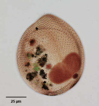
Stokesia (stoke-see-a) vernalis, the only member of the genus; this is an obliquely cone-shaped planktonic ciliate. The oral aperture is located on the flat ventral side and is formed like a furrow. The furrow is equipped an undulating membrane and two membranelles. There are symbiotic green algae in the cytoplasm. There are 3 - 4 micronuclei attached to the ellipsoidal macronucleus. The contractile vacuole is located dorsally and has very obvious pores and collecting canals. This species can be 100 - 220 microns long. This specimen was collected in freshwater ponds near Konstanz, Germany. Focal plane on the pores and collecting canals of the contractile vacuole. Differential interference contrast.
-
Stokesia (stoke-see-a) vernalis, the only member of the genus; this is an obliquely cone-shaped planktonic ciliate. The oral aperture is located on the flat ventral side and is formed like a furrow. The furrow is equipped an undulating membrane and two membranelles. There are symbiotic green algae in the cytoplasm. There are 3 - 4 micronuclei attached to the ellipsoidal macronucleus. The contractile vacuole is located dorsally and has very obvious pores and collecting canals. This species can be 100 - 220 microns long. This specimen was collected in freshwater ponds near Konstanz, Germany. Focal plane on the pores and collecting canals of the contractile vacuole. Squashed specimen in which the oral apparatus and the contractile vacuole are visible. Differential interference contrast.
-
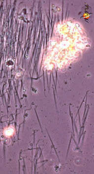
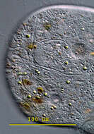
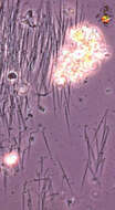
Frontonia (front-own-ee-a) is a peniculine ciliate and as such is closely related to the familiar Paramecium. It has many crystalline inclusions called trichocysts (a special form of extrusome). When stressed the crystalline structure of these changes, and they are expelled in large numbers and forceably from the cell. This action can force the cell away from the noxious stimulus. The expelled, the trichocysts look like little spears attached to the slide or to the substrate. Phase contrast.
-
Frontonia (front-own-ee-a) is a peniculine ciliate and as such is closely related to the familiar Paramecium. The mouth is supported by strong rods which assists Frontonia in ingesting its preferred food - diatoms and other moderate sized algae. A diatom can be seen inside the cell. The mouth is located at about 10 o clock. Like many peniculines the cell has many extrusomes lying just under the cell surface, and these are expelled when the cells are challenged. Large grey area is the nucleus. Phase contrast.