Ventral infraciliature
Description:
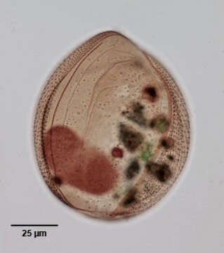
Infraciliature (ventral view) of the oligohymenophorean ciliate, Lembadion magnum (Stokes,1887;Kahl,1931). Cell outline is oval. The ventral surface is concave and the dorsum convex. The very large scoop-like peristome occupies most of the ventral surface. The cytostome is at the posterior end of the peristome. There is a long thin undulating membrane on the right margin of the peristome (seen here). A large sheet-like adoral membranelle arises from the left margin of the peristome (seen here as a broad light brown band on viewer's right). The longitudinal somatic kineties run between prominent pellicular striations visible here on either side of cytostome. This feature helps differentiate L. magnum from L. bullinum which has an areolate cortex. There is usually a tuft of longer caudal cilia. The single macronucleus and micronucleus are posterior (densely stained in this specimen). Collected from a freshwater pond near Boise, Idaho May 2004. stained by the silver carbonate technique (see Foissner, W.Europ. J. Protistol.27,313-330;1991).Brightfield.
Included On The Following Pages:
- Life (creatures)
- Cellular (cellular organisms)
- Eukaryota (eukaryotes)
- SAR (Stramenopiles, Alveolates, Rhizaria)
- Alveolata (alveolates)
- Ciliophora (ciliates)
- Intramacronucleata
- Oligohymenophorea
- Peniculida (Peniculid)
- Lembadionidae
- Lembadion
- Lembadion magnum
This image is not featured in any collections.
Source Information
- license
- cc-by-nc
- author
- William Bourland
- provider
- micro*scope
- original
- original media file
- visit source
- partner site
- micro*scope
- ID