Dorsal infraciliature
Description:
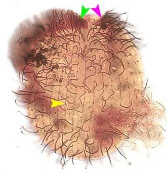
Portrait (ventral view) of the oligohymenophorean ciliate, Lembadion lucens (Maskell, 1887) Kahl, 1931. The cell outline is oval. The ventral surface is concave and the dorsum convex. The very large scoop-like peristome occupies most of the ventral surface. The cytostome is at the posterior end of the peristome. There is a small undulating membrane on the right margin of the peristome (pink arrowhead). A large sheet-like adoral membranelle arises from the left margin of the peristome (green arrowhead). There are 25-35 evenly spaced longitudinal somatic kineties (yellow arrowhead). The posterior 2/3 of the pellicle of L. lucens has an areolate pattern divided into small roughly rectangular depressions (similar to the pattern of the entire pellicle of L. bullinum). The dikinetids of the somatic kineties occupy the center of the rectangles. The anterior 1/3 of the pellicle has a longitudinal striate pattern (similar to the pattern of the entire pellicle of L. magnum). The somatic dikinetids lie in the center of these striae. There is usually a tuft of longer caudal cilia. The contractile vacuole connects with its excretory pore by a long curved canal. The single ovoid macronucleus and micronucleus are posterior. L. lucens is distinguished from L. magnum and L. bullinum by its smaller size and the structure of its pellicle. Stained by the silver carbonate technique (see Foissner, W.Europ. J. Protistol.27:313-330;1991).Brightfield.
Included On The Following Pages:
- Life (creatures)
- Cellular (cellular organisms)
- Eukaryota (eukaryotes)
- SAR (Stramenopiles, Alveolates, Rhizaria)
- Alveolata (alveolates)
- Ciliophora (ciliates)
- Intramacronucleata
- Oligohymenophorea
- Peniculida (Peniculid)
- Lembadionidae
- Lembadion
- Lembadion lucens
This image is not featured in any collections.
Source Information
- license
- cc-by-nc
- author
- William Bourland
- provider
- micro*scope
- original
- original media file
- visit source
- partner site
- micro*scope
- ID