About
Education
Discuss
TraitBank
Sign In
Sign Up
Language
Deutsch
English
Español
français
italiano
Nederlands
Piemontèis
Português do Brasil
suomi
Türkçe
Čeština
Ελληνικά
македонски
Українська
العربية
简体中文
繁體中文
names in breadcrumbs
vernacular
scientific
About
Education
Discuss
TraitBank
Sign In
Sign Up
en
Deutsch
English
Español
français
italiano
Nederlands
Piemontèis
Português do Brasil
suomi
Türkçe
Čeština
Ελληνικά
македонски
Українська
العربية
简体中文
繁體中文
names in breadcrumbs
vernacular
scientific
Life
»
…
»
Alveolates
»
Ciliates
»
…
Life
»
Cellular
»
Eukaryotes
»
SAR (Stramenopiles, Alveolates, Rhizaria)
»
Alveolates
»
Ciliates
»
Intramacronucleata
»
Oligohymenophorea
«
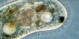
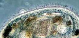
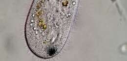
Peniculid
Peniculida
collect
overview
data
media
articles
maps
names
license
any license
CC-BY
CC-BY-NC
CC-BY-NC-SA
CC-BY-SA
No copyright
type
any type
image
video
provider
any provider
iNaturalist
Wikimedia Commons
Flickr Group
BioImages, the virtual fieldguide, UK
Freshwater and Marine Image Bank U Washington
micro*scope
protisten.de
vimeo
1
2
3
4
5
…
Last »
cc-publicdomain
trusted
cc-by-4.0
trusted
cc-by-4.0
trusted
cc-publicdomain
trusted
cc-publicdomain
trusted
cc-publicdomain
trusted
cc-by-nc-4.0
trusted
cc-by-nc-4.0
trusted
cc-by-nc-4.0
trusted
cc-by-nc-4.0
trusted
cc-by-nc-4.0
trusted
cc-by-nc-4.0
trusted
cc-by-nc-4.0
trusted
cc-by-nc-4.0
trusted
cc-publicdomain
trusted
cc-by-nc-4.0
trusted
cc-by-nc-4.0
trusted
cc-by-nc-4.0
trusted
cc-by-nc-4.0
trusted
cc-by-nc-4.0
trusted
cc-by-nc-4.0
trusted
cc-by-nc-4.0
trusted
cc-by-nc-4.0
trusted
cc-by-nc-4.0
trusted
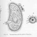
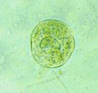
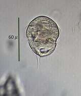
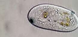
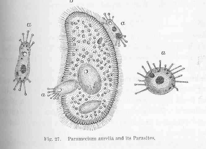
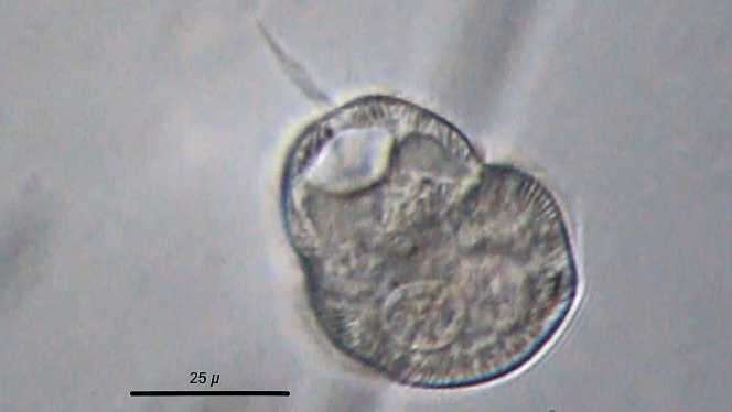
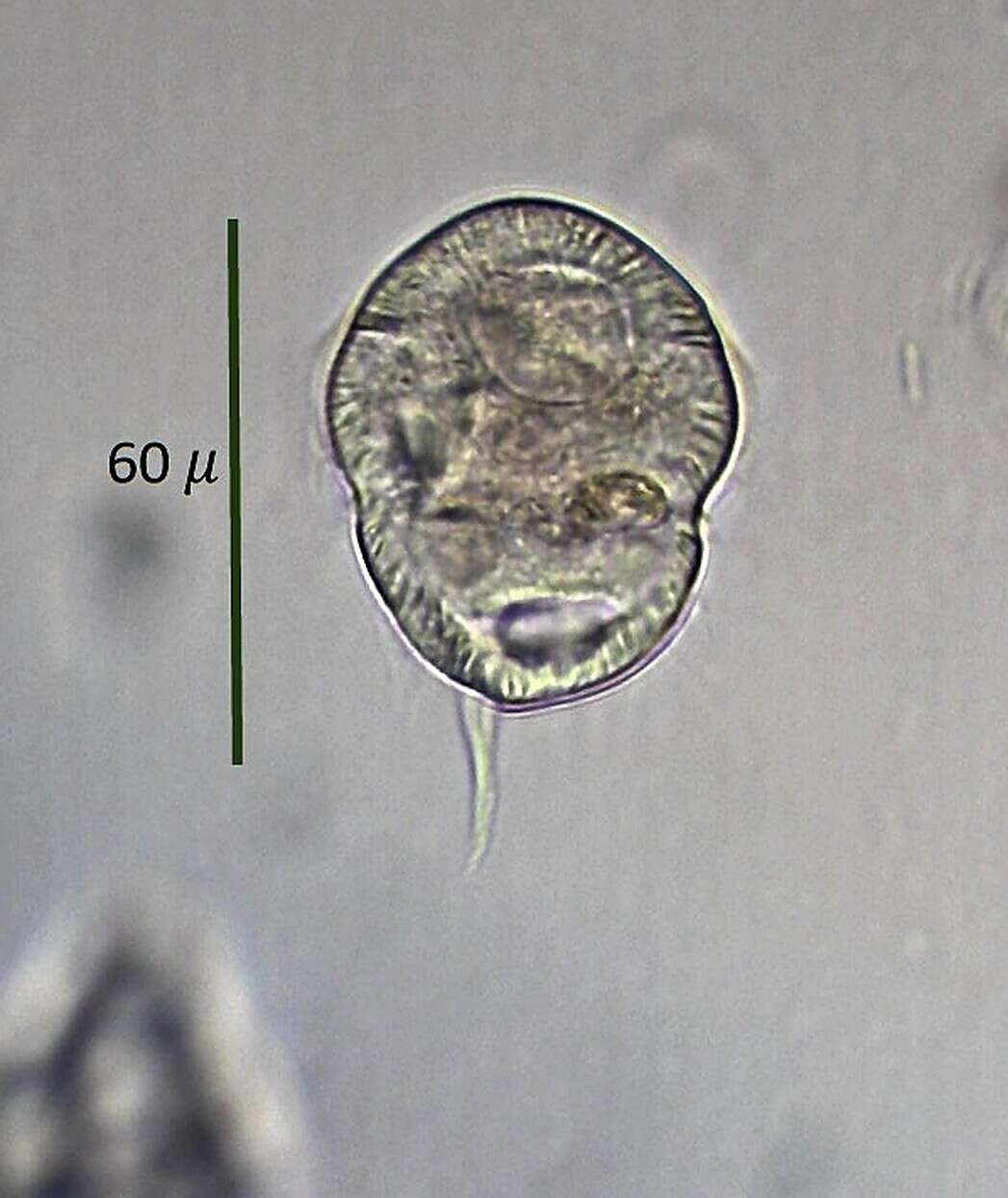
Paramecium aurelia and its Parasites. 1868. Paramecium aurelia.
cc-publicdomain
Freshwater and Marine Image Bank U Washington
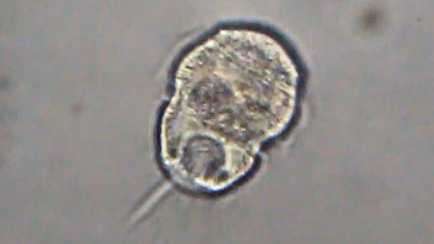
Paramecium aurelia and its Parasites.
"
cc-by-4.0
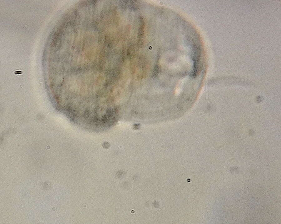
Don Loarie
iNaturalist
"
cc-by-4.0
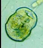
Don Loarie
iNaturalist
"
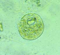
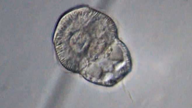
cc-publicdomain
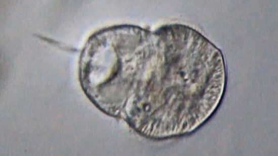
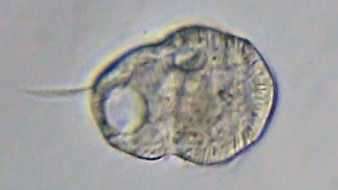
Ken Kneidel
iNaturalist
"
cc-publicdomain
Ken Kneidel
iNaturalist
"
cc-publicdomain
Ken Kneidel
iNaturalist
"
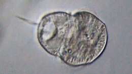
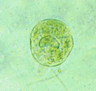
cc-by-nc-4.0
Brad Heath
iNaturalist
"
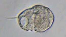
cc-by-nc-4.0
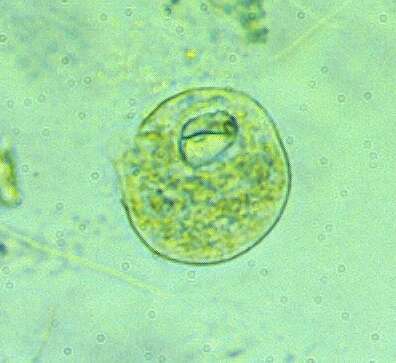
pierrelfr
iNaturalist
"
cc-by-nc-4.0
pierrelfr
iNaturalist
"
cc-by-nc-4.0
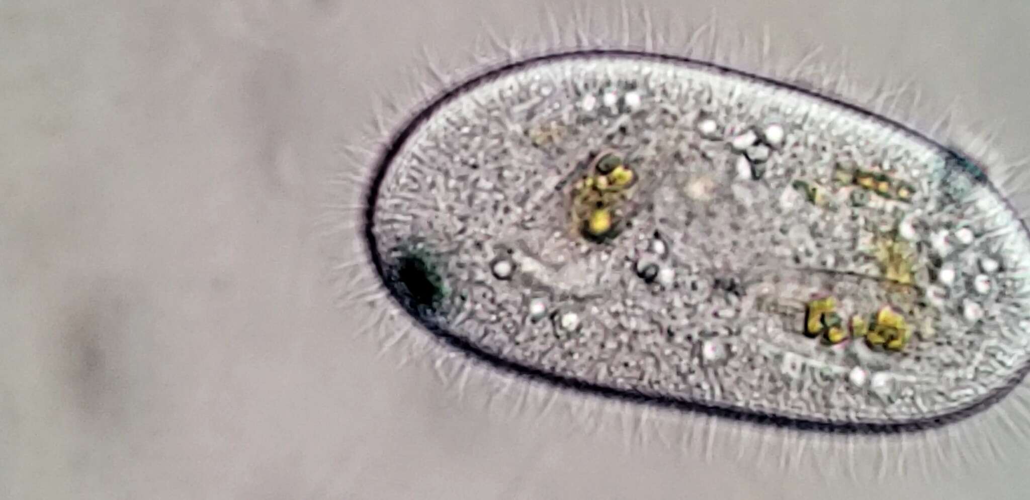
Logan Shaver
iNaturalist
"
cc-by-nc-4.0
pierrelfr
iNaturalist
"
cc-by-nc-4.0
Jane Trembath
iNaturalist
"
cc-by-nc-4.0
pierrelfr
iNaturalist
"
cc-by-nc-4.0
pierrelfr
iNaturalist
"
cc-publicdomain
Ken Kneidel
iNaturalist
"
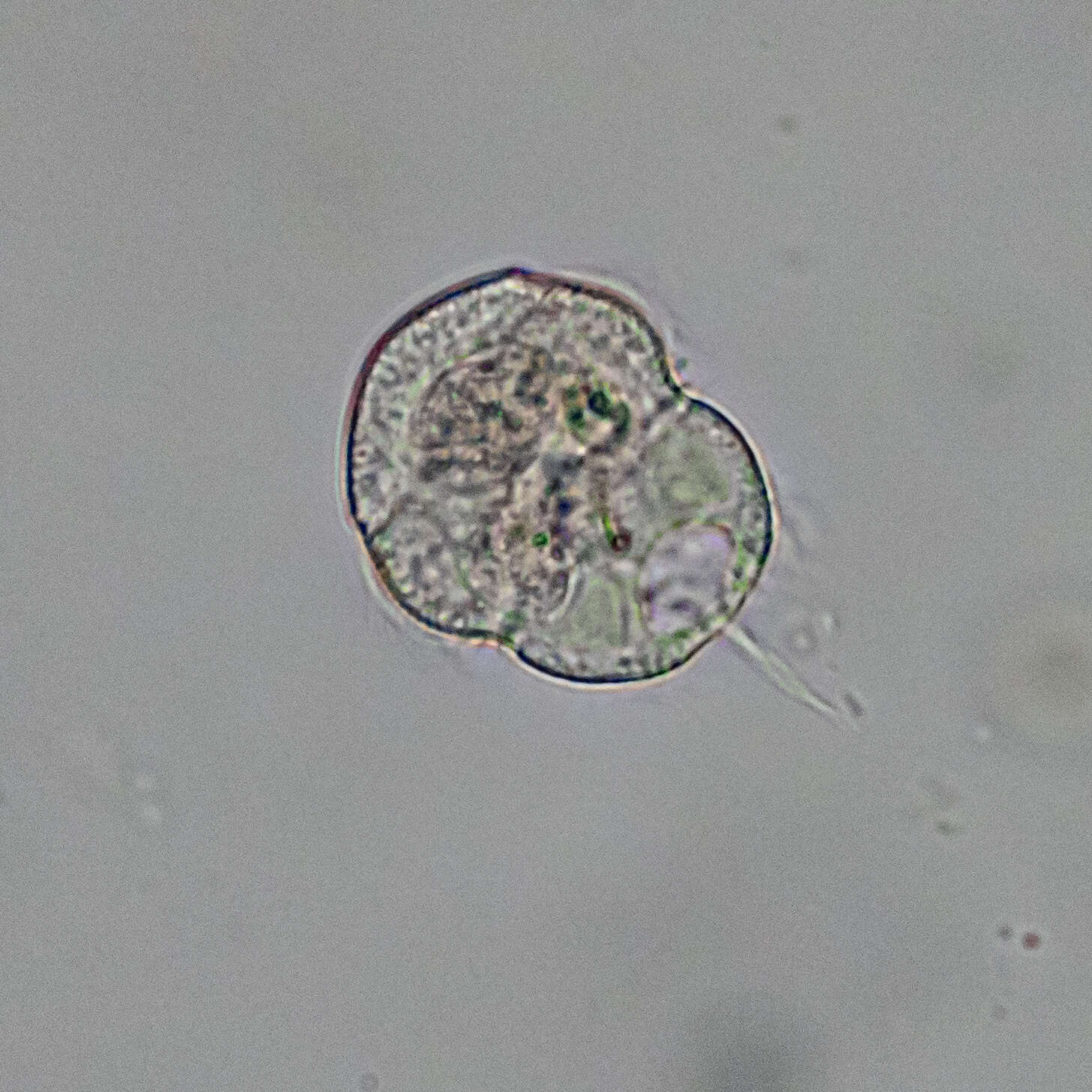
cc-by-nc-4.0
MikeN
iNaturalist
"
cc-by-nc-4.0
peptolab
iNaturalist
"
cc-by-nc-4.0
peptolab
iNaturalist
"
cc-by-nc-4.0
peptolab
iNaturalist
"
cc-by-nc-4.0
peptolab
iNaturalist
"
cc-by-nc-4.0
peptolab
iNaturalist
"
cc-by-nc-4.0
peptolab
iNaturalist
"
cc-by-nc-4.0
peptolab
iNaturalist
"
cc-by-nc-4.0
peptolab
iNaturalist
1
2
3
4
5
…
Last »