-
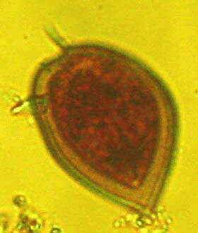
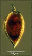
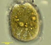
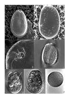
Prorocentrum rhathymum cells are asymmetrical oval in valve view, with the left side of the right valve slightly longer at the apical end. Valve length 29 - 40 microns, valve width 18 - 27microns, length to width ratio 1.4 -1.6. The apical area is a small indentation, covered by many small pores. A spine (approximately 2 microns long) is present to the right of the apical area on the right valve. Cell surface smooth, with large (0.5 microns ) pores, which are arranged in rows radiating from the periphery of the valve. Very small pores (approximately 0.1 microns ) also present. Extrusomes (3 - 5 microns ) are present in the anterior part of the valve, pointing in the direction of the apical area. Nucleus 10 microns diameter, in the posterior part of the valve. Plastid yellow-brown, diffuse.
-
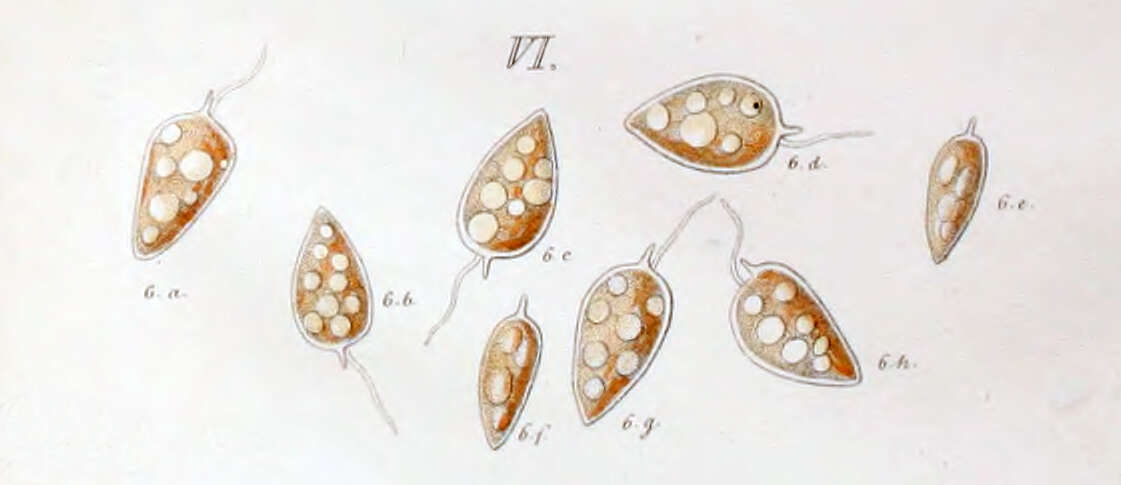
Members of this species have a fusiform body. The apex bears a prominent winged spine. In valve view the cell will have one arched and one convex side.
-
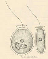
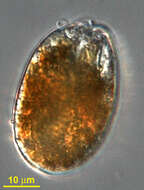
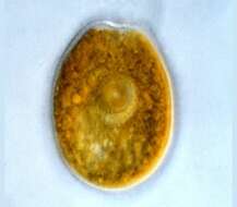
Exuviaella lima.
-
-
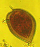
Prorocentrum rhathymum. Collected by ATOL team at Oyster Pond near to Woods Hole, Massachusetts for the Protistology Workshop at MBL. October-November 2005. Isolation and art by Adrian Reyes-Prieto.
-
Prorocentrum (pro-row-sent-rum) clipeus Hoppenrath 2000. The image shows one of the two valves of a cell. The nucleus is in the posterior of the cell. The plastids are yellow-brown. The cingulum is not visible. There is an apical spine present.
-
-
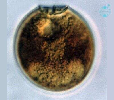
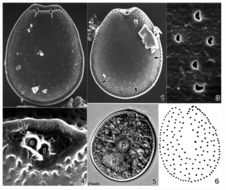
Prorocentrum clipeus cells are round, 37 - 44 long, 35 - 42 microns wide, length to width ratio 1.0-1.1. Apical area a wide rounded indentation, with a small list/collar present to the left side of the apical region. Small spine present, projecting from the apical region. Very small pores, approximately 0.1 microns diameter, present around the periphery of the valve and in short rows radiating towards the centre of the cell. Intercalary region with a horizontal banding pattern. Large yellow-brown plastid fills the cytoplasm. Large extrusomes (12 - 13 microns ) present in the anterior part of the valve, pointing towards the apical area. Nucleus large, approximately 20 microns by 10 microns , in the posterior part of the valve.
-
Prorocentrum emarginatum, showing plastids, observed in marine muds and sandy sediments in the vicinity of Broome, Western Australia in September 2003. This image was taken using differential interference contrast optics. This work was supported by the Australian Biological Resources Study.
-
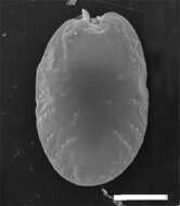
Prorocentrum emarginatum, an empty valve, observed in marine muds and sandy sediments in the vicinity of Broome, Western Australia in September 2003. This image was taken using phase contrast optics. This work was supported by the Australian Biological Resources Study.
-
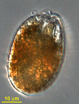
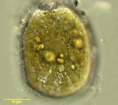
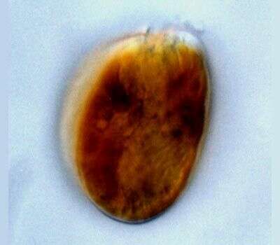
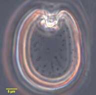
Prorocentrum (pro-row-sent-rum) lima (Ehrenberg) Dodge 1975. The image shows one of the two valves of a cell. The cingulum is not visible. The plastids are yellow-brown, and surround a large circular pyrenoid in the centre of the cell.
-
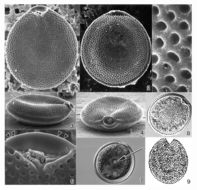
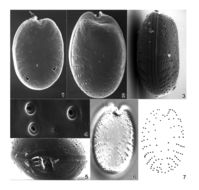
Prorocentrum lima valves are oval, 40 - 45 microns long, 27-33 microns wide , length to width ratio 1.3 - 1.5. Valve surface smooth. Valve pores scattered except in the centre, 58 - 80 per cell, approximately 0.3 microns diameter, round, oval to slightly kidney shaped. Marginal pores around periphery of cell, 51-74 per cell, round to oval approximately 0.3 microns diameter. Intercalary band smooth. Apical area consists of a small triangular indentation. A small flange (apical collar) is present. Plastids large, orange-brown. A pyrenoid, 7 - 8 microns diameter, is situated centrally. Nucleus in the posterior of the valve.
-
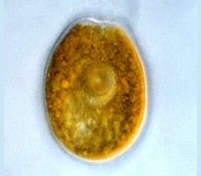
Prorocentrum (pro-row-sent-rum) mexicanum Tafall 1942. The image shows the valve of a cell. The plastid is yellow-brown. There is a small apical spine present. The cingulum is not visible.
-
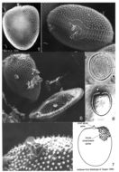
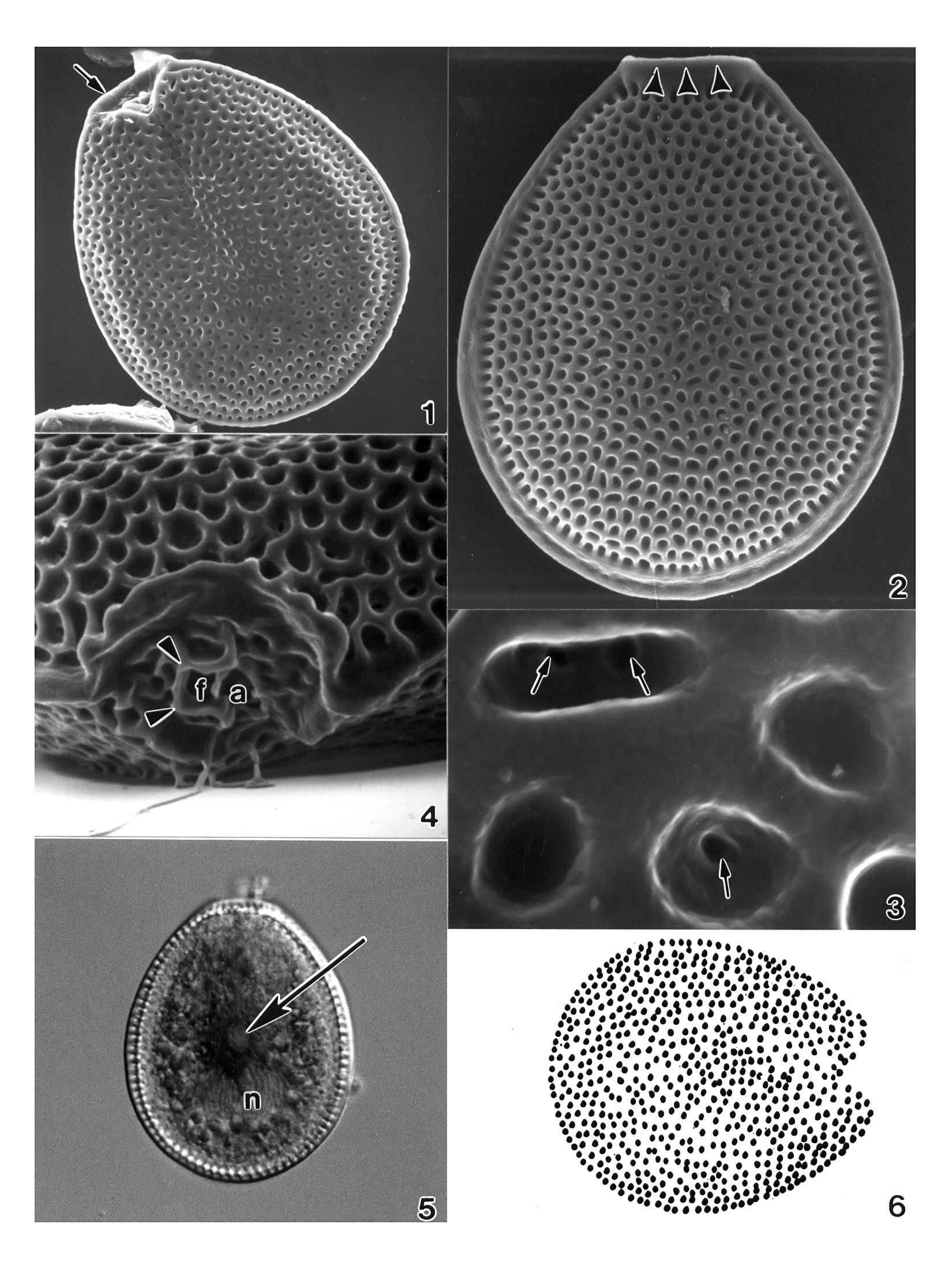
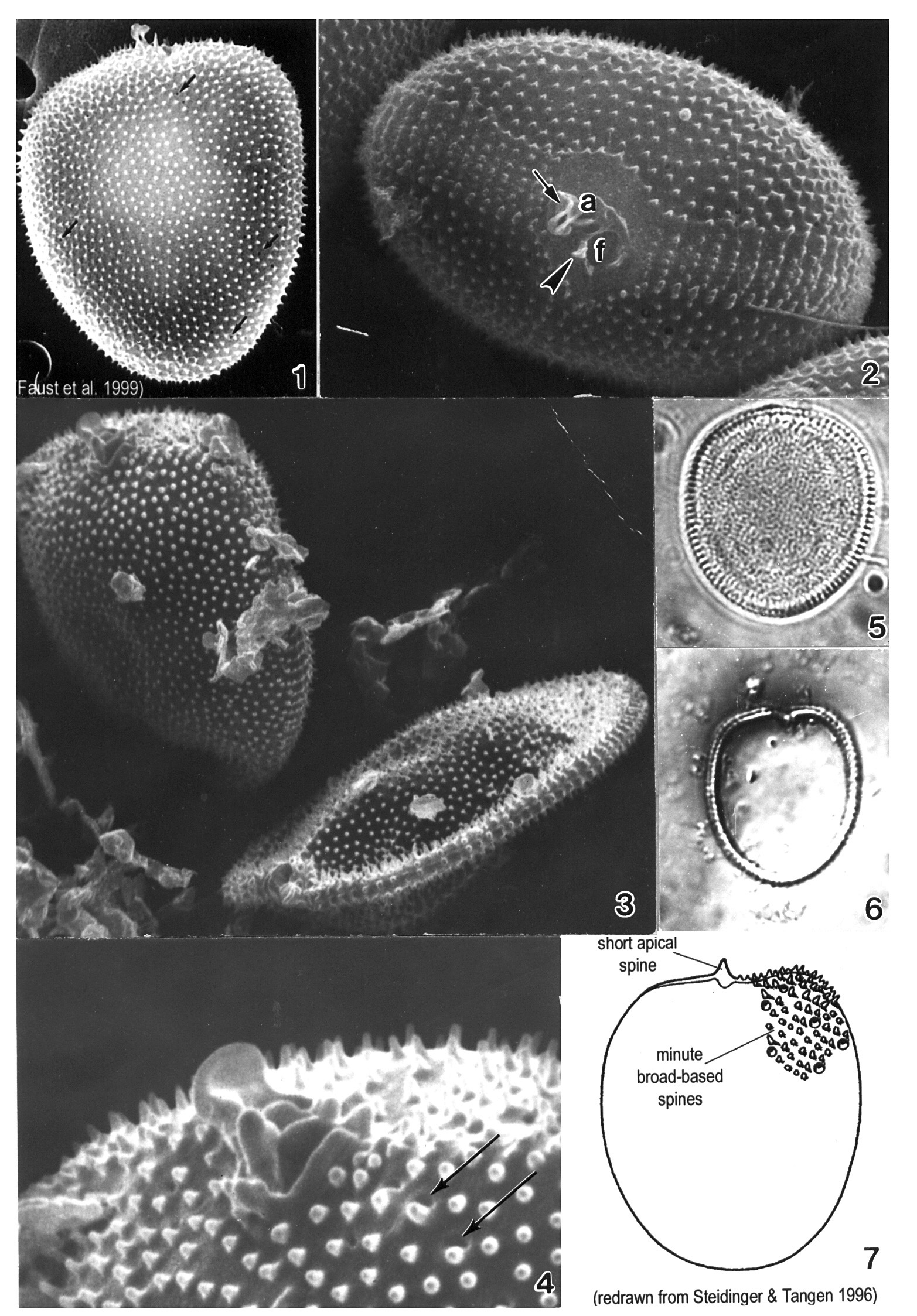
Plate 37. Prorocentrum arenarium. Figs. 1-5. SEM. Fig. 1. Right valve: cells round to ovoid. Periflagellar area is a broad, V-shaped depression. Short longitudinal flagellum visible (arrowhead). Marginal poroids present (arrows). Fig. 2. Left valve: surface smooth, with scattered valve and marginal poroids (arrows). Fig. 3. Lateral view: intercalary band smooth; marginal poroids evenly spaced (arrowheads). Fig.4. Marginal poroids oblong to kidney-shaped. Fig. 5. Periflagellar area: triangular and unornamented with large flagellar pore (f) and smaller auxiliary pore (a). Fig. 6. LM. Right valve: posterior nucleus (n) and prominent central pyrenoid (arrow).
-
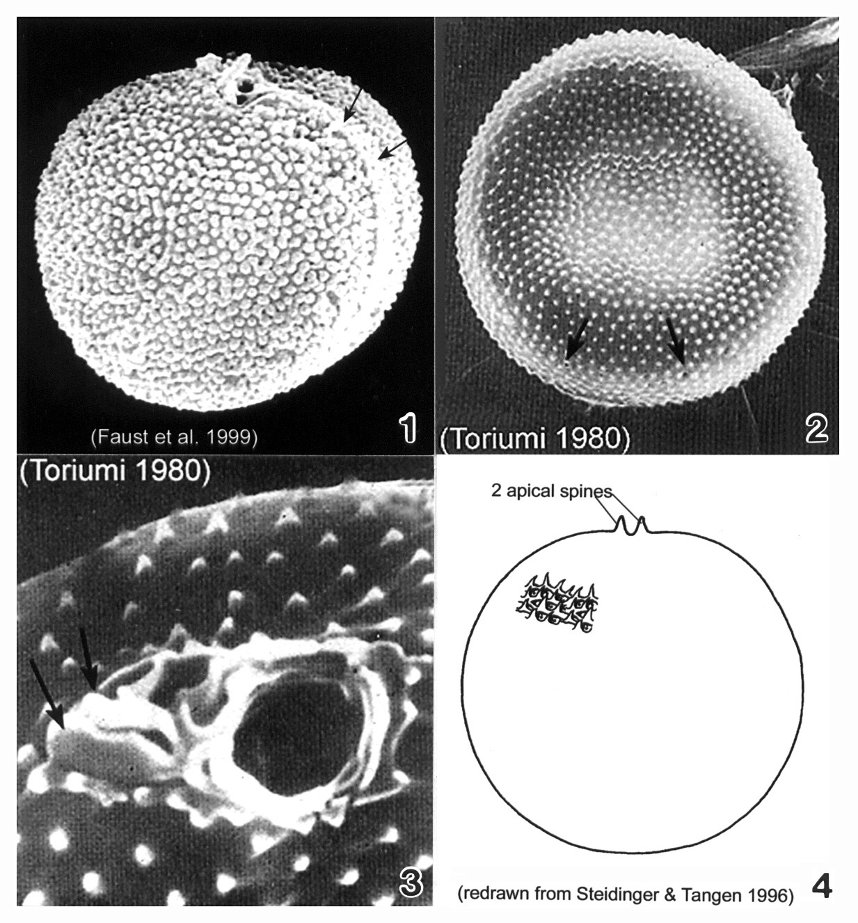
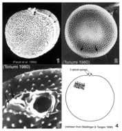
Plate 38. Prorocentrum balticum. Figs. 1-3. SEM. Fig. 1. Valve view: cell round to spherical, covered with many tiny spines. Apical spine apparent. Intercalary band broad, transversely striated (arrows). Fig. 2. Surface with scattered rimmed pores (arrows). Fig. 3. Periflagellar region: two different sized pores and two small apical projections (arrows). Fig. 4. Line drawing.
-
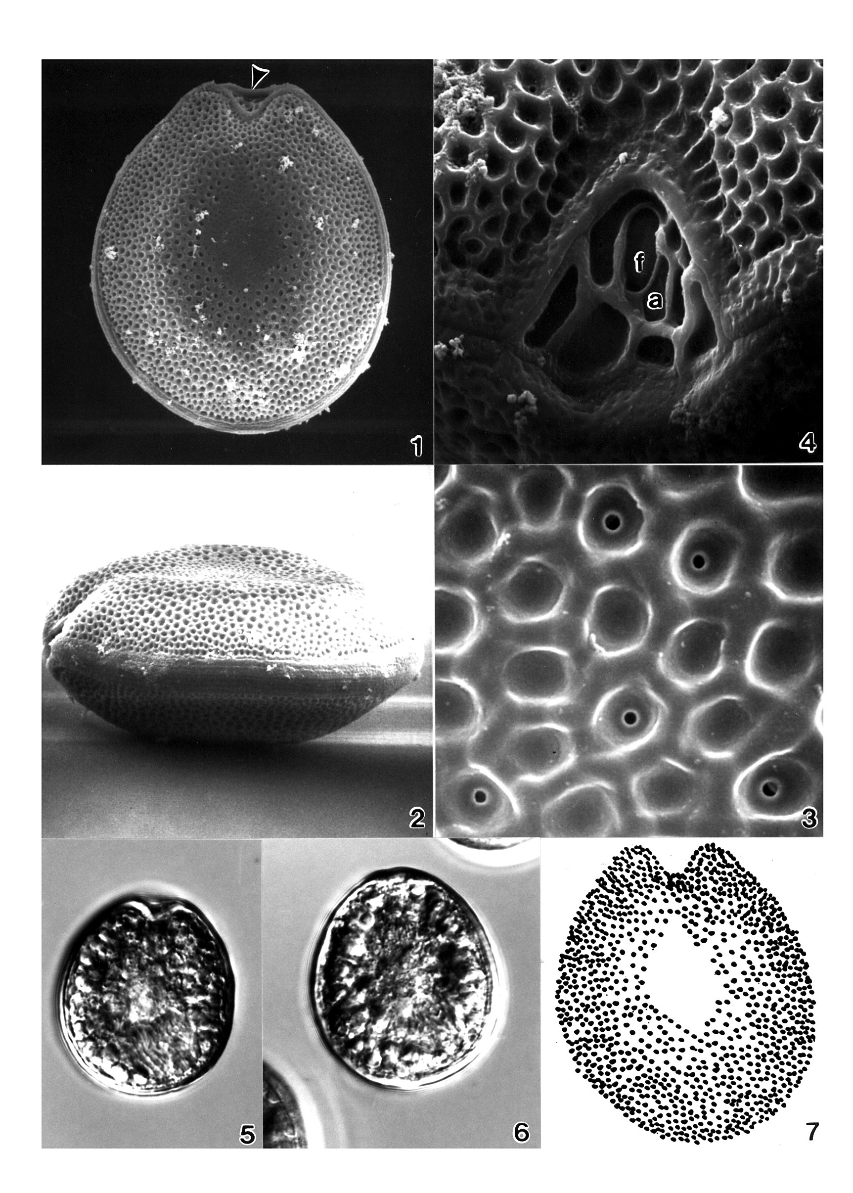
Plate 39. Prorocentrum belizeanum. Figs. 1-6. SEM. Fig. 1. Right valve: cell round to oval; surface heavily areolated. Fig. 2. Left valve: anterior margin with flared curved apical collar. Marginal areolae visible. Fig. 3. Lateral view: valve center concave; intercalary band smooth and wide. Fig. 4. Apical view: apical area with rounded lip; both valves excavated. Fig. 5. Areolae round to ovoid with smooth margins; some with pores. Fig. 6. Periflagellar area: auxiliary pore (a) surrounded by curved periflagellar collar (arrows); adjacent to flagellar pore (f). Left valve with flared apical collar (arrowheads). Fig. 7. Left valve: central pyrenoid (arrow) and posterior nucleus (n). Fig. 8. LM: right valve; flagella present. Fig. 9. Line drawing: areolae arrangement (after Faust 1993a).
-
Plate 40. Prorocentrum concavum. Figs. 1-4. SEM. Fig. 1. Right valve. Cell ovate and heavily areolate. Valve center devoid of areolae. Left valve with anterior apical ridge (arrowhead). Fig. 2. Lateral view. Valve center concave and flattened. Intercalary band granulated and horizontally striated. Fig. 3. Valve areolae round to oval with smooth edges; some with small central pores. Fig. 4. Periflagellar area a V-shaped depression. Two pores: small auxiliary pore (a); large flagellar pore (f). Figs. 5-6. LM (M.A. Faust). Fig. 5. Right valve. Fig. 6. Left valve. Fig. 7. Line drawing: areolae arrangement. (Figs. 1-4,7 after Faust 1990b)
-
Plate 41. Prorocentrum faustiae. Figs. 1-4. SEM. Fig. 1. Right valve. Cells broadly ovate to rotundate with slightly concave center. Valve surface rugose. Periflagellar area situated apically. Fig. 2. Left valve: apical region slightly excavated. Fig. 3. Intercalary band wide and transversely striated. Small marginal pores evenly spaced along cell perifery (arrows). Fig. 4. Periflagellar area: apical view. Broad V-shaped depression; larger flagellar pore (f) adjacent to smaller auxiliary pore (a). (All figures donated by S.L. Morton)
-
Plate 42. Prorocentrum hoffmannianum. Figs. 1-4. SEM. Fig. 1. Right valve: cell ovoid, tapering slightly apically. Valve surface areolated, slightly concave. Curved apical collar (arrow). Fig. 2. Left valve: distinct flared apical collar bordering periflagellar area (arrowheads). Marginal areolae large. Intercalary band smooth. Fig. 3. Areolae round to ovoid with smooth margins. Some with small pores (arrows). Fig. 4. Periflagellar area: flagellar pore (f) surrounded by flared periflagellar collar (arrowheads), adjacent to auxiliary pore (a); pores equal in size. Fig. 5. LM. Left valve: central pyrenoid (arrow); posterior nucleus (n). Intercalary band appears striated (M.A. Faust). Fig. 6. Line drawing: areolae arrangement. (Figs. 1-4,6 after Faust 1990b)
-
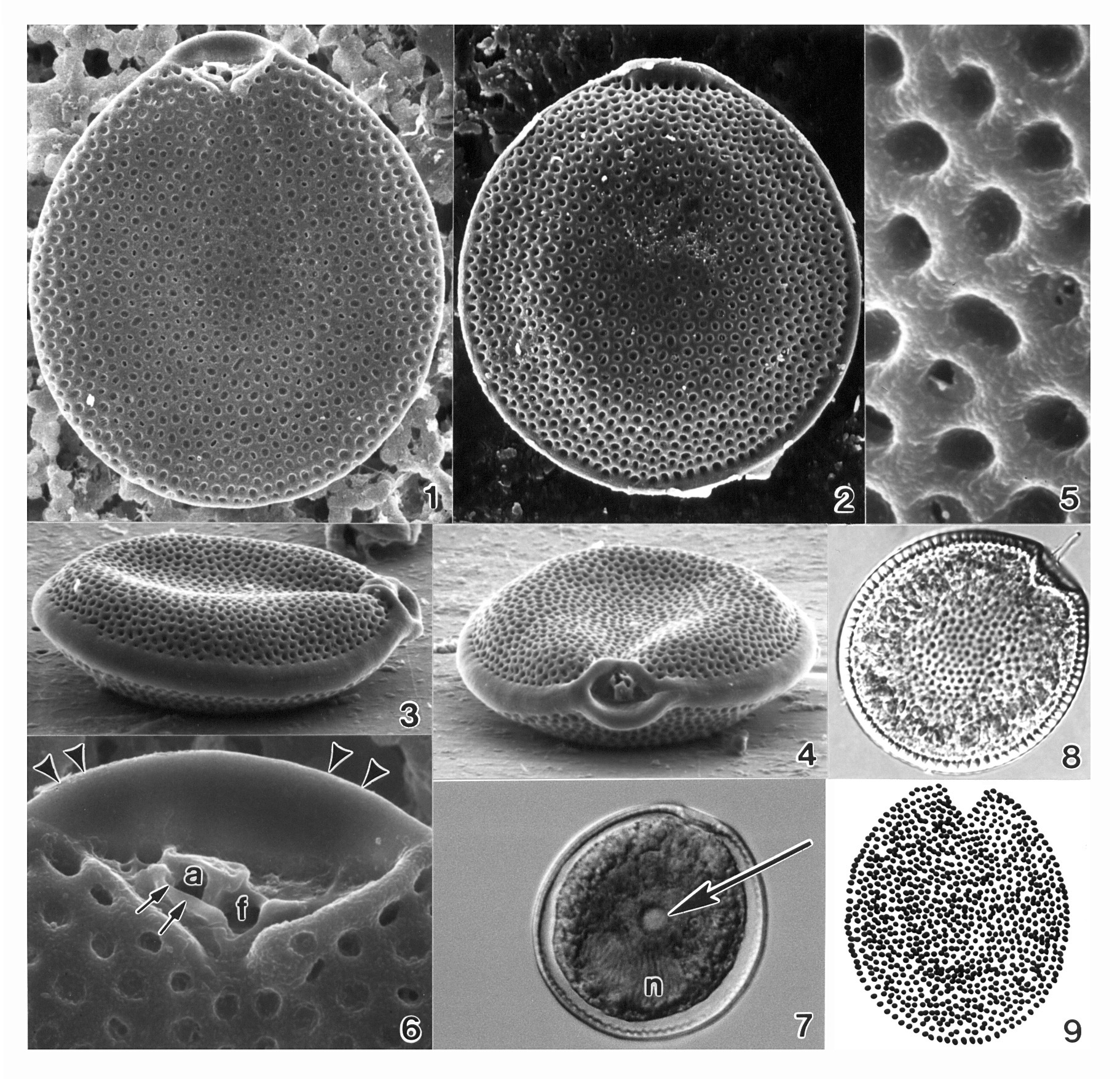
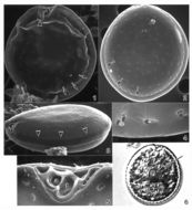
Plate 43. Prorocentrum lima. Figs. 1-3. SEM. Fig. 1. Right valve. Cells oblong to ovate with narrowed anterior. Marginal pores and scattered surface pores present; valve center devoid of pores. Intercalary band smooth and wide. Fig. 2. Left valve; bacteria attached (arrows). Fig. 3. Periflagellar area: shallow, broad, V-shaped depression on right valve. Flared periflagellar collar encircles auxiliary (a) pore (arrow); larger flagellar pore (f) adjacent (after Faust 1991). Figs. 4-7. LM. Fig. 4. Thecal pore arrangement. Fig. 5. Right valve with central pyrenoid (arrow). Fig. 6. Left valve and posterior nucleus (n). Fig. 7. Triple-layered resting cyst. (Figs. 1,2,4-7 after Faust 1993c)
-
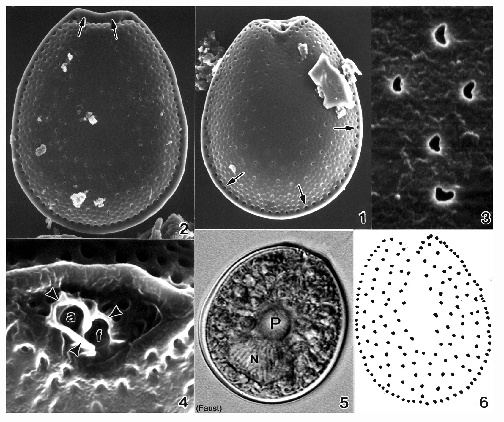
Plate 44. Prorocentrum maculosum. Figs. 1-4. SEM. Fig. 1. Right valve: cell broadly ovate, narrowing apically. Valve surface rugose with scattered poroids; valve center devoid of poroids. Marginal pores evenly spaced (arrows). Fig. 2. Left valve: anterior end flat to slightly concave with raised apical ridge (arrows). Valve margins appear as a flange around cell. Fig. 3. Valve poroids: unevenly distributed on valve surface; circular to oblong or kidney-shaped. Fig. 4. Periflagellar area: broad V-shaped depression on right valve. Apical ridge (raised margin) on left valve. Flagellar (f) and auxiliary (a) pores surrounded by protuberant periflagellar collar (arrowheads); equal in size. Fig. 5. LM. Right valve: central pyrenoid (P) and large posterior nucleus (N) (M.A. Faust). Fig. 6. Line drawing: valve poroid and marginal pore arrangement (Figs. 1-4,6 after Faust 1993b)
-
Plate 45. Prorocentrum mexicanum. Figs. 1-5. SEM. Fig. 1. Right valve: cell oval. Periflagellar collar curved and prominent (arrow). Trichocyst pores radially arranged (arrowheads). Fig. 2. Left valve. Apical area excavated (M.A. Faust). Fig. 3. Lateral view: cell ovate to convex; intercalary band broad and transversely striated. Cell surface rugose. Fig. 4. Trichocyst pores round with smooth edge, within deep furrowed depressions. Fig. 5. Periflagellar area: small, V-shaped shallow depression. Prominent curved periflagellar collar (double arrows) adjacent to auxiliary pore; protuberant periflagellar plate (single arrow) opposite and adjacent to flagellar pore. Fig. 6. LM. Right valve: radial pore arrangement visible (M.A. Faust). Fig. 7. Line drawing: trichocyst pore arrangement. (Figs. 1,3-5,7 after Faust 1990b)
-
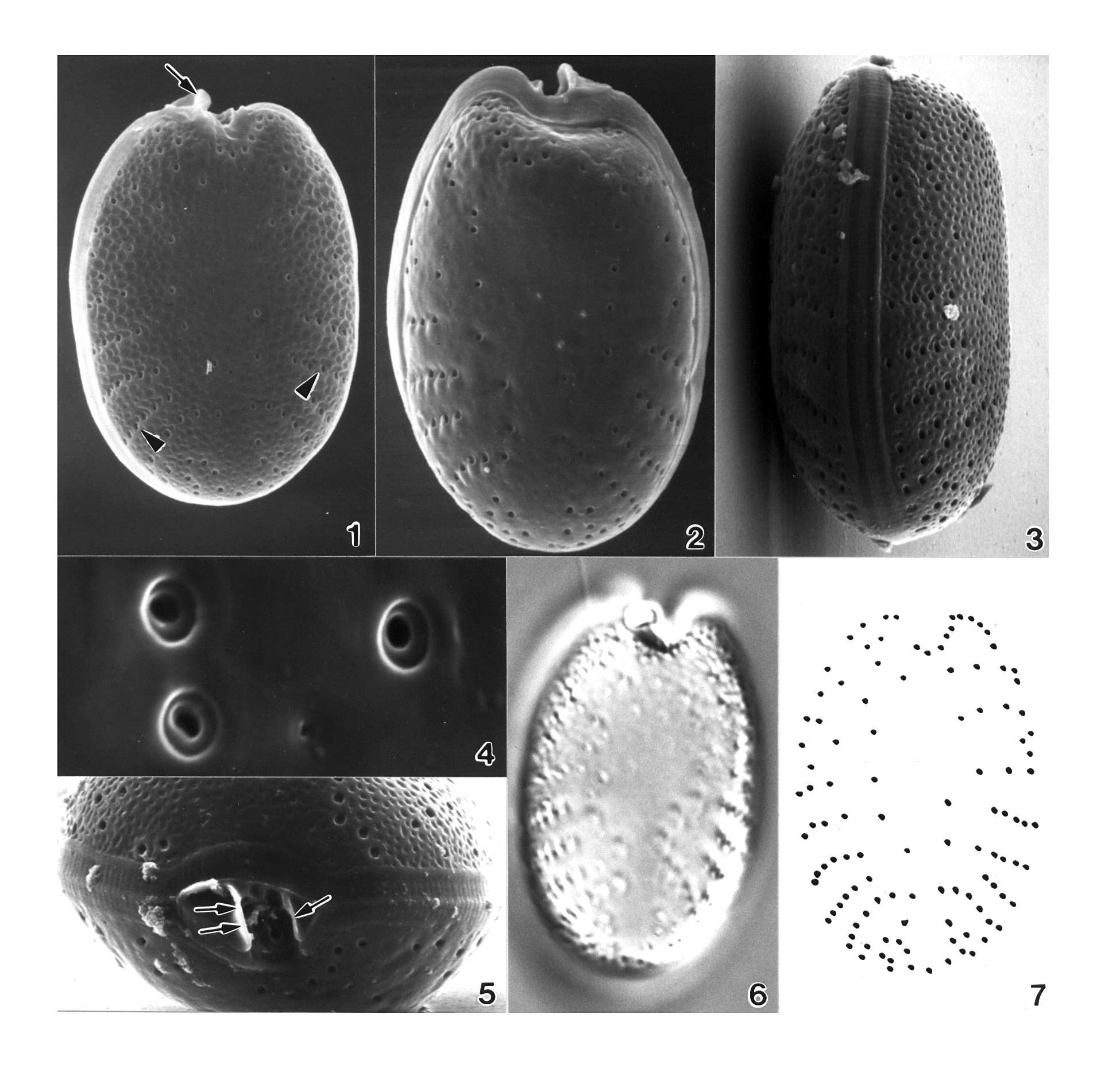
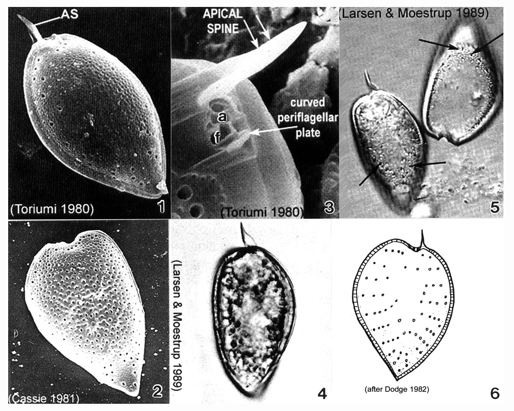
Plate 46. Prorocentrum micans. Figs. 1-3. SEM. Fig. 1. Right valve: cell tear-drop shaped; rounded anteriorly, pointed posteriorly, broadest in the middle. Apical spine (AS) winged. Rugose thecal surface. Intercalary band smooth and wide. Fig. 2. Heart-shaped cell. Apical spine missing. Fig. 3. Periflagellar area: small, shallow triangular depression on right valve. Flagellar (f) and auxiliary (a) pores present; curved periflagellar plate adjacent to f. Large winged AS directly opposite. Figs. 4-5. LM: Left valve. Winged AS visible. Fig. 5. Empty theca with visible trichocyst pores (arrows). Fig. 6 Line drawing: trichocyst pore arrangement.
-
Plate 47. Prorocentrum minimum. Figs. 1-4. SEM. Fig. 1. Right valve. Cell oval; broad truncate apical region. Thecal surface with numerous short broad spines. Small scattered pores (arrows). Fig. 2. Lateral apical view. Periflagellar area with 2 pores: large flagellar (f) and small auxiliary (a). Small apical spine (arrowhead) adjacent to f; small curved forked periflagellar collar (arrow) adjacent to a. Intercalary band wide; transversely striated. Fig. 3. Cells oval; ventrally flattened. Fig. 4. Apical view. Short thecal spines and small scattered pores (arrows). Figs. 5-6. LM. Surface features and intercalary band visible. Fig. 7. Line drawing. (Figs. 1-6 after Faust 1974)