-
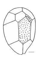
Fig 1: Coolia monotis Schematic diagram (hypothecal view) redrawn from Tomas et al. 1997.
-
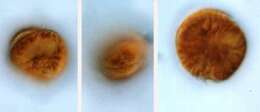
Coolia (coo-lee-a) monotis Meunier 1919. The images show swimming cells. The cells are flattened in the anterior-posterior plane, slightly asymmetrically. The cell on the left is in posterior-lateral view. The nucleus is visible. The cell in the middle is in ventral-lateral view. The cingulum is visible in the middle of the cell. The cell on the right is in posterior view. The cells contain yellow-brown plastids.
-
Fig 2: Coolia monotis Schematic diagram (epithecal view) redrawn from Tomas et al. 1997
-
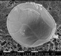
Ostreopsis lenticularis, scanning electron microscope image. This image was taken by Shauna Murray of a sample from Raine Island, northern Great Barrier Reef, Australia. This work was supported by the Australian Biological Resources Study.
-
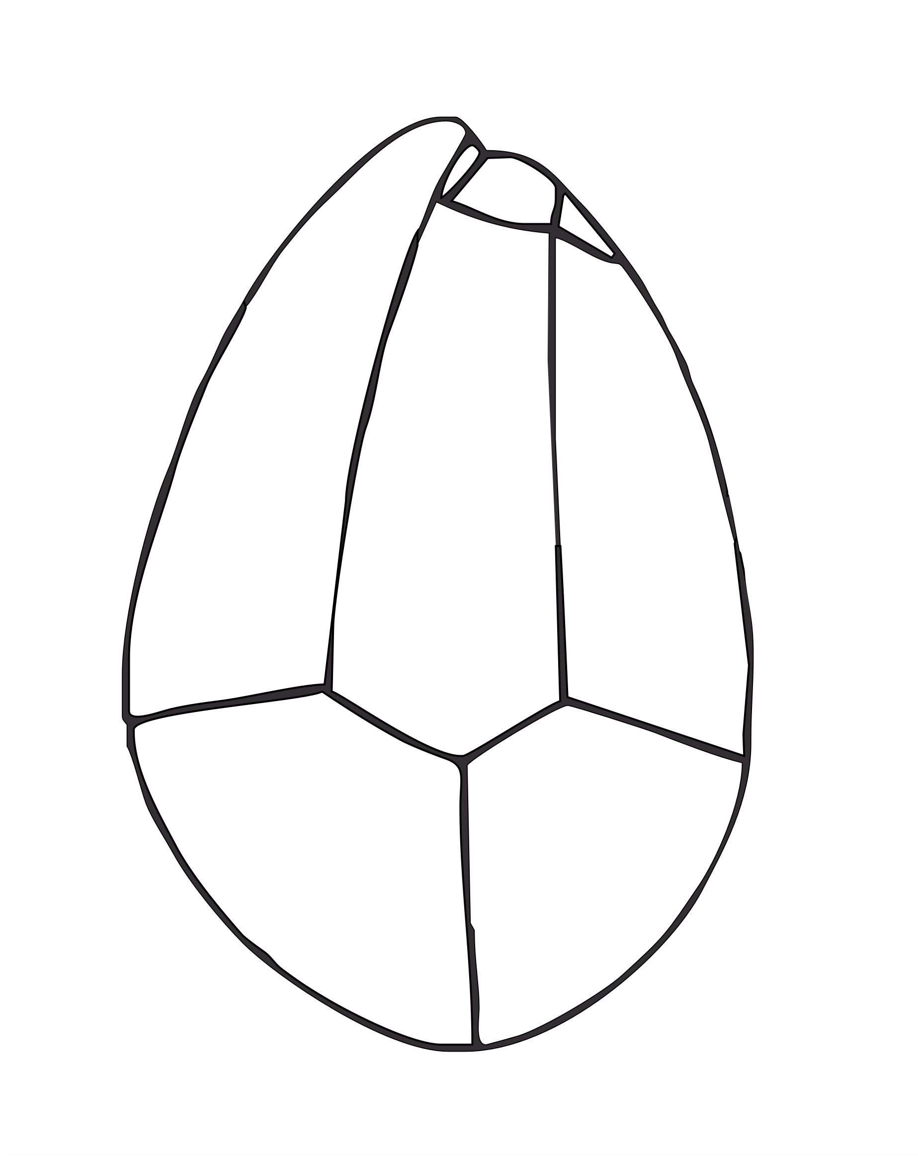
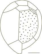
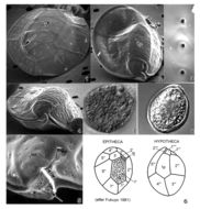
Fig 1: Schematic diagram of Ostreopsis ovata in hypothecal view. Scale bar = 10 ¦#181;m. Redrawn from Tomas et al. 1997
-
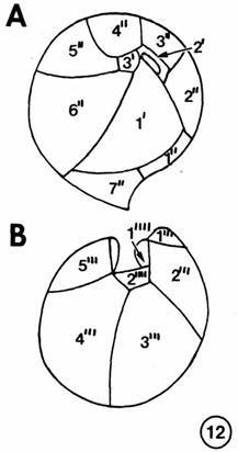
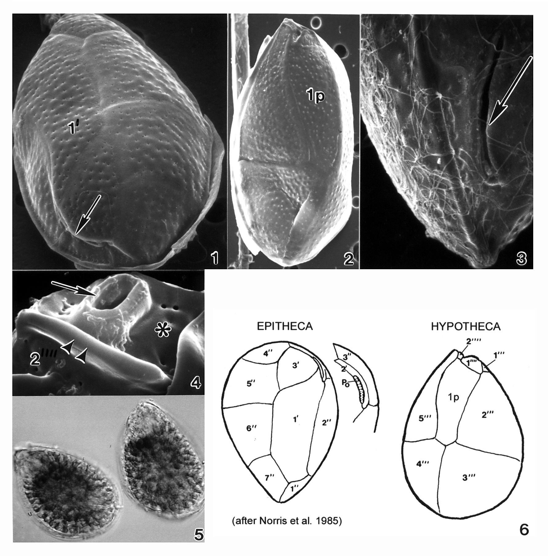
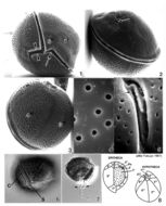
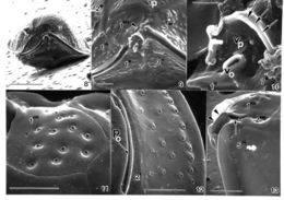
Plate 10. Coolia monotis: Figs. 1-5. SEM. Fig. 1. Ventral view: spherical shape. Cingulum lipped and equatorial. Sulcus with flexible lists (arrowheads). Ventral pore present (arrow). Fig. 2. Dorsal view: apical pore plate (arrow), Po, located off-center on epitheca. Fig. 3. Antapical view: hypothecal plates. Fig. 4. Smooth edged thecal pores unevenly distributed. Fig. 5. Po about 12 _ long, slightly curved and narrow with a slit-like apical pore. Two supporting rib-like costae (arrows) and evenly spaced round pores surround the pore. Figs. 6,7. LM. Fig. 6. Ventral view of lipped cingulum and sulcus. Fig. 7. Planozygote with two longitudinal flagella (arrows). Fig. 8. Line drawing: thecal plate arrangement.
-
Fig 2: Schematic diagram of Ostreopsis ovata in epithecal view. Scale bar = 10 ¦#181;m. Redrawn from Tomas et al. 1997
-
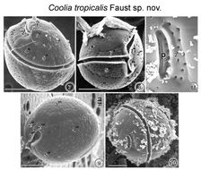
FIGS. 7-11. Scanning Electron micrographs of the surface morphology of Coolia tropicalis sp. nov. FIG. 7. Oblique dorsal view of C. tropicalis shows the apical pore and the equatorially located lipped cingulum. Cell surface is smooth with large scattered pores. FIG. 8. Cell is spherical in equatorial view shoving a deep cingulum and sulcus. Detritus adheres to the epitheca. FIG. 9. Antapical view of a cell show large unequal plates. FIG. 10. Apical pore is a narrow opening located in the epitheca. Fine detrital particles partially cover the thecal surface. FIG. 11. The apical pore is about 7 μm long straight and narrow slits with two supporting costae and evenly spaced round pores. Detritus attached to surface of apical pore plate. EMu:HOLOTYPE SEM NEGATIVE #166029; SEM STUB # 166; FIELD # 728-93;ACCESSION # 408431: CATALOG # 997; FIGURE # 7.
-
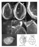
Fig 1: Ostreopsis siamensis Schematic diagram (hypothecal view) redrawn from Tomas et al. 1997.
-
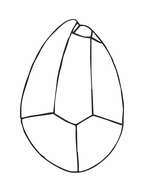
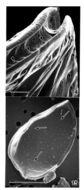
FIG. 12. Coolia tropicalis sp. nov. A) apical view of epitheca, and B) antapical hypotheca.
-
Fig 2: Ostreopsis siamensis Schematic diagram (epithecal view) redrawn from Tomas et al. 1997.
-
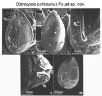
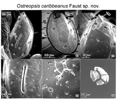
Figs 6-10. Cells of Ostreopsis belizeanus sp. nov. Figs 6-9. Scanning electron microscopy. Fig. 6. Morphology of epithecal plates and position of apical pore plate (Po). Fig. 7. Hypothecal plates. Fig. 8. In the cingulum, the ventral opening (Vo) is located adjacent to a ridged plate (Rp). Fig. 9. Apical pore plate includes a narrow apical pore (Po) located off-center. Thecal surface laced with round pores (arrows). Fig. 10. Epifluorescence light microscopy of epithecal plates.
EMu: HOLOTYPE SEM NEGATIVE # 211053; SEM STUB # 211; FIELD # 1005-96; ACCESSION # 2002408; CATALOG # 1541; FIGURE # 6.
-
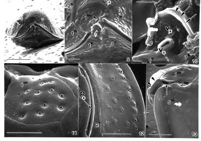
Figs 11-16. Cells of sp. nov. Figs. 11-15. Scanning electron microscopy. Fig. 11. Morphology of epithecal plates and position of apical pore plate (Po). Fig. 12. Hypothecal plates. Note long centrally situated Ip plate. Fig. 13. Cell in antapical view: antapical plate 1" is triangular; plate 2" is narrow and very small. The location of the ventral pore (Vo) and ridged plate (Rp) is illustrated. Fig. 14. Apical pore plate (Po) located off-center; note its morphology. Thecal surface smooth with round pores. Fig. 15. Ejected trichocyst emerges from thecal pores (arrowheads). Fig. 16. Epifluorescence microscopy of partially separated hypothecal plates.
EMu:HOLOTYPE SEM NEGATIVE # 174097; SEM STUB # 174; FIELD # Morton-Clones; ACCESSION # ; CATALOG # 1545 ; FIGURE # 11
-
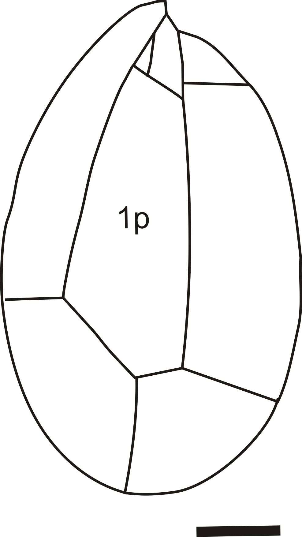
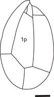
Plate 31. Ostreopsis heptagona. Figs. 1-4. SEM. Fig. 1. Epithecal view: cells broadly oval, oblong and pointed. Long curved apical pore plate, Po, off-center (arrow). Plate 1' heptagonal and distinctive. Fig. 2. Hypothecal view: plate 1p pentagonal and dorso-ventrally elongate. Fig. 3. Po long, narrow and curved. Narrow mucilage strands cover cell surface. Fig. 4. Ventral view: location of ventral opening (arrow), ventral plate (asterisk), and rigid plate (asterisk) within cingulum. Fig. 5. LM. Two cells. Fig. 6. Line drawing: thecal plate arrangement.
-
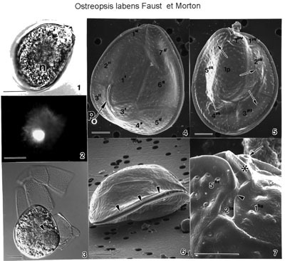
FIGS. 1-7. Ostreopsis labens sp. nov. FIGS. 1-3. Light microscope views. Scale bar = 25 μm. Cells contain chloroplasts and a spherical posterior nucleus (n). FIG.1. Cell is in epithecal view. FIG. 2. Location of nucleus stained with DAPI. FIG.3. Hypothecal plates partially separated with numerous pores. Cell with an engulfed prey organism (arrowhead); red color not detected on a black and white print. FIGS. 4-7. Cells viewed with SEM. FIG. 4. Cell is broadly ovoid in epithecal view. Note the curved, long apical pore (Po) located off-center (arrow). Scale bar =10 µm. FIG. 5. Cell is in hypothecal view. Cell is smooth with scattered pores (arrows). Scale bar =10µm. FIG. 6. Cell is slightly convex in lateral view. Note lipped, equatorial cingulum (arrowheads). Scale bar =10 µm. FIG. 7. Antapical plate 1"is with a slightly curved list (arrowhead). The sulcus narrow, recessed and hidden adjacent to plate 2". The ventral opening (arrow) is situated on the ventral surface adjacent to a ridged plate (Rp) (asterisk). Scale bar = 5 μm. EMu: : HOLOTYPE SEM NEGATIVE # 170058; SEM STUB # 170; FIELD # 745-94; ACCESSION # 410840; CATALOG # 984; FIGURE # 4
-
FIGS. 8-13. Ostreopsis labens sp. nov. FIG. 8. Ventral view is showing bi-convexity of the cell. Scale bar = 25 µm. FIG. 9. The 2" is very small (arrowhead). The ventral opening with a protuberant ridge and a curved plate (Rp) are situated in the cingulum adjacent to plate 1" and plate 1". Cingulum is smooth. Scale bar = 5 µm. FIG. 10. The ventral opening (Vo) is situated on the ventral plate (Vp). Scale bar = 5 µm. FIG. 11. Thecal surface is smooth; evenly spaced around trichocyst pores with smooth edges. Scale bar = 5 µm. FIG. 12. The apical pore (Po) is long, curved, and narrow associated with plate 2'. Row of marginal pores similar in size to thecal pores. Scale bar = 5 µm. FIG. 13. Right ventral view is unusual, recessed in sulcus. Flagellar pore opening is narrow (arrow). EMu: : HOLOTYPE SEM NEGATIVE # 170058; SEM STUB # 170; FIELD # 745-94; ACCESSION # 410840; CATALOG # 984; FIGURE # 4
-
FIGS. 14, 15. Ostreopsis labens sp. nov. FIG. 14. Surface of cingulum is smooth, deep and narrow with equally spaced round pores (arrowheads). Scale bar = 2 μm. FIG. 15. The inner cell surface is smooth; thecal plate relatively thick with round trichocyst pores (arrows). Scale bar = 2 µm. EMu: : HOLOTYPE SEM NEGATIVE # 170058; SEM STUB # 170; FIELD # 745-94; ACCESSION # 410840; CATALOG # 984; FIGURE # 4
-
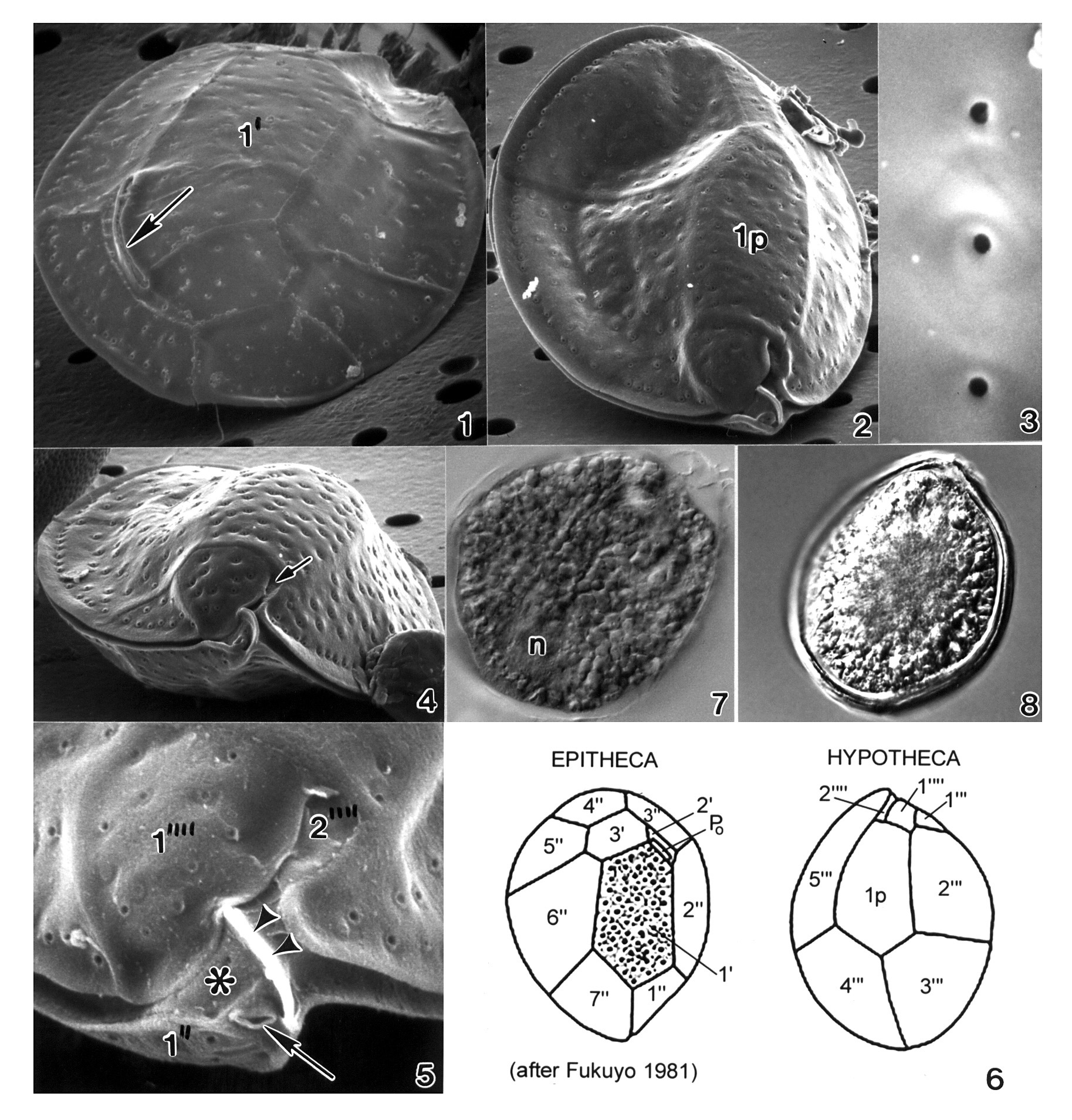
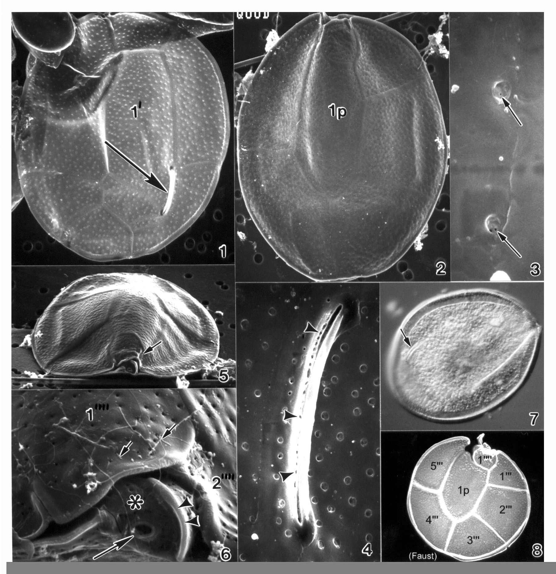
Plate 32. Ostreopsis lenticularis. Figs. 1-5. SEM. Fig. 1. Epithecal view: cell lenticulate to broadly oval. Curved off-center apical pore plate with a slit-like apical pore (arrow). Plate 1' irregularly pentagonal. Fig. 2. Hypothecal view: plate 1p central and pentagonal. Fig. 3. Smooth thecal surface. Round pores with smooth raised edges. Fig. 4. Hypothecal ventral view: cell anterio-posteriorly compressed. Shallow cingulum with smooth edge. Small sulcus hidden (arrow). Fig. 5. Location of ventral opening (arrow), ventral plate (asterisk), and rigid plate (arrowheads) within cingulum. Fig. 6. Line drawing: thecal plate arrangement. Figs. 7,8. LM. Fig. 7. Cytoplasma granulated; posterior nucleus (n). Fig. 8. Distinct cingular list.
-
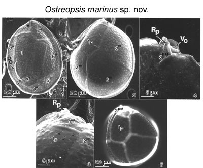
Figs 1-5. Cells of Ostreopsis marinus sp. nov. Figs 1-4. Scanning electron microscopy. Fig. 1. Morphology of epithecal plates and position of apical pore plate are shown (Po). Fig. 2. Hypothecal plates. Fig. 3. Antapical plate 1" is larger; plate 2" is tiny. Thecal surface is smooth with small, evenly distributed pores (arrows). Fig. 4. The ventral opening (Vo) is situated in the cingulum adjacent to a ridged plate (Rp). Fig. 5. Epifluorescence light microscopy of hypothecal plates; Ip plate is in the center.
EMu: HOLOTYPE SEM NEGATIVE # 212055; SEM STUB # 212; FIELD # 96/10; ACCESSION # 2002799; CATALOG #1537; FIGURE # 1.
-
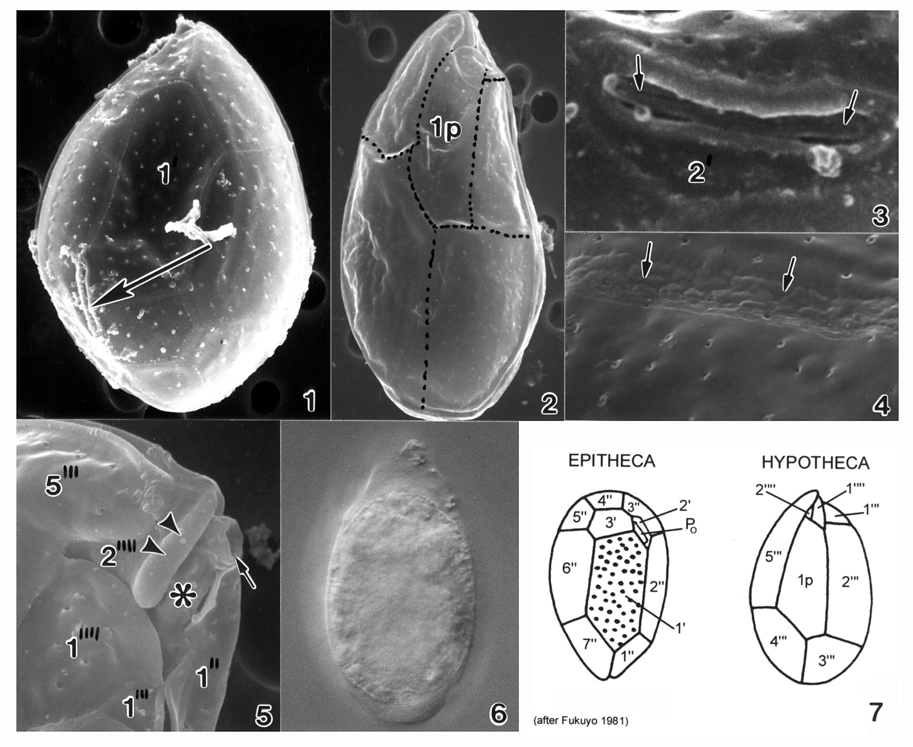
Plate 33. Ostreopsis mascarenensis. Figs. 1-5. SEM. Fig. 1. Epitheca: inner thecal surface. Cell very large, broadly ovate, large plates. Plate 1' elongate and hexagonal. Apical pore plate (Po) nearly straight. Fig. 2. Hypotheca: plate 1p long and wide. Fig. 3. Smooth cell surface with round pores; pores with two small openings (arrows). Fig. 4. Po with long narrow apical pore; small pores line the opening (arrowheads). Figs. 5-6. Ventral view of epitheca. Fig. 5. Cell compressed anterio-posteriorly; cingulum narrow with smooth edge. Small sulcus hidden (arrow). Fig. 6. Location of ventral opening (large arrow), ventral plate (asterisk), and rigid plate (arrowheads) within cingulum. Pores with ejected trichocysts (small arrows). Fig. 7. LM. Epitheca: Po (arrow) and cingulum in focus. Fig. 8. Line drawing: hypotheca plate arrangement.
-
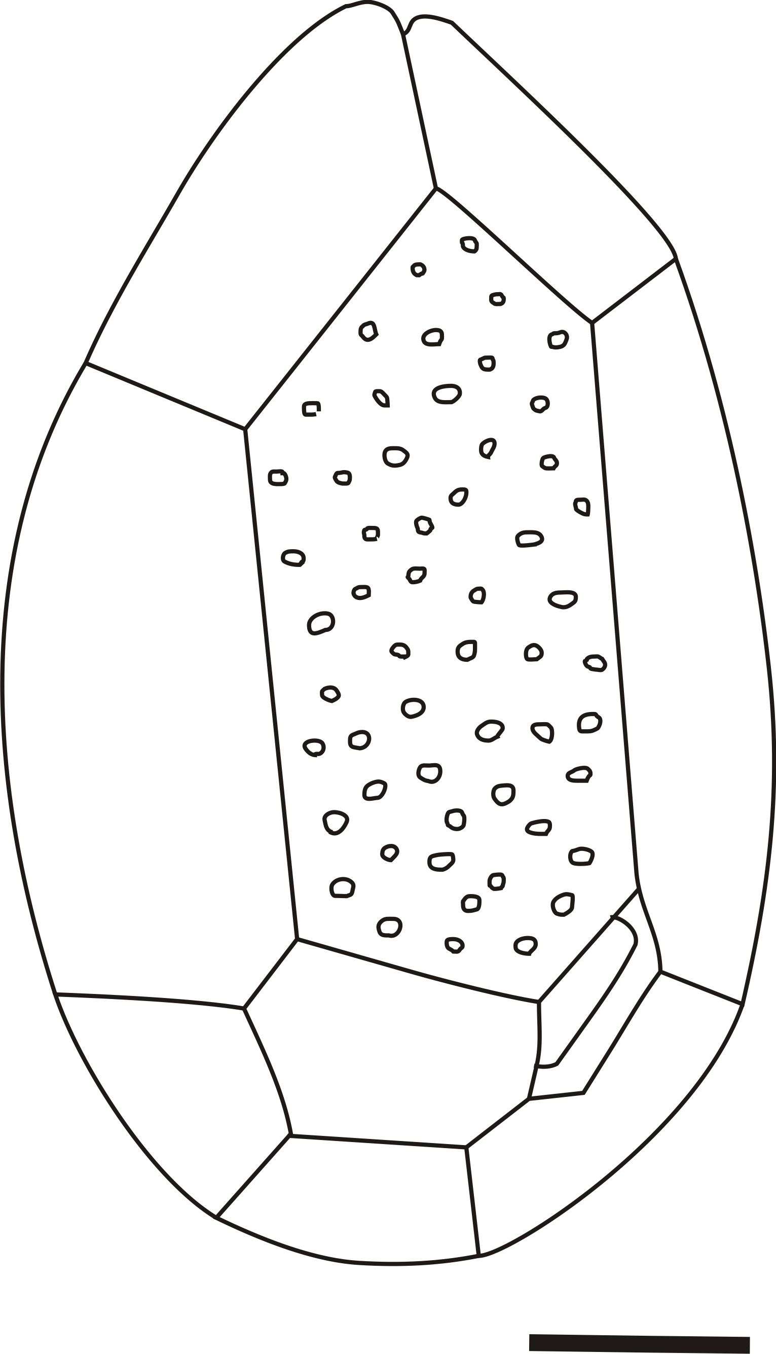
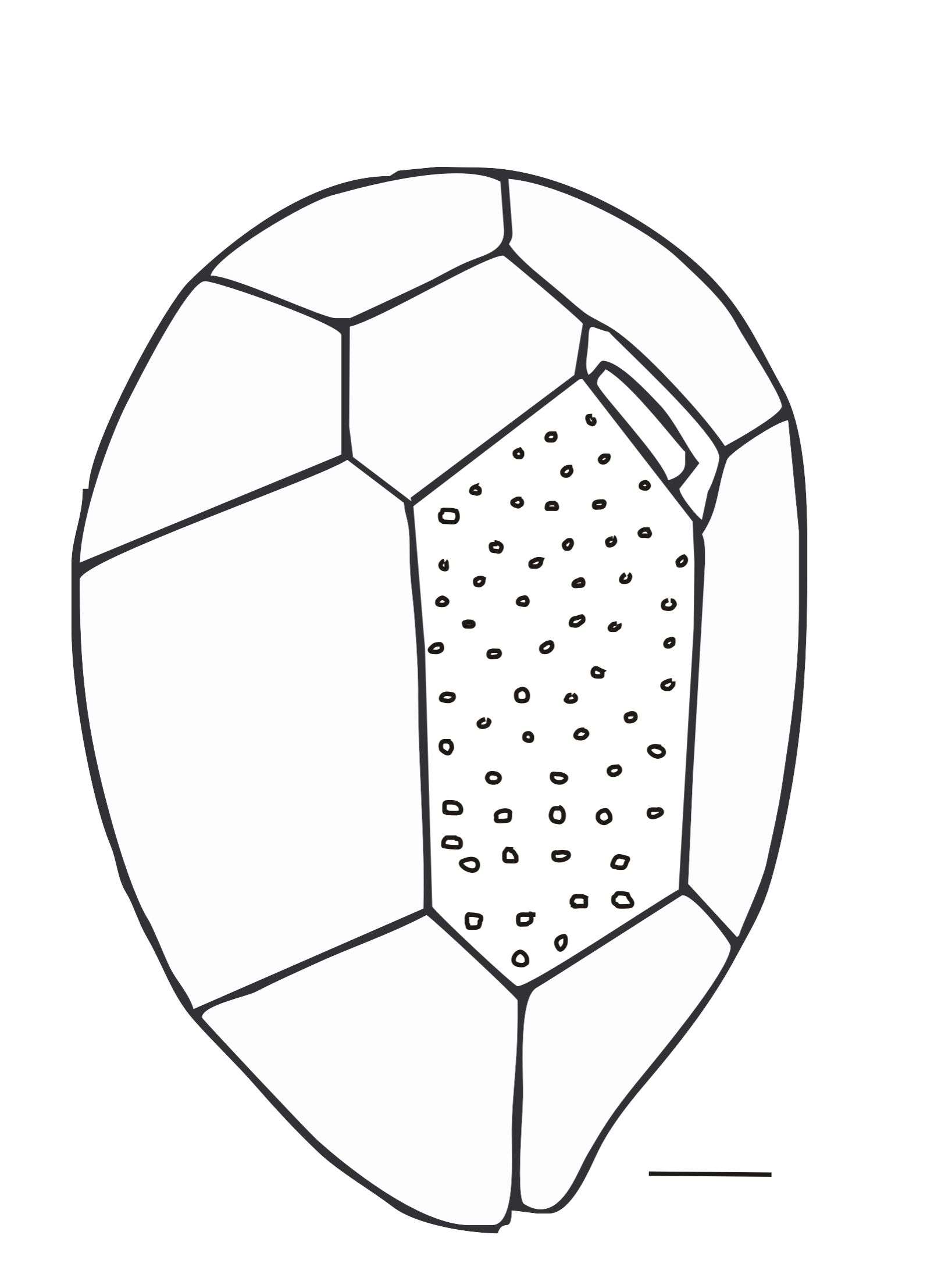
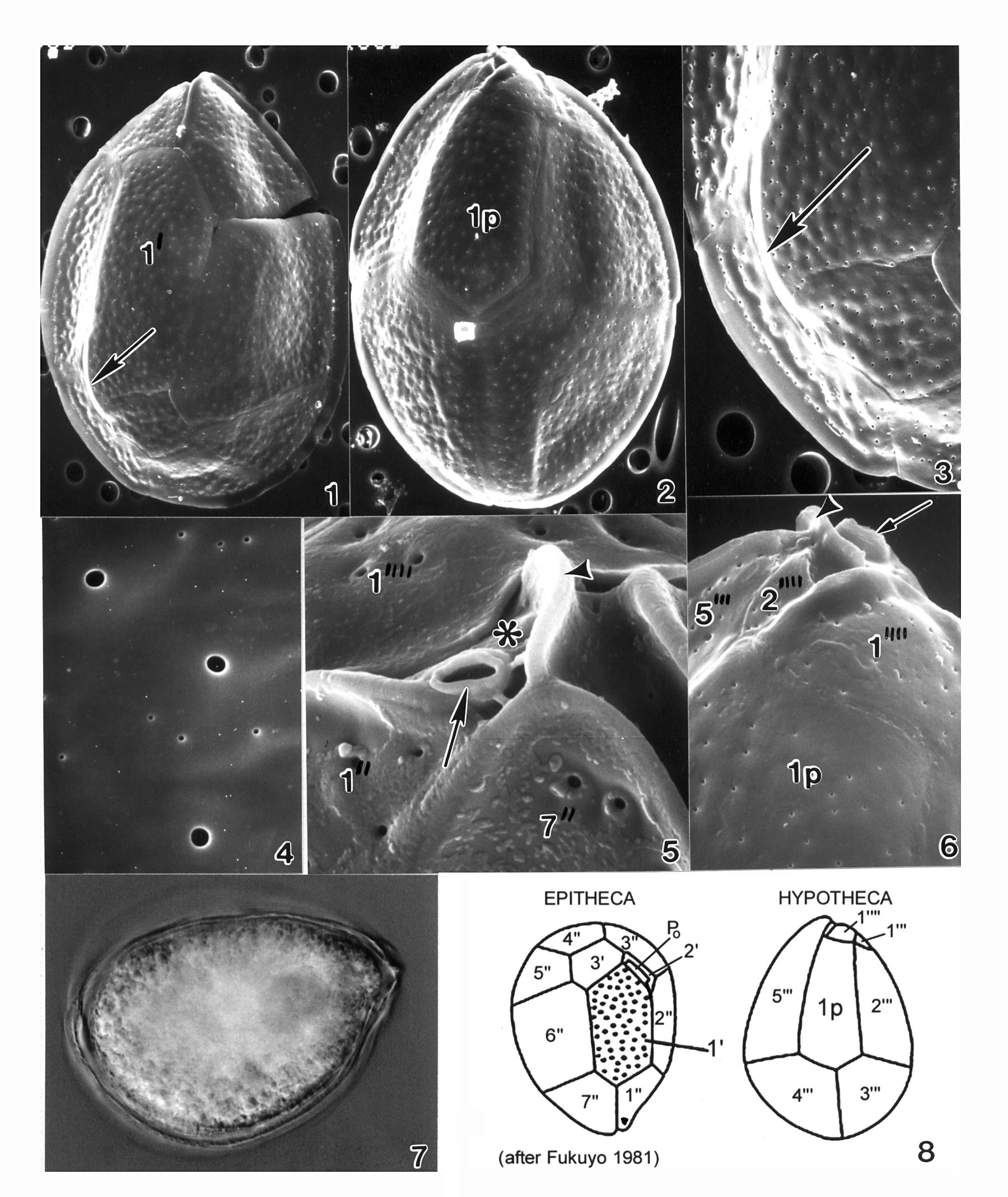
Plate 34. Ostreopsis ovata. Figs. 1-5. SEM. Fig. 1. Epithecal view: cell slender and tear-shaped. Apical pore plate (Po) off-center (arrow). Plate 1' large and hexagonal. Cingulum wide with narrow lists. Fig. 2. Hypothecal view: plates delicate. Plate 1p long and narrow. Fig. 3. Po: short and straight, adjacent to plate 2'. Fig. 4. Thecal surface smooth with scattered small pores. Suture line uneven and bumpy (arrows). Fig. 5. Hypothecal view: ventral opening (arrow), ventral plate (asterisk), and rigid plate (arrowhead) on cingulum. Fig. 6. LM. Large posterior nucleus. Fig. 7. Line drawing: thecal plate arrangement.
-
Plate 35. Ostreopsis siamensis. Figs. 1-6. SEM. Fig. 1. Epithecal view: cell broad and tear-shaped. Thecal surface smooth with scattered pores. Apical pore plate (Po) off-center (arrow). Narrow cingulum with smooth edge. Plate 1' narrow and pentagonal. Fig. 2. Hypothecal view: plate 1p long and pentagonal. Fig. 3. Po: long, curved and narrow. Fig. 4. Large and small pores on thecal surface. Fig. 5. Ventral view: location of ventral opening (arrow), ventral plate (asterisk), and rigid plate (arrowhead) on cingulum. Fig. 6. Hypothecal view: Vo (arrow) and Rp (arrowhead). Fig. 7. LM. Hypotheca. Fig. 8. Line drawing: thecal plate arrangement.