NMNH Gambierdiscus toxicus type specimen
Description:
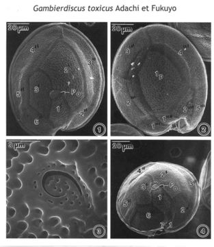
Figs. 1-4. Scanning electron micrographs illustrate the surface morphology of Gambierdiscus toxicus (GTT-91). Fig. 1. In epithecal view. The cell shape is round, and the apical pore plate (Po) oriented ventrally. Fig. 2. In hypothecal view. Posterior intercalary plate (Ip) broad and pentagonal, centrally located, occupying about 1/3 of cell's width. Fig.3. Po plate ellipsoid with a fish-hook-shaped apical pore surrounded by rows of 28 evenly distributed pores. Fig. 4. Cell in central view, shape compressed and bordered by a cingular list. The cell surface is smooth with small scattered pores. NOTE: This is the apotype of the genus Gambierdiscus. It is an important toxic species. I would like to add this species to the dinoflagellate ‘Types’ since the SEM plate of G. toxicus is the only record. Adachi and Fukuyo (1979) described G. toxicus sp. nov. only in line drawing to illustrate the morphology of plates.
Included On The Following Pages:
- Life (creatures)
- Cellular (cellular organisms)
- Eukaryota (eukaryotes)
- SAR (Stramenopiles, Alveolates, Rhizaria)
- Alveolata (alveolates)
- Dinophyceae
- Gonyaulacales
- Goniodomataceae
- Gambierdiscus
- Gambierdiscus toxicus
- Dinoflagellata (dinoflagellates)
This image is not featured in any collections.
Source Information
- license
- cc-by-nc-sa-3.0
- copyright
- National Museum of Natural History, Smithsonian Institution
- original
- original media file
- visit source
- partner site
- NMNH Marine Dinoflagellates
- ID