-
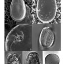
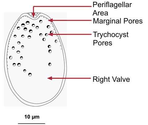
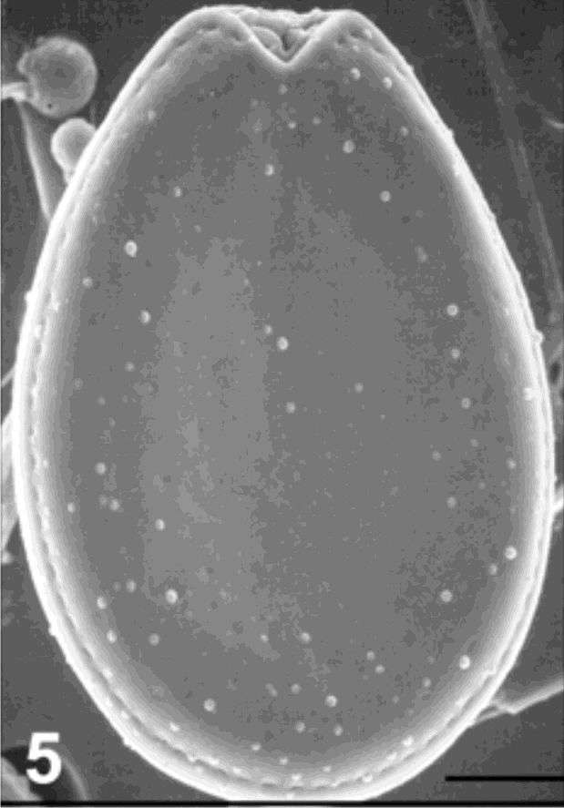
Plate 43. Prorocentrum lima. Figs. 1-3. SEM. Fig. 1. Right valve. Cells oblong to ovate with narrowed anterior. Marginal pores and scattered surface pores present; valve center devoid of pores. Intercalary band smooth and wide. Fig. 2. Left valve; bacteria attached (arrows). Fig. 3. Periflagellar area: shallow, broad, V-shaped depression on right valve. Flared periflagellar collar encircles auxiliary (a) pore (arrow); larger flagellar pore (f) adjacent (after Faust 1991). Figs. 4-7. LM. Fig. 4. Thecal pore arrangement. Fig. 5. Right valve with central pyrenoid (arrow). Fig. 6. Left valve and posterior nucleus (n). Fig. 7. Triple-layered resting cyst. (Figs. 1,2,4-7 after Faust 1993c)
-
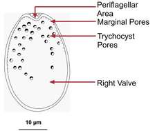
Fig 1: Schematic drawing of Prorocentrum lima
-
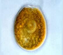
Fig 5:Prorocentrum lima SEM image of cell in right valve view
-
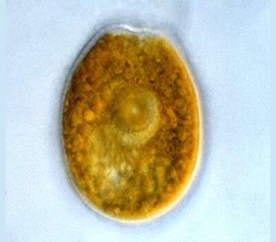
Prorocentrum (pro-row-sent-rum) lima (Ehrenberg) Dodge 1975. The image shows one of the two valves of a cell. The cingulum is not visible. The plastids are yellow-brown, and surround a large circular pyrenoid in the centre of the cell.
-
Prorocentrum lima valves are oval, 40 - 45 microns long, 27-33 microns wide , length to width ratio 1.3 - 1.5. Valve surface smooth. Valve pores scattered except in the centre, 58 - 80 per cell, approximately 0.3 microns diameter, round, oval to slightly kidney shaped. Marginal pores around periphery of cell, 51-74 per cell, round to oval approximately 0.3 microns diameter. Intercalary band smooth. Apical area consists of a small triangular indentation. A small flange (apical collar) is present. Plastids large, orange-brown. A pyrenoid, 7 - 8 microns diameter, is situated centrally. Nucleus in the posterior of the valve.