Błaszkowski, J. and B. Czerniawska, 2008.
EOL staff
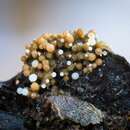
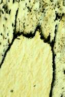
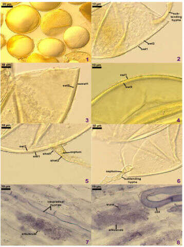
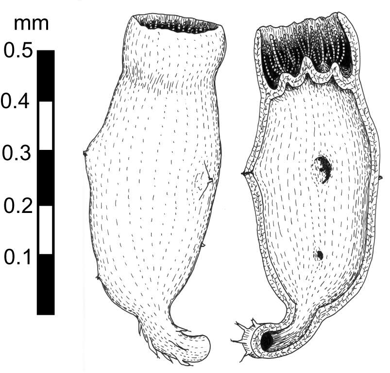
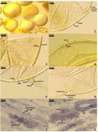
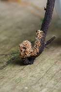
Glomus eburneumFigures 1-8 from Błaszkowski, J. and B. Czerniawska, 2008. Glomus eburneum and Scutellospora fulgida, species of arbuscular mycorrhizal fungi (Glomeromycota) new for Europe. Acta Mycologica 43(1): 57-65.1. Intact spores. 2. Spore wall layers 1 (swl1) and 2 (swl2) and subtending hypha. 3 and 4. Spore wall layers 1 (swl1) and 2 (swl2). 5. Spore wall layers 1 (swl1) and 2 (swl2), subtending hyphal wall layers 1 (shwl1) and 2 (shwl2), and curved septum. 6. Subtending hypha occluded by curved septum. 7 and 8. Arbuscules, trunk, intraradical hyphae, and coil of mycorrhizae stained in 0.1% trypan blue. Fig. 1, spores in lactic acid. Figs 2,3,4-8, spores crushed in PVLG. Fig. 4, spore crushed in PVLG+Melzer's reagent. Fig. 1, bright field microscopy. Figs 2-8, differential interference contrast.
Błaszkowski, J. and B. Czerniawska, 2008.
EOL staff
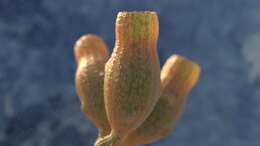
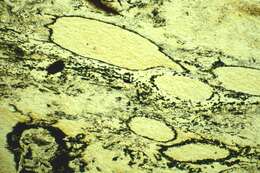
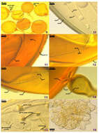
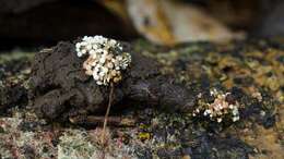
Scutellospora fulgidaFigures 9-16 from Błaszkowski, J. and B. Czerniawska, 2008. Glomus eburneum and Scutellospora fulgida, species of arbuscular mycorrhizal fungi (Glomeromycota) new for Europe. Acta Mycologica 43(1): 57-65. Figure 9-16. Scutellospora fulgida. 9. Intact spores with bulbous sporogenous cells. 10-13. Spore wall layers 1 (swl1) and 2 (swl2) and inner germination wall layers1 (gwl1 and 2 (gwl2). 14. Spore wall layers (swl1) and 2 (swl2) and sporogenous cell wall layers 1 (scwl1) and 2 (scwl2). 15. Germination shield with germ tube and two germ tube initials. Fig. 9, spores in lactic acid. Figs 10 and 16, spores crushed in PVLG. Figs 11-15 spores crushed in PVLG+Melzer[s reagent. FIg. 9, bright field microscopy. Figs 10-16, differential interference contrast.
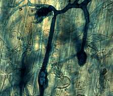
: This file has been superseded by Réseau de mycorhize à l'intérieur d'une racine.tif. It is recommended to use the other file. Please note that deleting superseded images requires consent. : . Description: Français : Réseau mycélien du champignon Rhizophagus irregularis inséré au milieu des cellules de son hôte végétal (ici Vicia fabae). Date: 22 May 2014, 03:25:32. Source: Own work. Author: Mylène Durant.
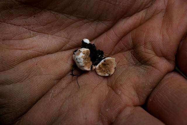
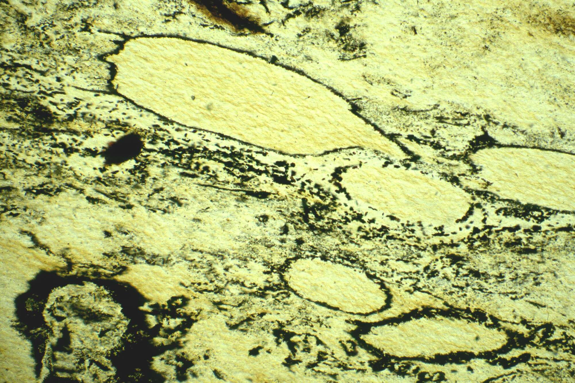
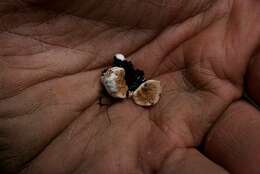
Description: English: apex of holotype of problematic fossil Diskagma buttonii from 2200 million year old Waterval Onder paleosol, South Africa. Date: 5 September 2013, 13:19:46. Source: Own work. Author: Retallack.
Description: English: holotype of problematic fossil Diskagma buttonii from 2200 million year old Waterval Onder paleosol, South Africa. Date: 5 September 2013, 13:19:41. Source: Own work. Author: Retallack.
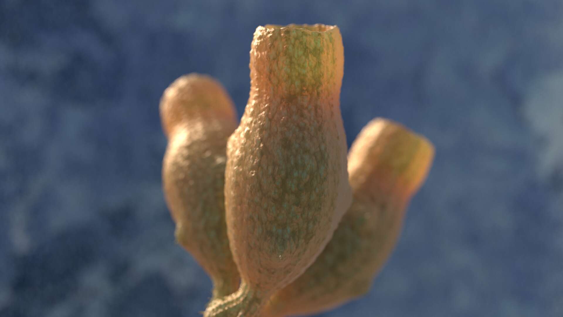
Description: English: reconstruction of problematic fossil Diskagma buttonii from 2200 million year olf Waterval Onder paleosol, South Africa. Date: 5 September 2013, 13:19:48. Source: Own work. Author: Retallack.
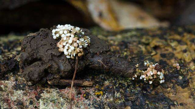
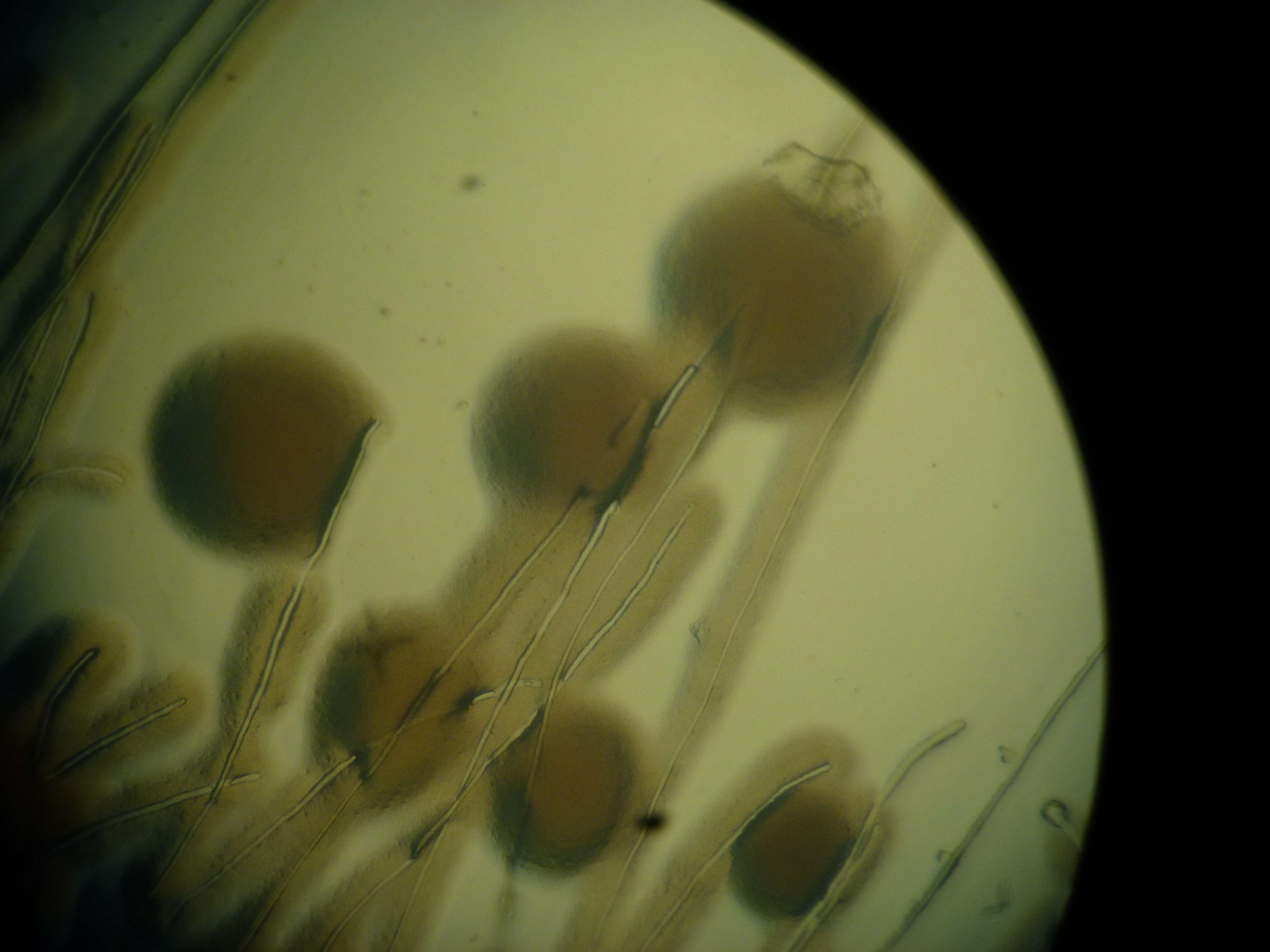
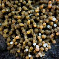
Description: English: New produced spores of Glomus mosseae in a dual culture with tomato on MM medium. Date: 15 January 2014, 11:34:35. Source: Own work. Author: Samson90.
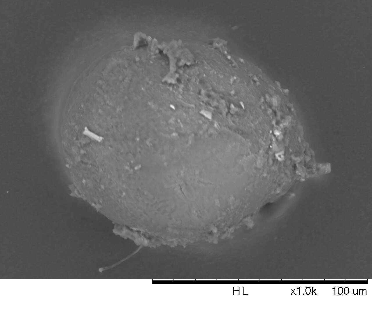
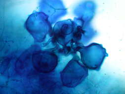
Description: English: 1000x electon microscopy of a mycorrhiza spore, probably Funneliformis mosseae (former 'Glomus mosseae'), wetsieved out of soil. Spore is coated with bacteria, soil and other fungi. Kindly provided by: Dr. rer. nat. tech. Tobias Sieberer, Matthias Salomon. Department 'Biotechnologie gartenbaulicher Kulturen', WZW, Technical University Munich. Date: 23 May 2014, 21:06:23. Source: Own work. Author: Samson90.


