-
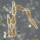
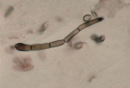
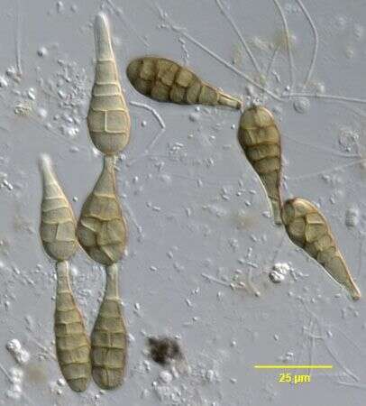
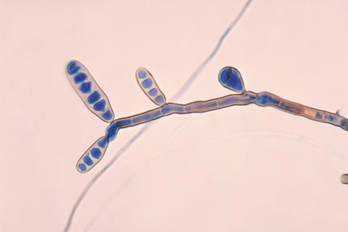
Conidia (spores) of the deuteromycotan fungus, Alternaria alternata (FRIES,1832) KEISSLER,1912. The conidia are obclavate (shaped like a bowling pin) and form single file chains as seen here. The spores have both longitudinal and horizontal septae. Each conidium tapers into a narrow rounded protuberance. Alternaria digests cellulose and is commonly found on dead grasses. Some species are plant pathogens causing "early" potato and tomato blight and leaf rot. The Irish potato famine of 1845-1849 was due to inection by a different fungus, Phytophthora infestans which causes "late" blight.These specimens of A. alternata were found at the margins of a slow-moving freshwater stream in Boise, Idaho.Since A. alternata is terrestrial and not aquatic, the water was probably contaminated by airborne conidia.Phase contrast.
-
Centers for Disease Control/Division of Parasitic Diseases and Malaria
EOL staff
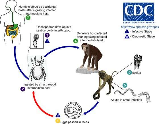
Life cycle of Bertiella tapeworms The life cycle of Bertiella species is not completely understood. Bertiella are believed to have two-host life cycles, with an arthropod intermediate host (usually a mite, likely an oribatid mite) and a vertebrate definitive host (usually non-human primates for the species implicated in human infections). Bertiella studeri (which is found in Africa and Asia) usually infects monkeys in the genera Anthropithecus, Cercopithecus, Cynomologus, and Macaca. Bertiella mucronata (which is found in South America and Cuba) usually infects monkeys in the genera Callicebus and Alouatta. Bertiella eggs and proglottids are passed in the feces of the definitive host (1). Oncospheres (which contain the tapeworm larvae) are ingested by the arthropod intermediate host (2) and within this host the oncospheres develop into cysticercoid larvae (3). The definitive hosts become infected when they ingest arthropod intermediate hosts (4) infected with cysticercoids. Adult Bertiella reside in the small intestine of the definitive host (5), where they attach to the mucosa with the aid of an unarmed scolex (6) (the anterior end of a tapeworm's head). Humans can occasionally serve as definitive hosts for both B. studeri and B. mucronata, usually after accidentally ingesting infected mites (7).From
Centers for Disease Control Parasites and Health website
-
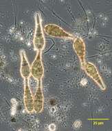
Conidia (spores) of the deuteromycotan fungus, Alternaria alternata (FRIES,1832) KEISSLER,1912. The conidia are obclavate (shaped like a bowling pin) and form single file chains as seen here. The spores have both longitudinal and horizontal septae. Each conidium tapers into a narrow rounded protuberance. Alternaria digests cellulose and is commonly found on dead grasses. Some species are plant pathogens causing "early" potato and tomato blight and leaf rot. The Irish potato famine of 1845-1849 was due to inection by a different fungus, Phytophthora infestans which causes "late" blight.These specimens of A. alternata were found at the margins of a slow-moving freshwater stream in Boise, Idaho.Since A. alternata is terrestrial and not aquatic, the water was probably contaminated by airborne conidia.DIC
-
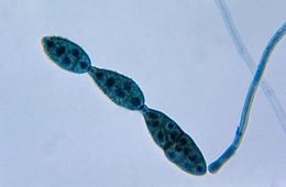
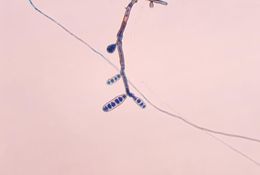
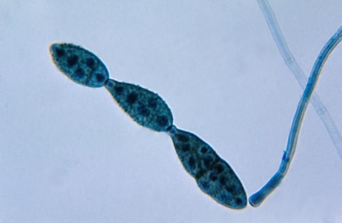
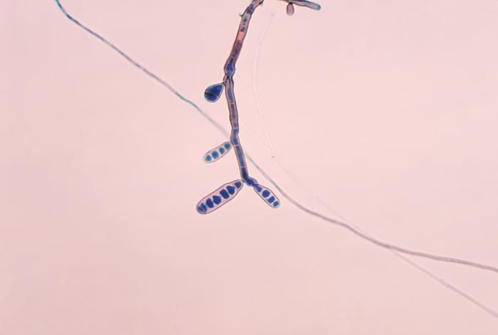
This photomicrograph shows a chain of conidia of a Alternaria sp. fungus, which can be a cause of phaeohyphomycosis.Created: 1955
-
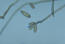
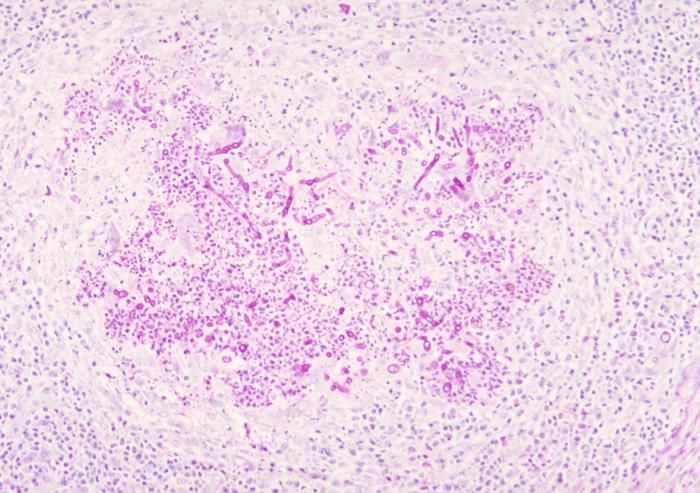
This was a case of phaeohyphomycosis of subcutaneous tissue due to the fungus Curvularia harveyi.Created: 1973
-
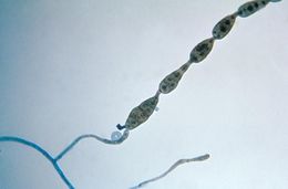
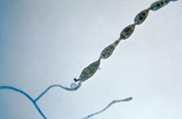
This photomicrograph shows a chain of conidia of a Alternaria sp. fungus, which can be a cause of phaeohyphomycosis.Created: 1955
-
This was a case of phaeohyphomycosis of subcutaneous tissue due to the fungus Curvularia harveyi.Created: 1973
-
This photomicrograph shows a chain of conidia of a Alternaria sp. fungus, which can be a cause of phaeohyphomycosis.Created: 1955
-
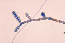
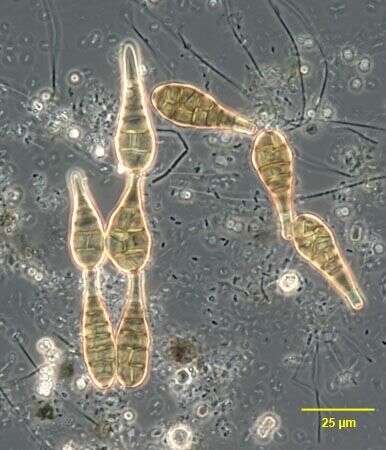
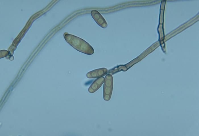
This photomicrograph shows conidiophores and conidia of the fungus Curvularia harveyi.Created: 1973
-
This photomicrograph shows a chain of conidia of a Alternaria sp. fungus, which can be a cause of phaeohyphomycosis.Created: 1955
-
This photomicrograph shows conidiophores and conidia of the fungus Curvularia harveyi.Created: 1973
-
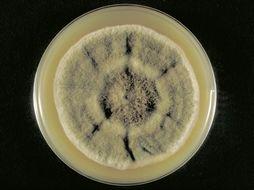
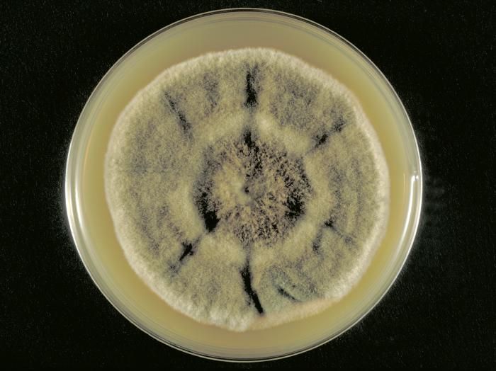
This was a Sabourauds dextrose agar plate culture growing the fungus Curvularia harveyi.Created: 1973
-
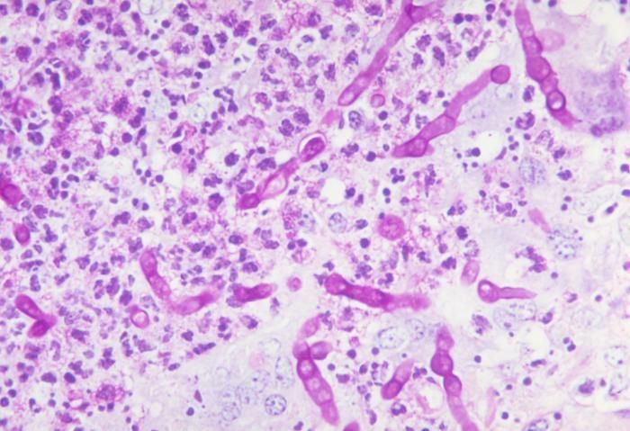
This was a case of phaeohyphomycosis of subcutaneous tissue due to the fungus Curvularia harveyi.Created: 1973
-
This was a case of phaeohyphomycosis of subcutaneous tissue due to the fungus Curvularia harveyi.Created: 1973
-
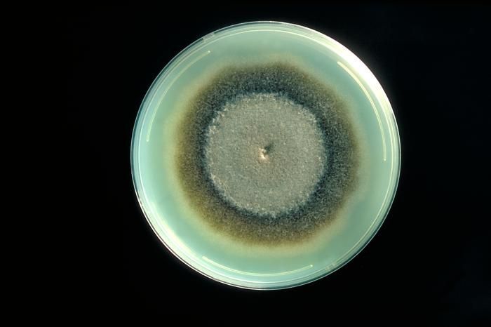
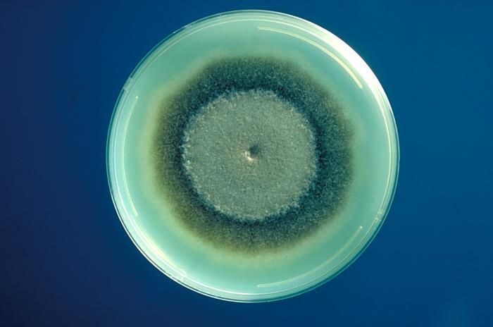
This was a plate culture of Exserohilum rostratum, a fungus, which causes Phaeohyphomycosis.Created: 1977
-
This was a plate culture of Exserohilum rostratum, a fungus, which causes Phaeohyphomycosis.Created: 1977
-
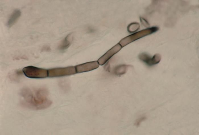
Note the fine branching tubes of the fungus Exserohilum rostratum, which is the cause of Phaeohyphomycosis.Created: 1978
-
This image demonstrates the fine branching tubes of Exserohilum rostratum.Created: 1978
-
Photomicrograph showing fine branching tubes of Exserohilum rostratum.Created: 1978
-
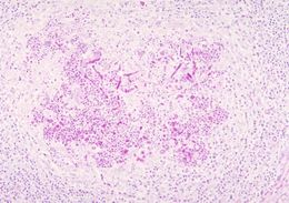
A micrograph revealing histopathologic changes in phaeohyphomycosis due to Exserohilum rostratum.Created: 1980
-
This was a plate culture of Exserohilum rostratum, a fungus, which causes Phaeohyphomycosis.Created: 1977
-
This was a plate culture of Exserohilum rostratum, a fungus, which causes Phaeohyphomycosis.Created: 1977
-
Note the fine branching tubes of the fungus Exserohilum rostratum, which is the cause of Phaeohyphomycosis.Created: 1978
-
This image demonstrates the fine branching tubes of Exserohilum rostratum.Created: 1978