-
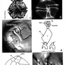
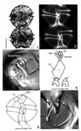
Plate 8. Alexandrium tamiyavanichi. Figs. 1-3. LM. Fig. 1. Two cell chain: cells medium-sized; round to slightly wider than long. Epitheca with shoulders. Fig. 2. Cells stained with calcofluor white: cingulum displaced 1X its width; sulcus widens posteriorly. Fig. 3. Apical view: apical pore plate (Po) houses comma-shaped foramen. First apical plate (1') with ventral pore (vp). Figs. 4-5. Line drawings. Fig. 4. 1' plate in direct contact with Po. Po with large central foramen surrounded by small pores. Anterior sulcal plate (s.a.) invades epitheca; an anterior projection of s.a. fits into a notch in the 1' plate (arrows). Fig. 5. Ventral view: sulcal lists project anteriorly (arrows). Fig. 6. Posterior sulcal plate (s.p.) with round posterior attachment plate (pap) in center (arrow).
-
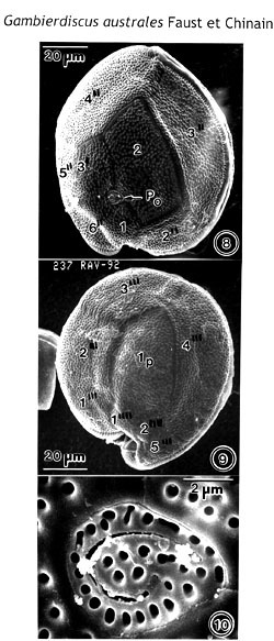
Figs. 8-10. Scanning electron micrographs of Gambierdiscus australes (RAV-92), sp. nov. Figs. 8, 9. Cells round to ellipsoid. The cell surface is smooth with scattered small pores. Fig. 8. Epithecal view. The Po plate is oriented ventrally. Fig. 9. Hypothecal view. The Ip plate, long and narrow, occupies 30% of the hypotheca width. Fig. 10. The Po plate is a broadly ellipsoid plate, with fish-hook-shaped apical opening surrounded by 31 pores.
EMu: Holotype SEM negative # 237047; SEM stub # 237; Field # RAV-92; Catalog # 1526; Figure #8.
-
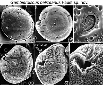
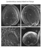
Figs. 1-6. Scanning electron micrographs of the surface morphology of Gambierdiscus toxicus and Gambierdiscus belizeanus are shown. FIGS.1-2. Scanning electron micrographs of the surface morphology of Gambierdiscus toxicus Adachi et Fukuyo. FIG.1. Cell in epithecal view. FIG.2. Cell in hypothecal view. Cell shape is round, compressed and ellipsoidal. Cell surface is smooth with scattered small pores. Thecal plate is large and quadrangular. FIGS.3-6. Gambierdiscus belizeanus sp. nov. FIG.3. Cell in epithecal view slightly damaged. Cell surface areolated and plates partially separated. FIG. 4. Cell is in hypothecal and ventral view. Plate is narrow. FIG.5. Apical pore complex is triangular with a fish-hook-shaped apical pore. A round pore is present in the areolae (arrowhead). FIG.6. Cingulum deep, ascending into a deep sulcal hollow.
EMu: Holotype SEM NEGATIVE # 132003B; SEM STUB # 152; FIELD # 682-93; ACCESSION # 407167; CATALOG # 798; FIGURE # 3.
-
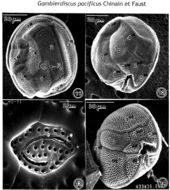
Figs. 11-14. Scanning electron micrographs of Gambierdiscus pacificus (HO-91), sp. nov., and Gambierdiscus belizeanus. Figs. 11-13. Gambler discus pacificus, sp. nov. Fig. 11, 12. Cells are round to ellipsoid. The Cell surface is smooth with scattered small pores. Fig. 11. Epithecal view. The Po plate is oriented ventrally. Fig. 12. The Ip plate, short and narrow, occupies 20% of the hypotheca width. Postcingular plates 2'" and 4'" are wide. Fig. 13. The Po plate is four-sided plate with a narrow fish-hook-shaped apical opening surrounded by 31 pores. Fig. 14. Gambierdiscus belizeanus. Cell in hypothecal view. The cell surface is areolated. The Ip plate, narrow and pentagonal, is wedged between very wide postcingular plates 2'", and 4'". The cingulum, deeply excavated, is ascending into a deep sulcal hollow.
EMu: Gambierdiscus pacificus
Holotype SEM negative # 241006; SEM stub # 241; Field # HO-91; Catalog # 1528; Figure #11.
-
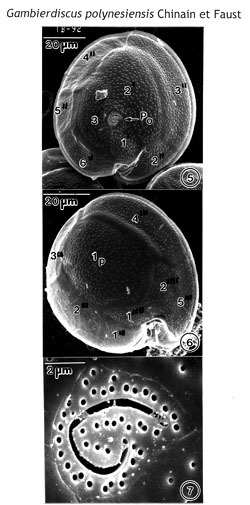
Figs. 5-7. Scanning electron micrographs of Gamblerdiscus polynesiensis (TB-92), sp. nov. Figs. 5, 6. Cells are round to ellipsoid. Cell surface is smooth with small scattered pores. Fig. 5. Epithecal view. The PO plate is oriented ventrally. Fig. 6. Hypothecal view. The Ip plate, broad and pentagonal, occupies 60% of the of hypotheca width. Postcingular plates 2'", 3'" and 4'" are narrow. The cingulum, deep, is ascending into a deep sulcal hollow. Fig. 7. The Po plate is triangular with fish-hook-shaped apical opening surrounded by 44 pores.
EMu: Holotype SEM negative # 242010; SEM stub # 242; Field # TB-92; Catalog # 1522; Figure #5.
-
Figs. 1-4. Scanning electron micrographs illustrate the surface morphology of Gambierdiscus toxicus (GTT-91). Fig. 1. In epithecal view. The cell shape is round, and the apical pore plate (Po) oriented ventrally. Fig. 2. In hypothecal view. Posterior intercalary plate (Ip) broad and pentagonal, centrally located, occupying about 1/3 of cell's width. Fig.3. Po plate ellipsoid with a fish-hook-shaped apical pore surrounded by rows of 28 evenly distributed pores. Fig. 4. Cell in central view, shape compressed and bordered by a cingular list. The cell surface is smooth with small scattered pores.
NOTE: This is the apotype of the genus Gambierdiscus. It is an important toxic species. I would like to add this species to the dinoflagellate ‘Types’ since the SEM plate of G. toxicus is the only record. Adachi and Fukuyo (1979) described G. toxicus sp. nov. only in line drawing to illustrate the morphology of plates.
-
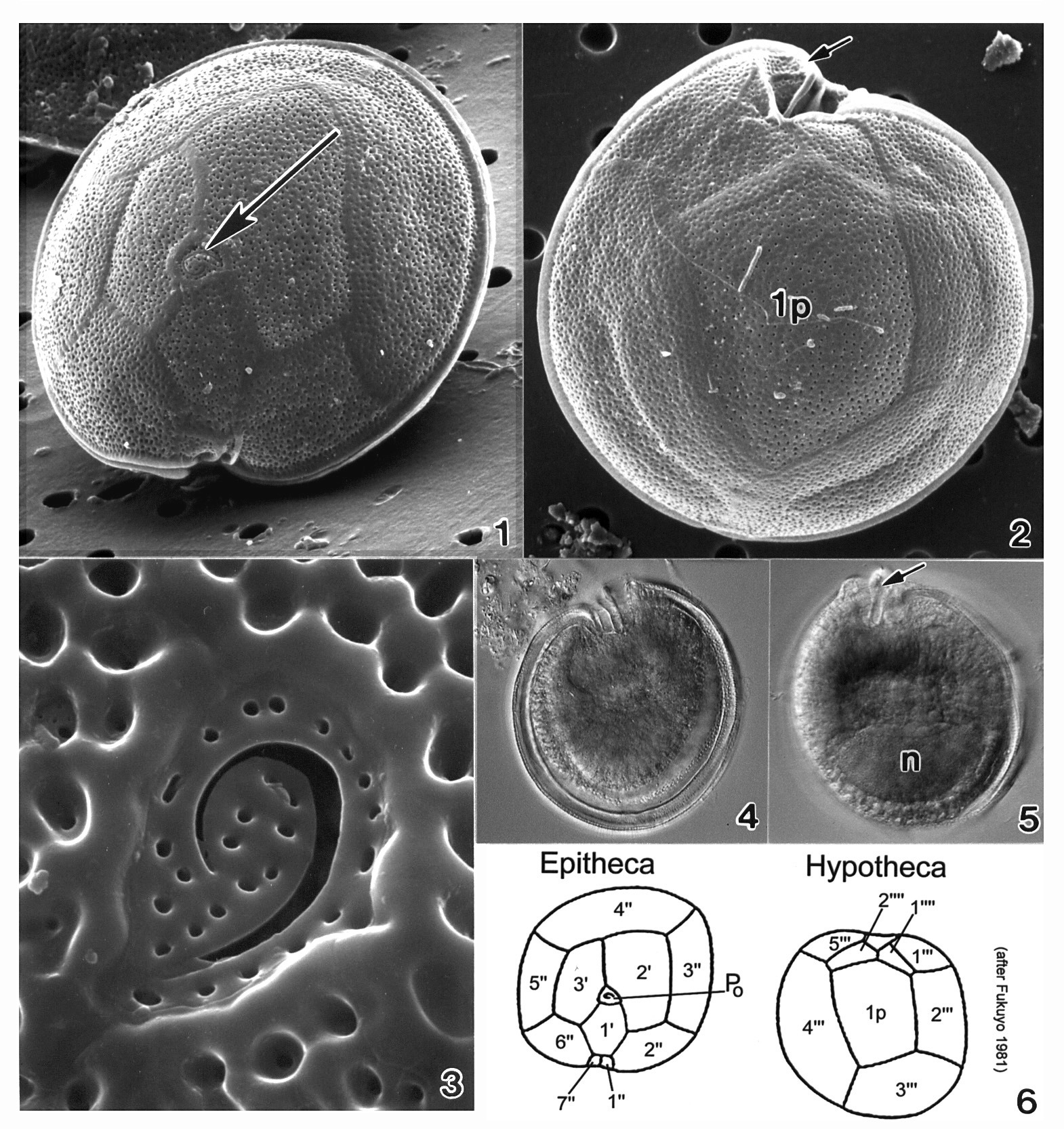
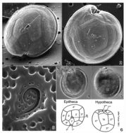
Plate 20. Gambierdiscus toxicus. Figs. 1-3. SEM. Fig. 1. Epitheca: cell round to ellipsoid; anterior-posteriorly compressed. Cell surface smooth with small scattered pores. Apical pore complex located at the apex (arrow). Fig. 2. Hypotheca: 1p plate large and pentagonal. Sulcal region deeply excavated (arrow). Fig. 3. Apical pore plate with characteristic fishhook shaped apical pore. Fig. 4. LM. Epitheca: cingulum and sulcal region in focus. Fig. 5. LM. Hypotheca: sulcal ridge (arrow); large nucleus (n). Fig. 6. Line drawing: thecal plate arrangement.
-
-
-
-
-
-
-
-
-
-
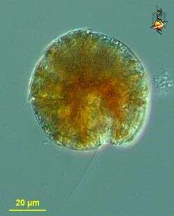
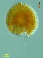
Gambierdiscus (gam-beer-disk-us) toxicus, a toxic dinoflagellate with chloroplasts. With two flagella, but only one (the trailing flagellum) visible here. Small lobes of the plastid extend to the surface of the cell. Armoured, with fairly thick thecal plates. Flattened and this view is more or less polar. Mostly associated with sediments of warmer waters. Differential interference microscopy.
data on this strain.
-
Gambierdiscus (gam-beer-disk-us) toxicus, a toxic dinoflagellate with chloroplasts. With two flagella, but only one (the trailing flagellum) visible here. Armoured, with fairly thick thecal plates. Flattened and this view is more or less polar. Mostly from warmer waters. Differential interference microscopy.
data on this strain.
-
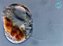
Gambierdiscus (gam-beer-disk-us) toxicus, a toxic dinoflagellate with chloroplasts (not shown in this image). Armoured, with fairly thick thecal plates. This image shows the plates and a large apical pore. Mostly from sediments of warmer waters. Differential interference microscopy.
data on this strain.
-
Thecadinium (theek-a-din-ee-um) inclinatum Balech 1956. The image shows a cell in right lateral view. The cell is laterally compressed. The cell contains no plastids, but several food particles are present. The cell is laterally compressed. The cell is thecate, but has no processes.