-
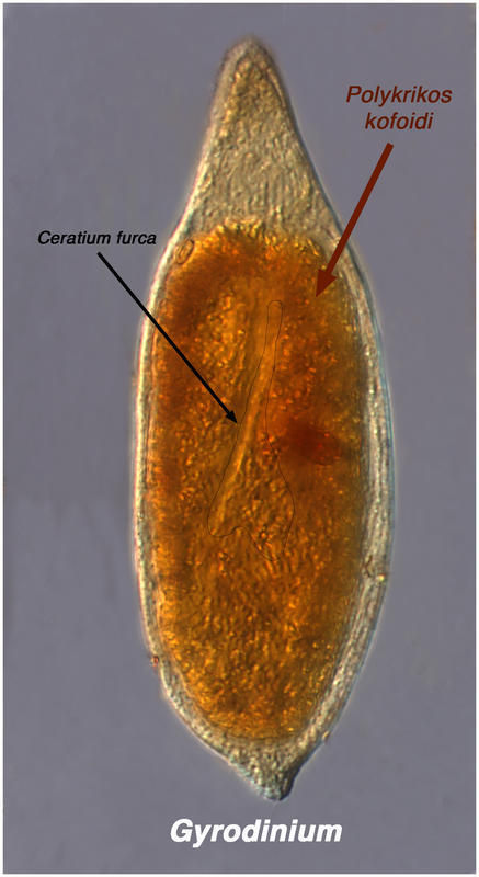
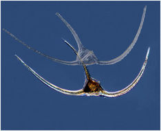
This probably is Gyrodinium eating Ceratium
-
Gyrodinium ate a Polykrikos which had eaten a Ceratium. Specimen from the Etang de Thau.
-
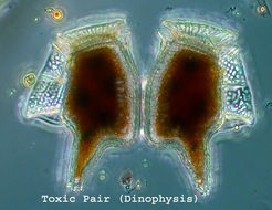
Arctic Dinophysis norvegica (empty!)from the Chukchi Sea in summer 2011.
-
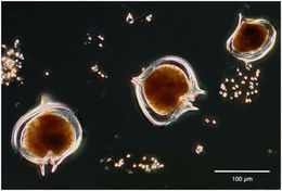
Ceratium arcticum specimens from the Chukchi Sea.
-
-
Dinophysis from the SW Pacific (Biosope Cruise)
-
-
A heterotrophic dinoflagellate found with Phaeocystis on which it likely feeds.
-
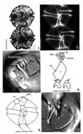
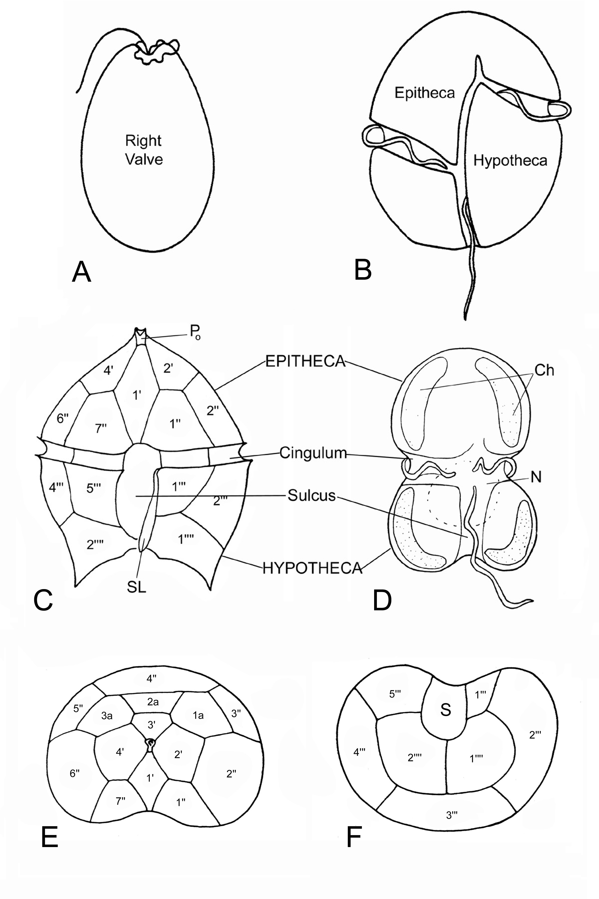
Fig. 1. Identifying dinoflagellates: a. lateral view of a desmokont cell type (two dissimilar flagella apically inserted)
-
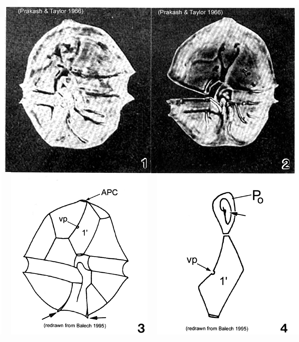
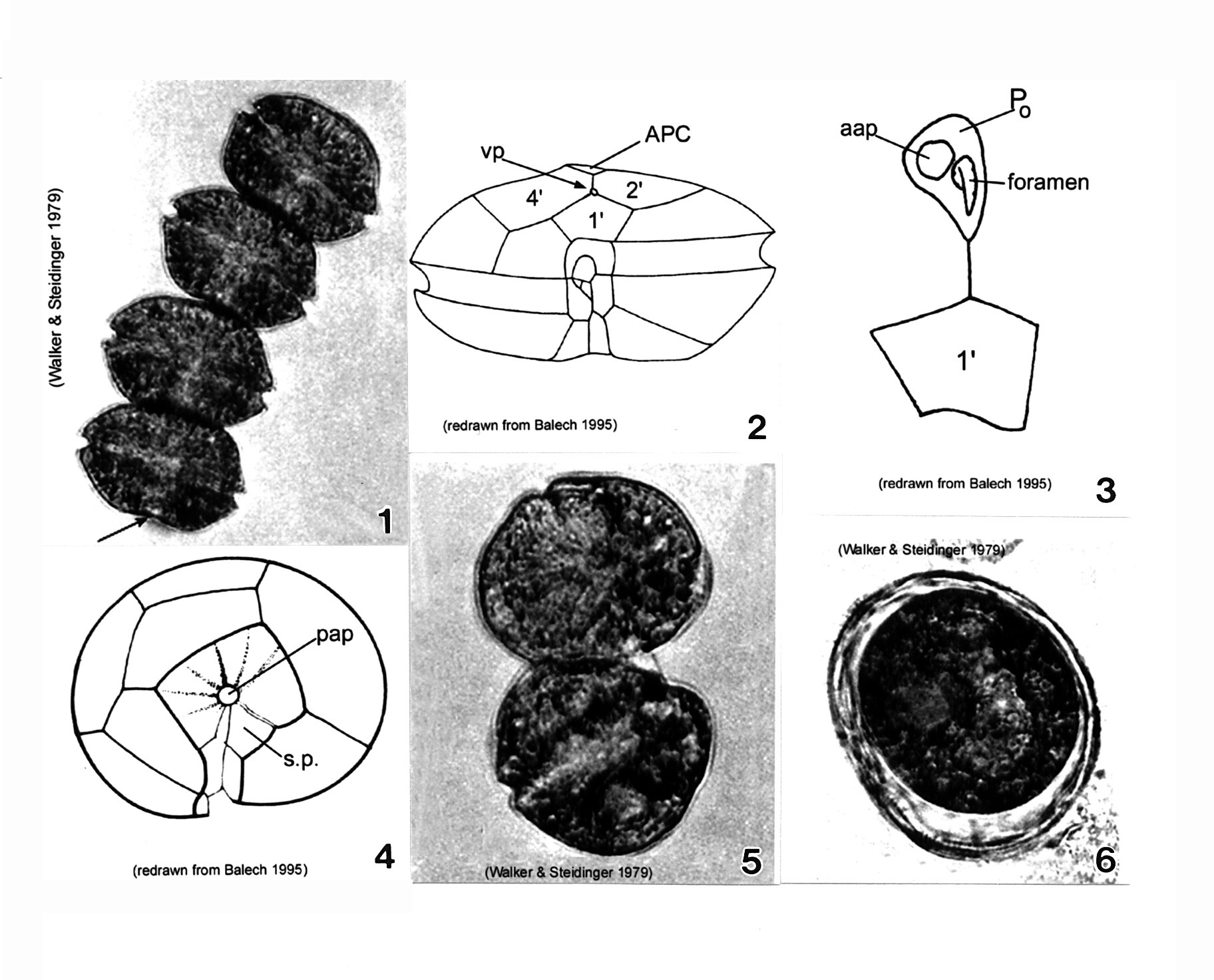
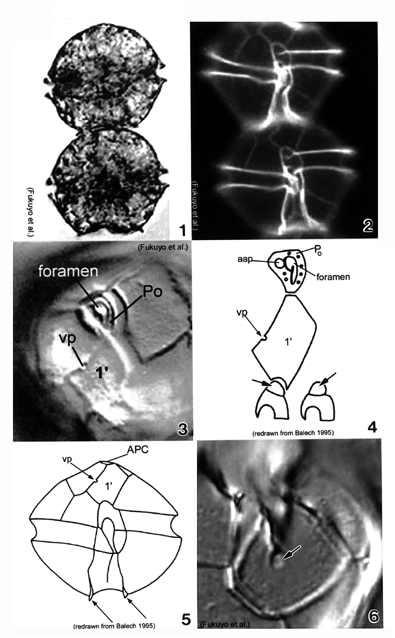
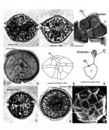
Plate 1. Alexandrium acatenella. Figs. 1-2. LM: ventral view of empty thecae. Cell small to medium, longer than wide, angular to round. Conical epitheca with shoulders; larger than hypotheca. Figs. 3-4. Line drawings. Fig. 3. Ventral view: 1' plate bears ventral pore (vp). Hypotheca with two antapical spines (arrows). Fig. 4. Po comes in direct contact with 1' plate. APC: comma-shaped foramen (arrow).
-
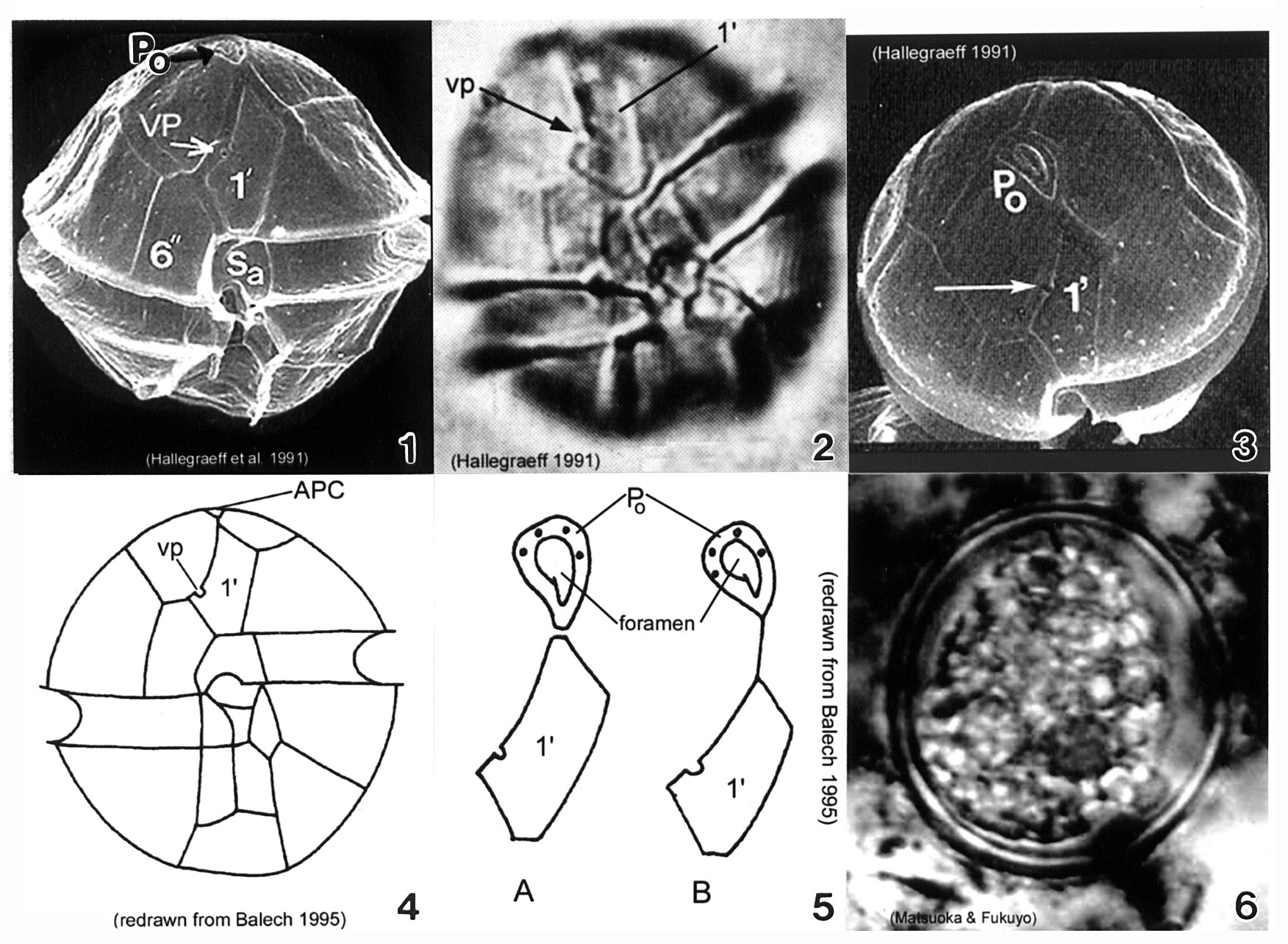
Plate 3. Alexandrium minutum. Fig. 1. SEM: ventral view. Cell small and ellipsoidal. Epitheca conical, larger than hypotheca. Hypotheca short and wide; antapex obliquely flattened. Intercalary bands present. Cingulum deep, lipped; displaced 1X its width. Sulcus shallow (sa=anterior sulcal plate). Apical pore plate (Po) in direct contact with 1' plate. Fig. 2. LM: ventral view. Ventral pore (vp) present on 1' plate. Fig. 3. SEM: apical view. Po large, narrow and oval; indirectly connected to 1' plate. Vp present (arrow). Figs. 4-5. Line drawing. Fig. 4. Ventral view. 1' plate slender and rhomboidal. Fig. 5. Po connection to 1' plate: a. direct; b. indirect via thin suture. Fig. 6. LM: cyst circular in apical view.
-
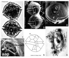
Plate 4. Alexandrium monilatum. Fig. 1. LM: four-cell chain. Cells large, wider than long, flattened anterio-posteriorly. Antapex slightly concave (arrow). Figs. 2-4. Line drawings. Fig. 2. Ventral pore (vp) depicted (Florida specimens) at anterior margin of 1' plate where it comes in contact with plates 2' and 4'. Cingulum (C) deeply excavated, wide, descending; displaced one time its width. Fig. 3. Apical pore plate (Po) does not come in contact with 1' plate. Anterior attachment pore (aap) large, round and dorsally situated in the APC. Foramen comma-shaped. Fig. 4. Antapical view: posterior sulcal plate (sp) large, rhomboid and concave with radial markings. Posterior attachment pore (pap) large and centrally located. Figs. 5-6. LM. Fig. 5. Two isogamous gametes fusing at oblique angles. Fig. 6. Mature resting cysts: dark and round, with a triple layered wall.
-
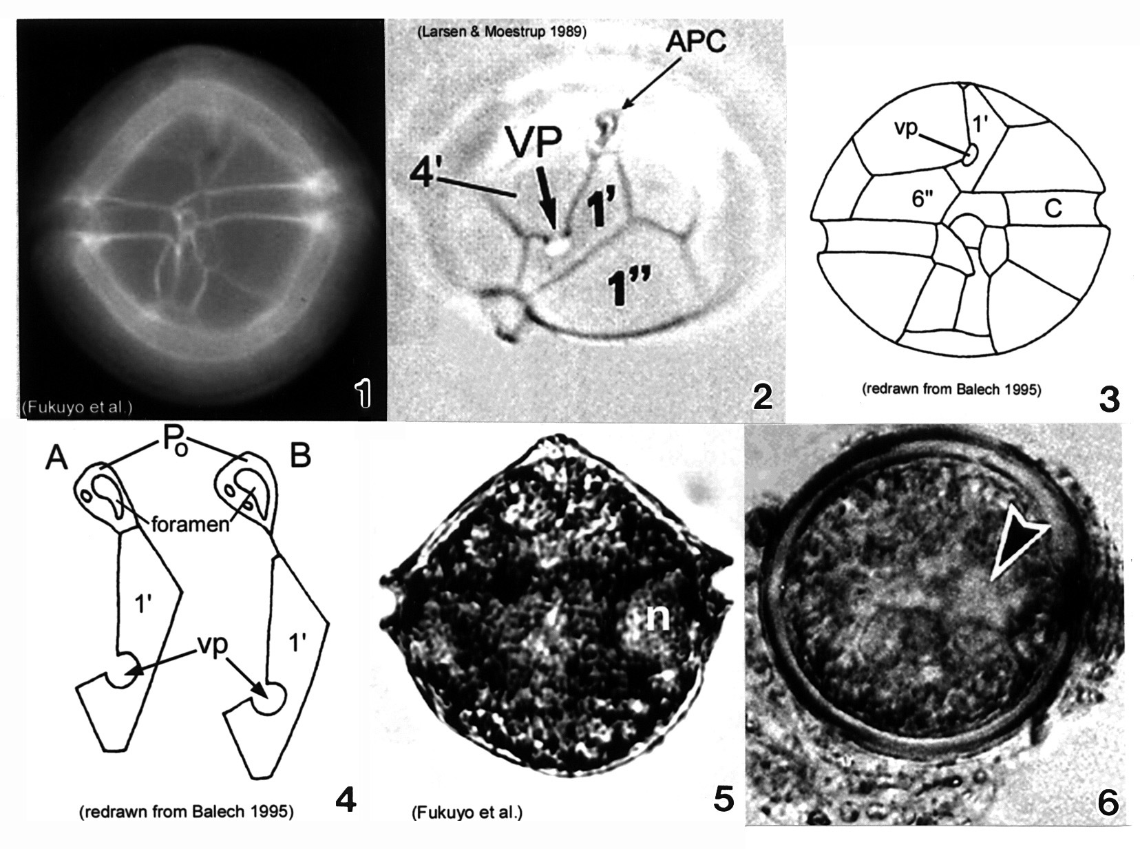
Plate 5. Alexandrium ostenfeldii. Figs. 1-3. LM. Fig. 1. Ventral view. Cell large and nearly spherical. Cingulum deeply excavated. Epitheca broad and convex-conical. Hypotheca hemispherical with an obliquely flattened antapex. Fig. 2. Epitheca: apical view. Ventral pore (vp) large and distinct. First apical plate (1') forms a 90 degree angle at the point where vp and 4' plate come in contact. Apical pore complex (APC) with comma-shaped foramen. Figs. 3-4. Line drawings. Fig. 3. Ventral view: 6'' plate wider than high. Cingulum (C) slightly excavated. Fig. 4. APC and 1' plate: a. Po in direct contact with 1'; b. Po in indirect contact with 1' via thin suture. Fig. 5. LM: vegetative cell. Small equatorial nucleus (n). Fig. 6. LM: temporary cyst large and spherical, covered in mucilage. Nucleus visible (arrowhead)(Mackenzie et al. 1996).
-
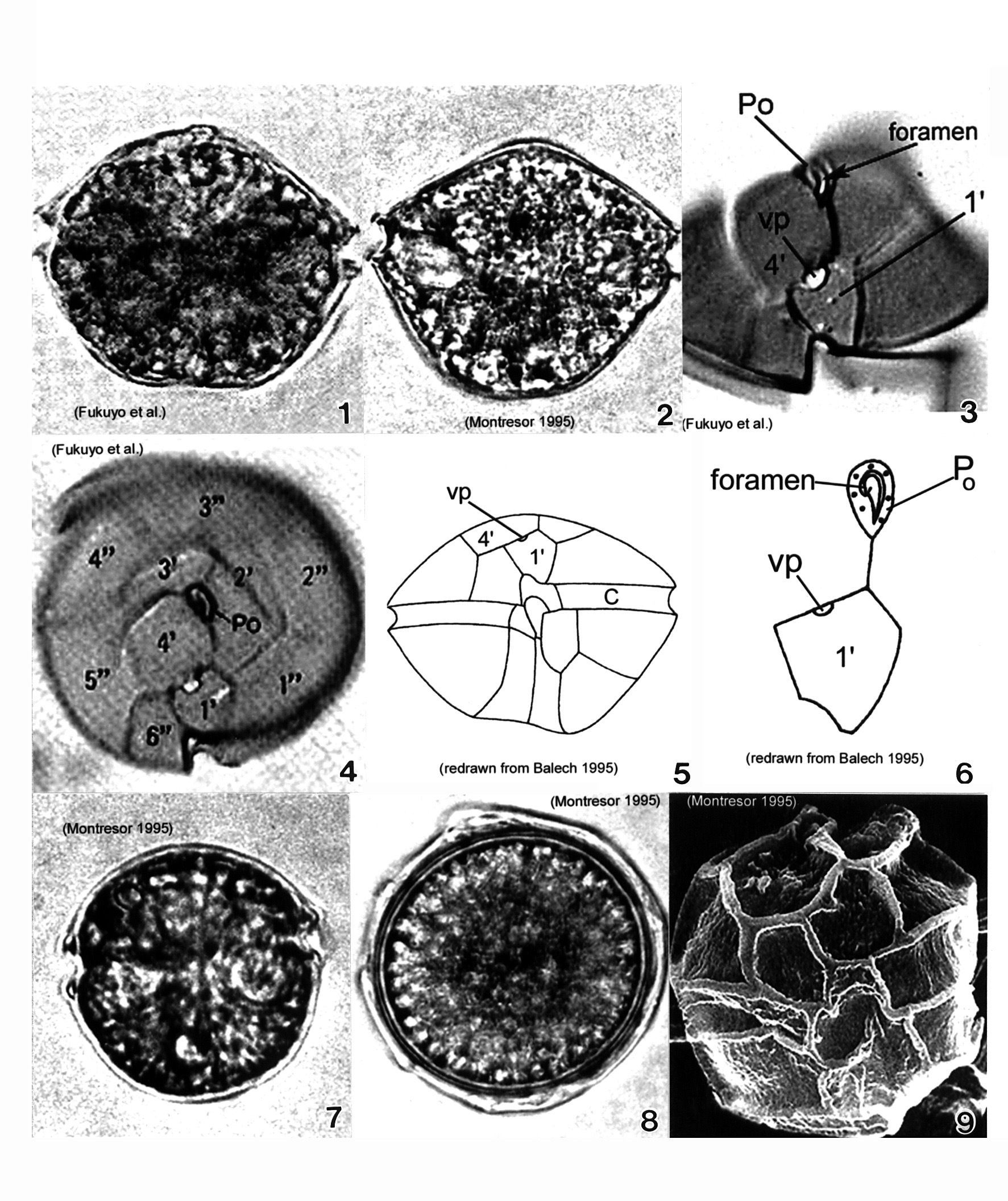
Plate 6. Alexandrium pseudogonyaulax. Figs. 1-4. LM. Fig. 1. Ventral view. Cell broadly pentagonal; wider than long. Epitheca short and dome-shaped. Hypotheca longer than epitheca. Cingulum shallow and barely displaced. Fig. 2. Dorsal view. Antapex obliquely concave. Fig. 3. Epitheca: ventral view. Apical pore plate (Po) with comma-shaped foramen. 1' plate pentagonal with large wide ventral pore (vp) on 4' plate margin. Fig. 4. Epitheca: apical view. 1' plate does not come in contact with Po. Po oval and longitudinal on apex. Figs. 5-6. Line drawings. Fig. 6. Po and 1' plate not in contact. Fig. 7. LM: isogamous gametes smaller and rounder than vegetative cells. Fig. 8. LM: round resting cyst. Fig. 9. SEM: paratabulate cyst.
-
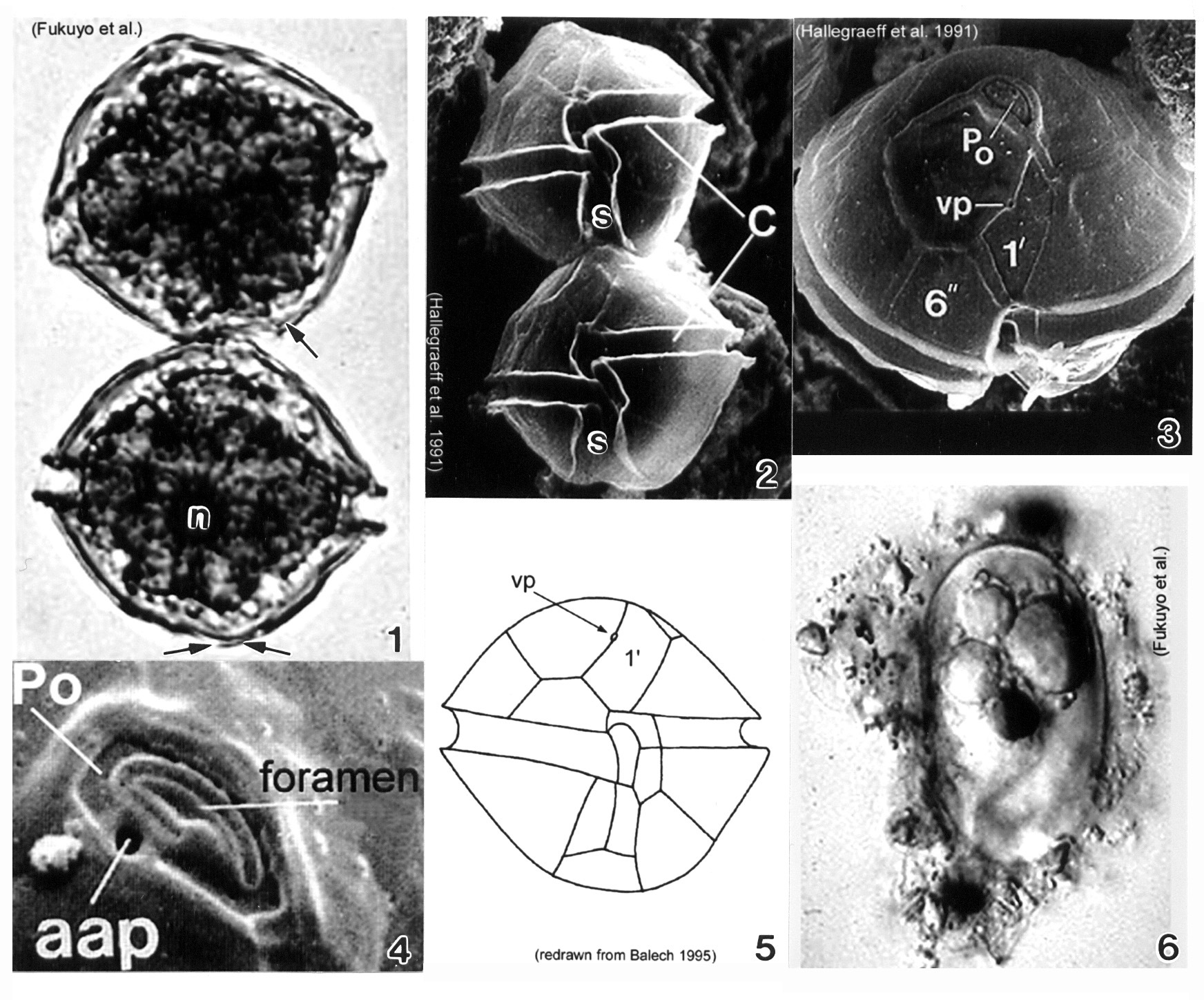
Plate 7. Alexandrium tamarense. Fig. 1. LM. Two cell chain: cells small to medium; slightly longer than wide, nearly spherical. Cingulum (C) deeply escavated and lipped. Left hypothcal lobe slightly larger than right. Nucleus (n) visible. Figs. 2-4. SEM. Fig. 2. Two cell chain: cingulum displaced 1X its width. Deep sulcus (s) widens posteriorly. Fig. 3. Epitheca: apical view. Apical pore plate (Po) rectangular; narrows ventrally. Po and first apical plate (1') in direct contact. Small ventral pore present on 1' plate. Fig. 4. Apical pore complex (APC): foramen large and fishhook shaped. Small round anterior attachment pore (aap) present (Hallegraeff 1991). Fig. 5. Line drawing. Fig. 6. LM. Oblong resting cyst with rounded ends, reddish lipid bodies; covered in mucilage.
-
Plate 8. Alexandrium tamiyavanichi. Figs. 1-3. LM. Fig. 1. Two cell chain: cells medium-sized; round to slightly wider than long. Epitheca with shoulders. Fig. 2. Cells stained with calcofluor white: cingulum displaced 1X its width; sulcus widens posteriorly. Fig. 3. Apical view: apical pore plate (Po) houses comma-shaped foramen. First apical plate (1') with ventral pore (vp). Figs. 4-5. Line drawings. Fig. 4. 1' plate in direct contact with Po. Po with large central foramen surrounded by small pores. Anterior sulcal plate (s.a.) invades epitheca; an anterior projection of s.a. fits into a notch in the 1' plate (arrows). Fig. 5. Ventral view: sulcal lists project anteriorly (arrows). Fig. 6. Posterior sulcal plate (s.p.) with round posterior attachment plate (pap) in center (arrow).
-
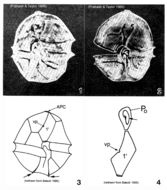
Figs 1-7. Lateral view of a cell with reticulated thecal surface, a conical epitheca, wide and deep, displaced cingulum, and a trapezoid hypotheca. The apical pore complex is situated ventrally. The apical plate 1' is asymmetric and pentagonal. The hypotheca is ventrally indented forming two lobes separating plates 2'" and 5'". The cingulum is displaced and finely striated with small pores aligned along the cingular lists. Fig. 2.The apical pore complex is a recessed chamber with a centrally located raised dome surrounded by a collar; it includes the apical pore plate (PO) and canal plate (X). Fig.3. Lateral view of a cell: a conical epitheca, wide and deep cingulum, and trapezoid hypotheca. Fig.4. Architecture of the epitheca including the position of the apical pore complex. Intercalary plates 2a and 3a are separated by plate 3'. The intercalary bands are striated.
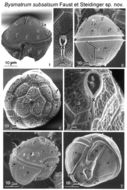
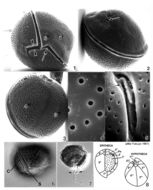
EMu: Holotype SEM negative # 23040; SEM stub # ?; Field # 78-87; Accession # 407159; Catalog #1730; Figure # 1.
-
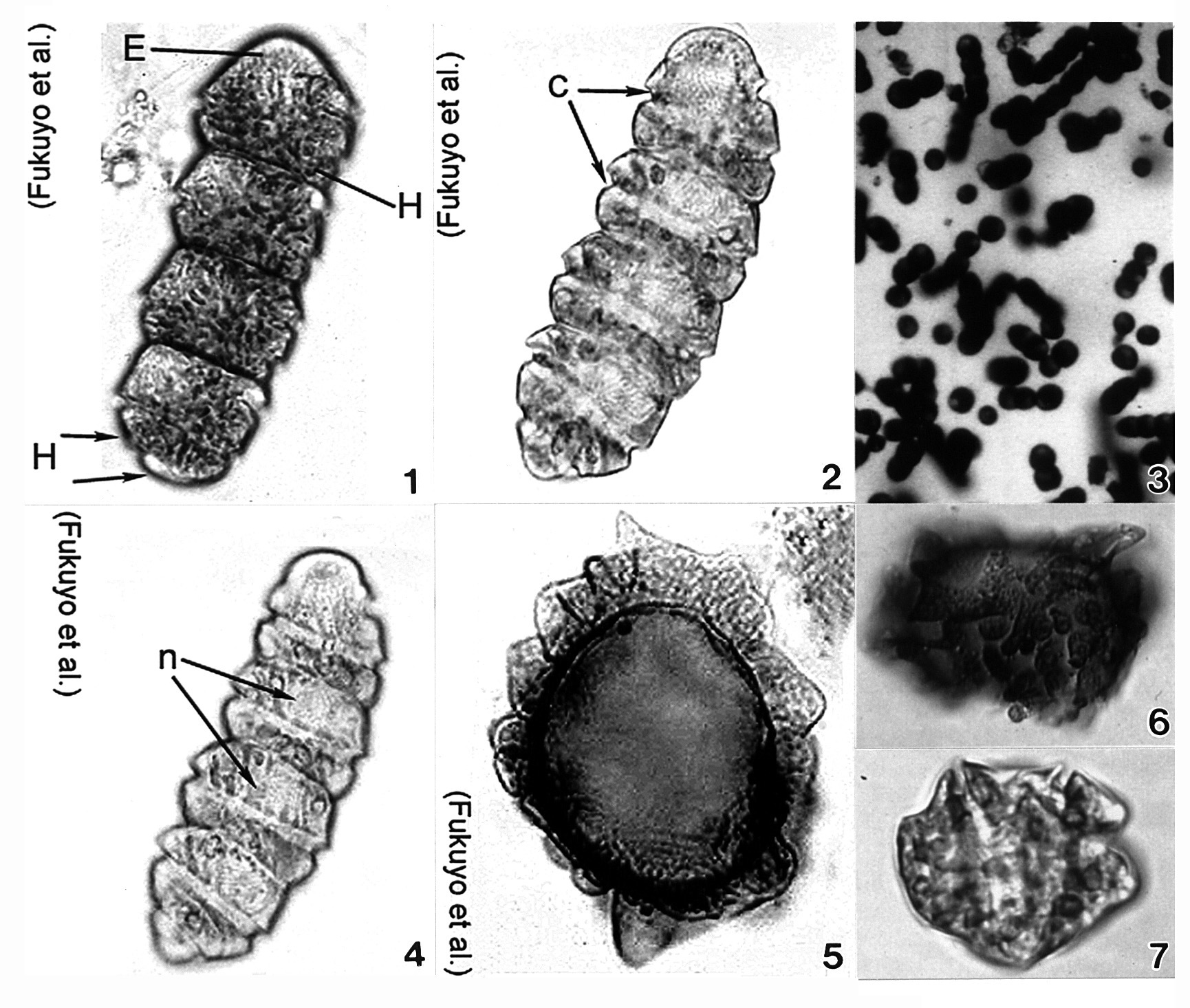
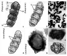
Plate 9. Cochlodinium polykrikoides. Figs. 1-7. LM. Fig. 1. Four cell chain. Single cell small and ellipsoid. Epitheca (E) rounded and conical. Hypotheca (H) divided into two posterior lobes (arrows). Numerous rod-shaped chloroplasts. Fig. 2. Cingulum (c) deeply excavated; circles cell 1.8-1.9 times. Fig. 3. Colony of single and chained cells. Fig. 4. Large nucleus (n) in epitheca. Figs. 5-7. Cysts. (Figs. 3,6,7 by Matsuoka & Fukuyo)
-
Plate 10. Coolia monotis: Figs. 1-5. SEM. Fig. 1. Ventral view: spherical shape. Cingulum lipped and equatorial. Sulcus with flexible lists (arrowheads). Ventral pore present (arrow). Fig. 2. Dorsal view: apical pore plate (arrow), Po, located off-center on epitheca. Fig. 3. Antapical view: hypothecal plates. Fig. 4. Smooth edged thecal pores unevenly distributed. Fig. 5. Po about 12 _ long, slightly curved and narrow with a slit-like apical pore. Two supporting rib-like costae (arrows) and evenly spaced round pores surround the pore. Figs. 6,7. LM. Fig. 6. Ventral view of lipped cingulum and sulcus. Fig. 7. Planozygote with two longitudinal flagella (arrows). Fig. 8. Line drawing: thecal plate arrangement.
-
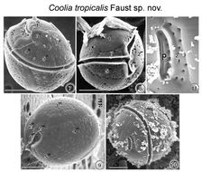
FIGS. 7-11. Scanning Electron micrographs of the surface morphology of Coolia tropicalis sp. nov. FIG. 7. Oblique dorsal view of C. tropicalis shows the apical pore and the equatorially located lipped cingulum. Cell surface is smooth with large scattered pores. FIG. 8. Cell is spherical in equatorial view shoving a deep cingulum and sulcus. Detritus adheres to the epitheca. FIG. 9. Antapical view of a cell show large unequal plates. FIG. 10. Apical pore is a narrow opening located in the epitheca. Fine detrital particles partially cover the thecal surface. FIG. 11. The apical pore is about 7 μm long straight and narrow slits with two supporting costae and evenly spaced round pores. Detritus attached to surface of apical pore plate. EMu:HOLOTYPE SEM NEGATIVE #166029; SEM STUB # 166; FIELD # 728-93;ACCESSION # 408431: CATALOG # 997; FIGURE # 7.
-
FIG. 12. Coolia tropicalis sp. nov. A) apical view of epitheca, and B) antapical hypotheca.
-
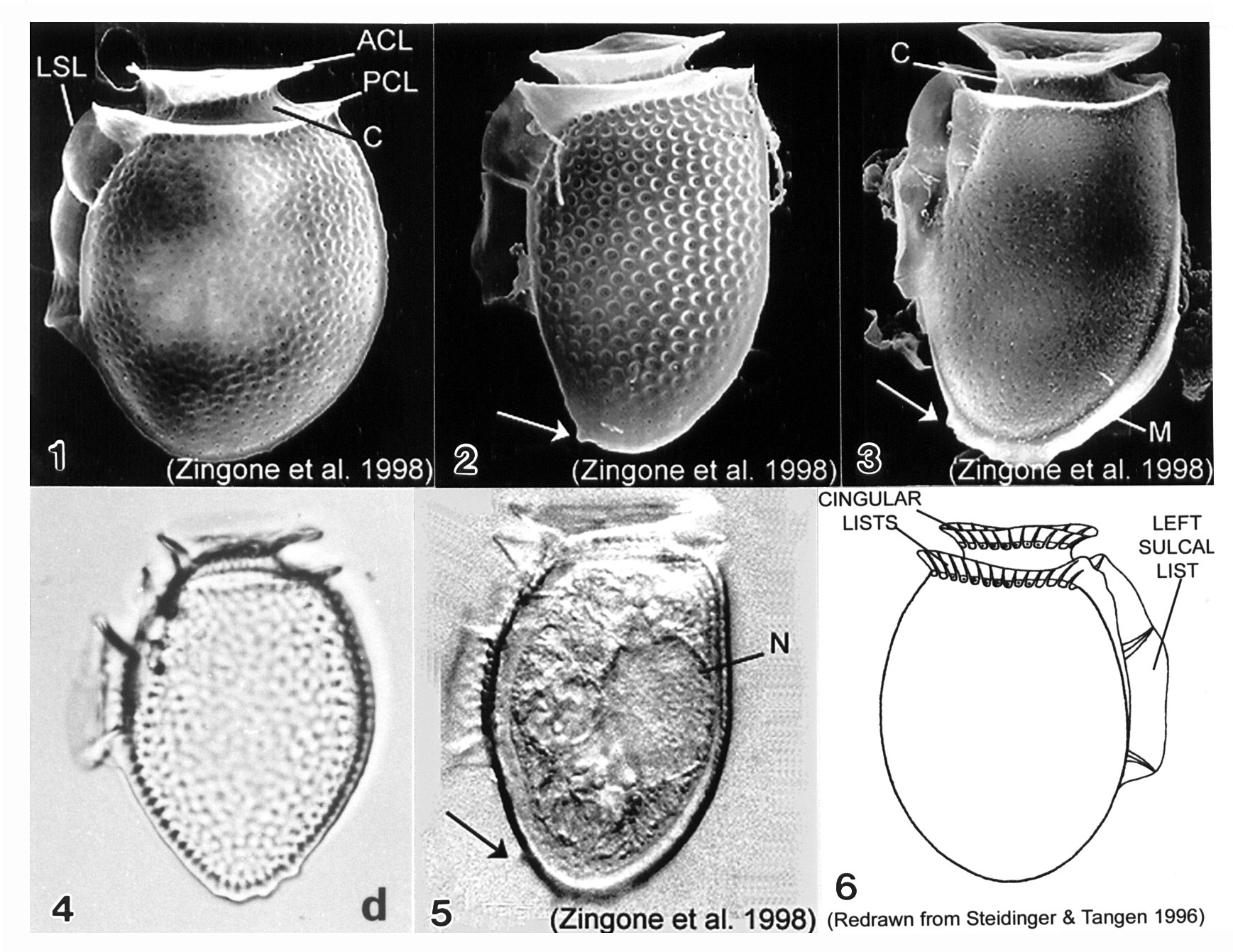
Plate 11. Dinophysis acuminata. Figs. 1-5. SEM: lateral view. Fig. 1. Cell oval and rotund; thecal surface with shallow depressions and scattered pores. Left sulcal list (LSL) extends beyond midpoint of cell. Well-developed cingular lists: anterior cingular list (ACL); posterior cingular list (PCL). C=cingulum. Fig. 2. Long and narrow cell with prominent surface areolae, each with a pore. Antapex tapered and ventrally off-center. Small posterior protrusion present (arrow). Fig. 3. Long and narrow cell. Thecal surface smooth with small scattered pores. Megacytic zone (M) void of pores. Posterior protrusions on antapex (arrow). Figs. 4-5. LM: lateral view. Fig. 4. Surface areolae and tapered antapex (from Larsen & Moestrup 1992: fig. 1d). Fig. 5. Large dorsal nucleus (N). Small, blunt projections on tapered antapex (arrow). Fig. 6. Line drawing.
-
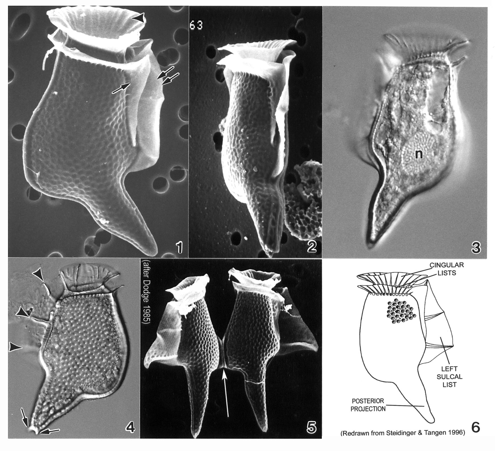
Plate 12. Dinophysis acuta. Fig. 1. SEM: lateral view. Cell oblong and robust; theca heavily areolated. Well developed cingular lists (CL) and left sulcal list (LSL). Pointed antapex. Figs. 2-3. LM: lateral view (from Larsen & Moestrup 1992: fig.s 2a,d; scale bars=20 _). Fig. 2. Large areolae, each with a pore (arrows). Fig. 3. Widest point below mid-section (dashed line) aligned with third sulcal rib (arrow). Fig. 4. Line drawing.
-
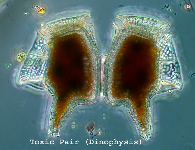
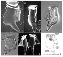
Plate 13. Dinophysis caudata. Figs. 1-2. SEM. Fig. 1. Large, long and distinctive cell with extended ventral hypothecal process. Cingulum narrow; lists supported by ribs (arrowhead). Strong left sulcal list (double arrows). Right sulcal list present (single arrow). Fig. 2. Ventral view: cell compressed laterally. Figs. 3-4. LM. Fig. 3. Large posterior nucleus (n). Fig. 4. Left sulcal list with three supporting ribs (arrowheads); posterior projection with small knob-like spines (arrows). Surface areolae evident. Fig. 5. SEM. Paired cells joined at dorsal expansion (arrow). Fig. 6. Line drawing.