-
Photo credits. A, B, C, D, G – David Emerson; E, Wood’s Hole Oceanographic Institution; F, Clara Cha
EOL staff
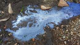
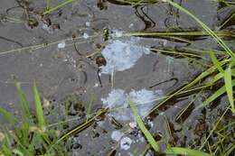
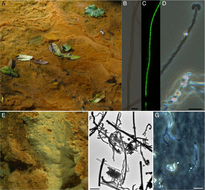
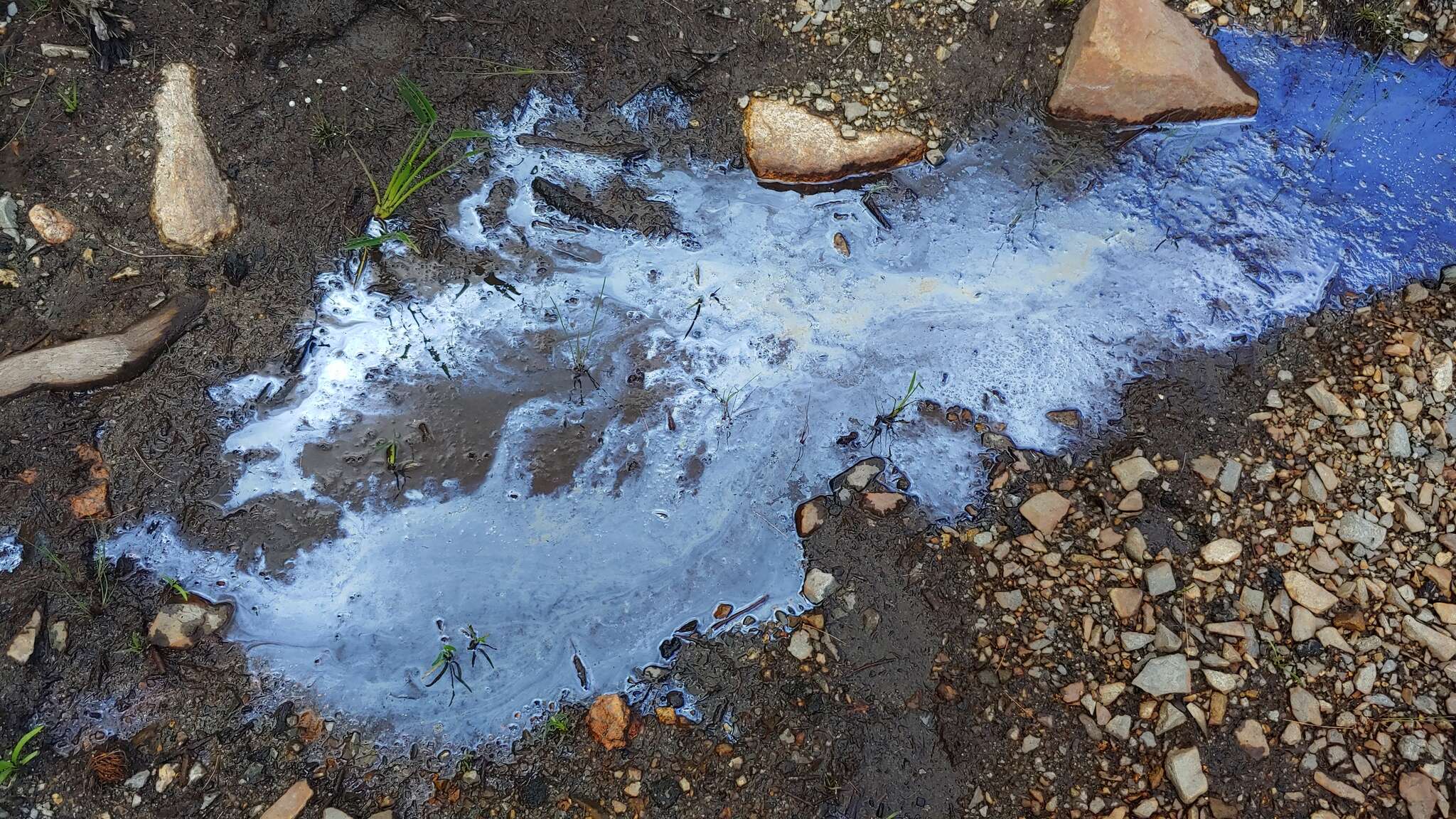
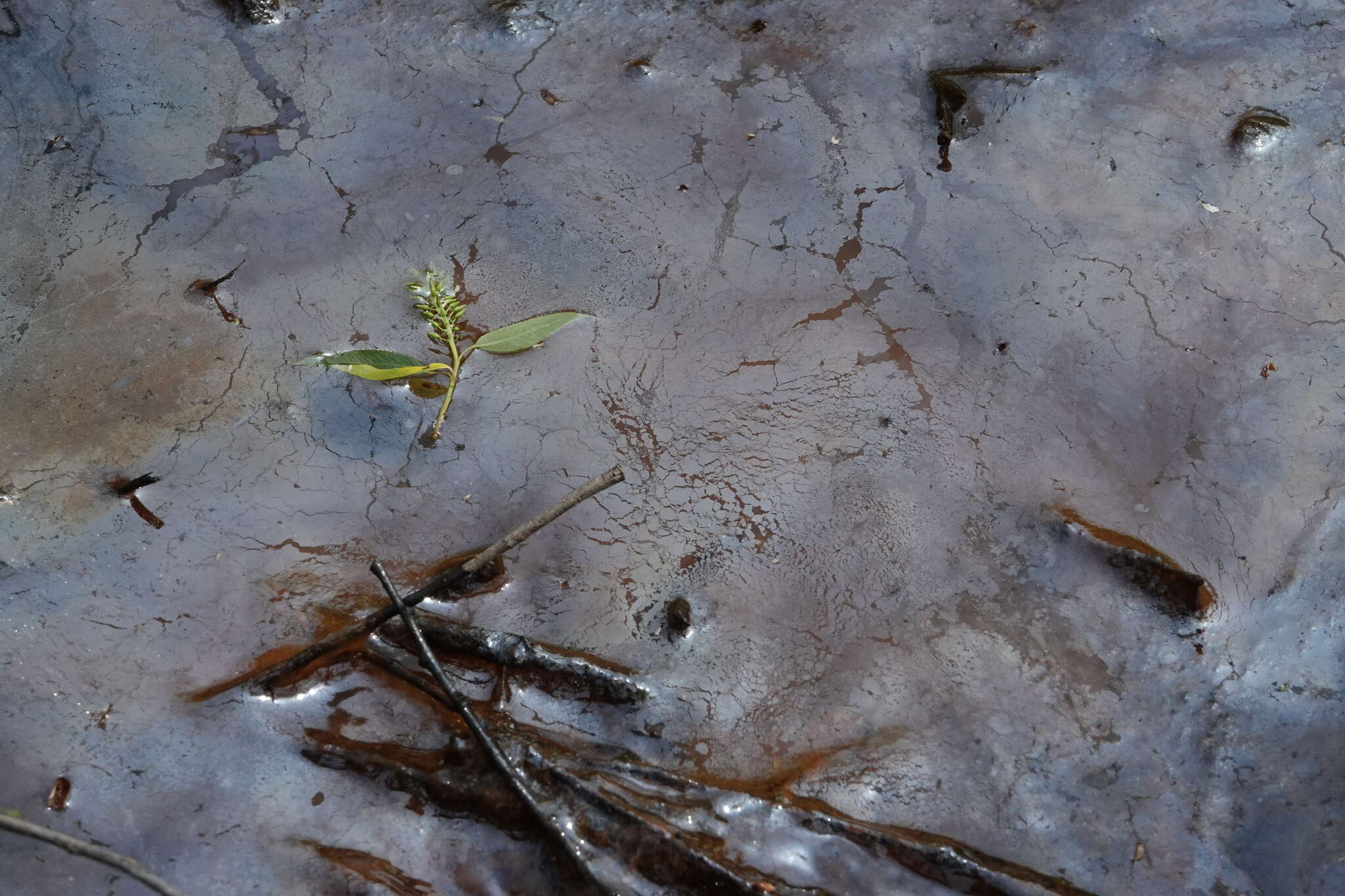
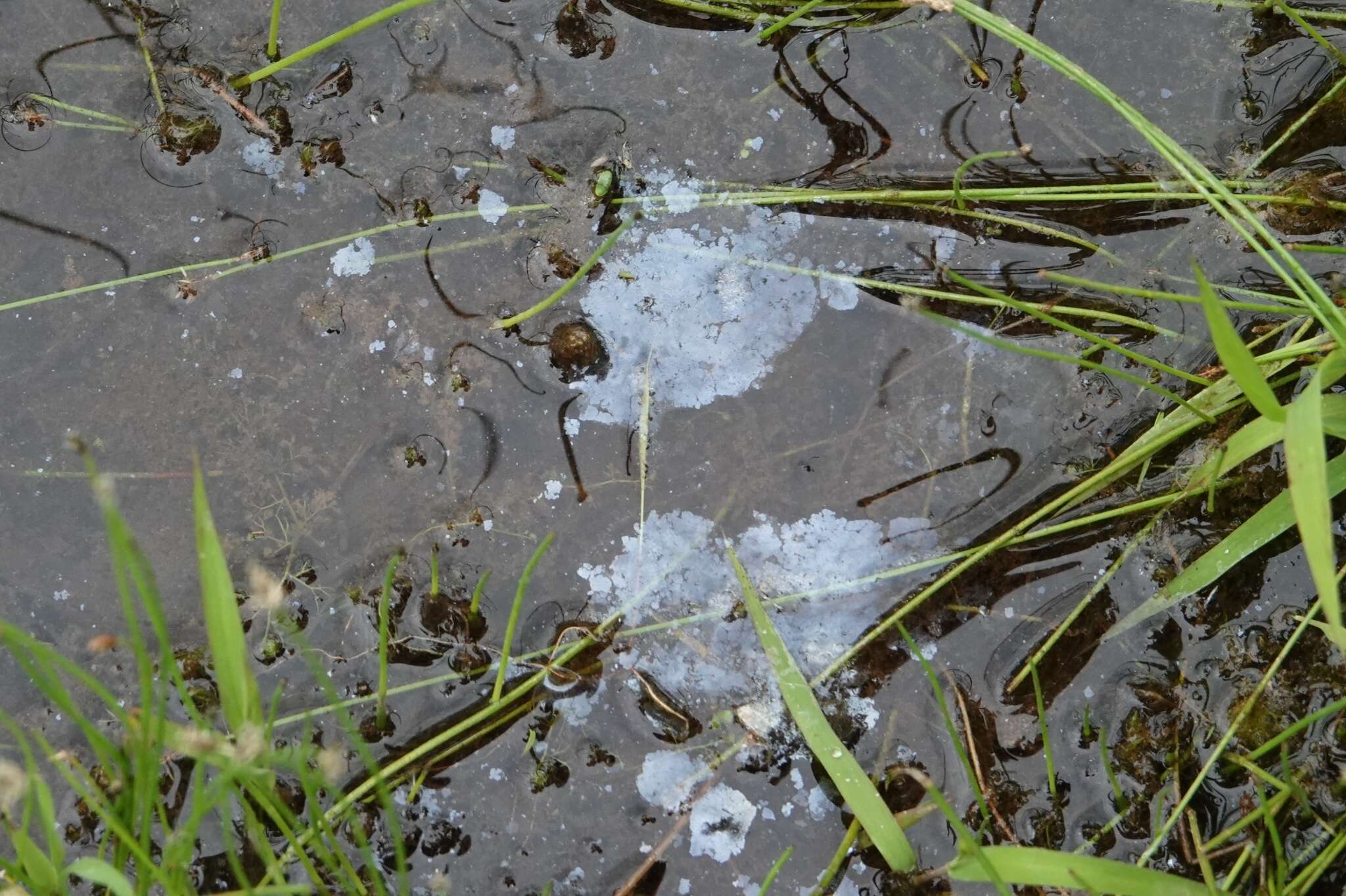
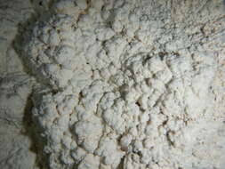
Fe-oxidizing microbial matsFe-oxidizing microbial mats. A. Atypical freshwater iron mat in a slow-moving stream where Fe(II)-enriched groundwater is mixing with oxygenated surface water, resulting in growth of Fe-oxidizing bacteria and precipitation of iron oxides; B & C. phase contrast and epiflouresence images of the common sheath-forming Fe-oxidizer Leptothrix ochracea (bar = 5 µm); D, the stalk-forming Fe-oxidizer Gallionella ferruginea, note the bean-shaped cells in the process of cell division at the end of the Fe-oxide encrusted stalk (bar = 5 µm); E, an iron mat associated with a deep-sea hydrothermal vent (1000 mbsl) at Loihi Seamount; F, TEM image of biogenic oxides produced at Loihi, note the variety of helical stalks and tubular sheath-like filaments (bar = 10 µm); G, phase contrast image of unidentified Zetaproteobacteria that are marine Fe-oxidizers growing at the ends of iron-oxide filaments (cells denoted by arrows) from an in-situ incubation at Loihi (bar = 5 µm). Photo credits. A, B, C, D, G – David Emerson; E, Wood’s Hole Oceanographic Institution; F, Clara Chan
-
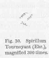
Spirillum Tournoyani (Ehr.), magnified....
-
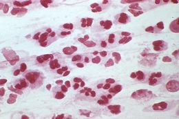
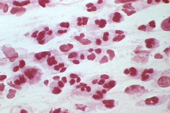
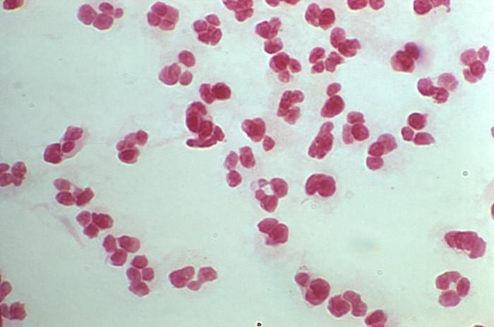
This is a photomicrograph of a Gram-stained urethral exudate sample from a male who presented with a case of urethritis. In this particular view, numbers of polymorphonuclear leukocytes (PMNs) were visible, however, no organisms were evident. This specimen proved to be negative for the presence of Gram-negative Neisseria gonorrhoeae bacteria.Created: 1974
-
Magnified 1125X, this photomicrograph revealed the presence of numerous Gram-negative bacilli, i.e., rod-shaped organisms, Eikenella corrodens, which in part, derives its name from the fact that when this organism is grown on agar medium, it appears to erode the medium. E. corrodens is a facultative anaerobic organism, which means that in the presence of environmental oxygen it creates ATP, but switches to fermentation in oxygens absence. As a commensal organism, E. corrodens is normally found in the human mouth and upper airways, and has been found to be the cause of infection in cancer patients, and patients injured through a bite injury.Created: 1972
-
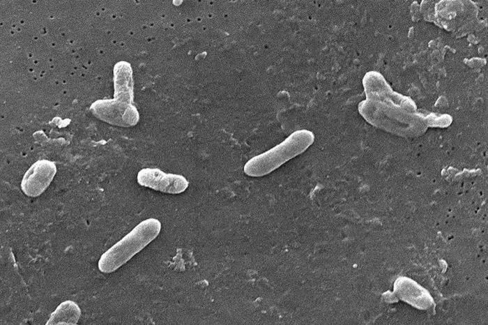
This scanning electron micrograph (SEM) depicted a number of Gram-negative Bordetella bronchiseptica coccobacilli bacteria. This organism is commonly found to be the cause of respiratory tract infections in dogs, as well as human beings whose immune system had been compromised including those who are infected by the HIV virus.Created:
-
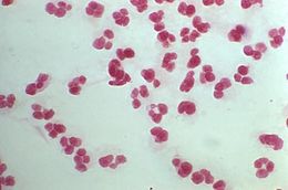
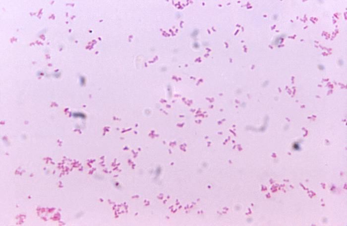
This is a photomicrograph of a Gram-stained urethral exudate sample from a male who presented with a case of urethritis. In this particular view, no Gram-negative diplococci were evident, however, in PHIL 1908 and 2307, which featured other views of this specimen, Neisseria gonorrhoeae bacteria, were found to be present.Created: 1974
-
Scanning Electron Micrograph of Burkholderia cepacia. See PHIL 10608 for a colorized version of this image.Created:
-
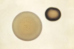
This 1972 image depicted the morphologic appearance of Neisseria gonorrhoeae colonies after having grown for a period of 24 hours on GC media base agar supplemented with IsoVitaleX. These were photographed here at a magnification of 50X. GC media base agar is used in the isolation of N. gonorrhoeae bacteria, and is often used in conjunction with various antibiotics, in order to determine N. gonorrhoeae antimicrobial sensitivity/selectivity.What are the signs and symptoms of gonorrhea?Some men with gonorrhea may have no symptoms at all. However, some men have signs or symptoms that appear two to five days after infection; symptoms can take as long as 30 days to appear. Symptoms and signs include a burning sensation when urinating, or a white, yellow, or green discharge from the penis. Sometimes men with gonorrhea get painful or swollen testicles.Created: 1972
-
Scanning Electron Micrograph of Burkholderia cepacia. See PHIL 10608 for a colorized version of this image.Created:
-
This 1972 image depicted the morphologic appearance of Neisseria gonorrhoeae colonies after having grown for a period of 24 hours on GC media base agar supplemented with IsoVitaleX. These were photographed here at a magnification of 50X. GC media base agar is used in the isolation of N. gonorrhoeae bacteria, and is often used in conjunction with various antibiotics, in order to determine N. gonorrhoeae antimicrobial sensitivity/selectivity.What is gonorrhea?Gonorrhea is a sexually transmitted disease (STD). Gonorrhea is caused by Neisseria gonorrhoeae, a bacterium that can grow and multiply easily in the warm, moist areas of the reproductive tract, including the cervix (opening to the womb), uterus (womb), and fallopian tubes (egg canals) in women, and in the urethra (urine canal) in women and men. The bacterium can also grow in the mouth, throat, eyes, and anus.Created: 1972
-
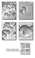
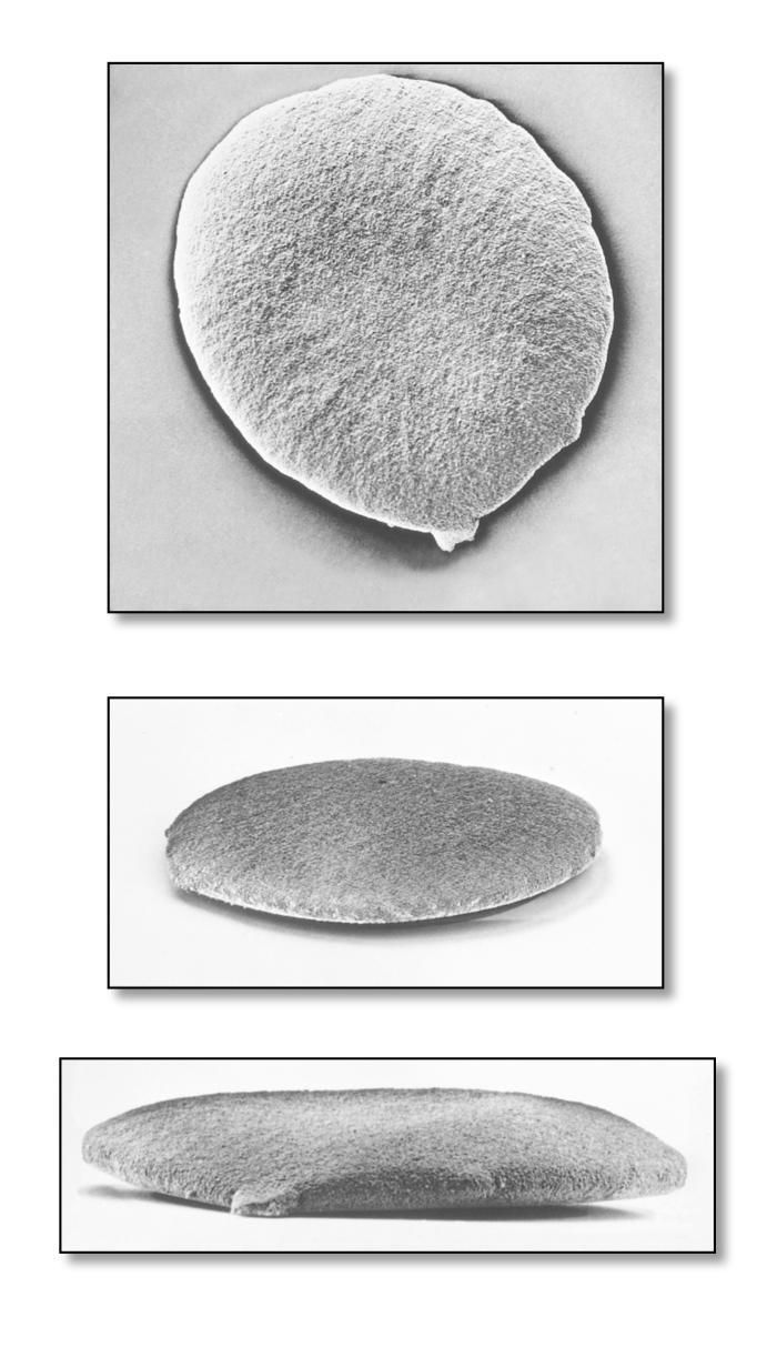
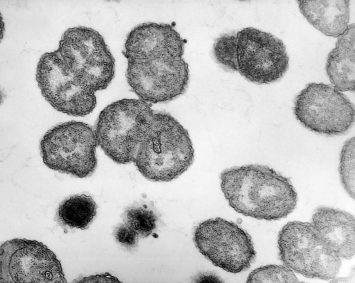
This scanning electron micrograph (SEM) depicted four views of Gram-negative Neisseria gonorrhoeae bacteria. Under this highly-magnified view, details of the protective, adhesive matrix, which this colony had secreted is revealed. This molecular matrix consists of polymeric constituents known as the extracellular polymeric substance, or EPS. Typically, these cocci appear as paired diplococci, which have been outlined in color. See PHIL 10248 for a black and white version of this image.Created: 1973
-
This scanning electron micrograph (SEM) depicted four views of Gram-negative Neisseria gonorrhoeae bacteria. Under this highly-magnified view, details of the protective, adhesive matrix, which this colony had secreted is revealed. This molecular matrix consists of polymeric constituents known as the extracellular polymeric substance, or EPS. See PHIL 10250 for an enlarged view of the colorized inset, which distinguishes the diplococcal pairs.Created: 1973
-
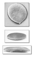
This scanning electron micrograph (SEM) depicted three views of a single Gram-negative Neisseria gonorrhoeae bacterium. Under this highly-magnified view, the roughened texture of the bacteriums cell wall is made visible. As a Gram-negative bacterium, N. gonorrhoeae possess a thinner cell wall than its Gram-positive cousins, composed of peptidoglycan molecular layers that are sandwiched between a lipid membrane layer.Created: 1973
-
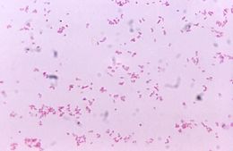
Electron micrograph of Neisseria gonorrhoeae bacteria, the causative agent of gonorrhea; magnification 100,000X.Created: 1971
-
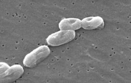
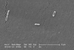
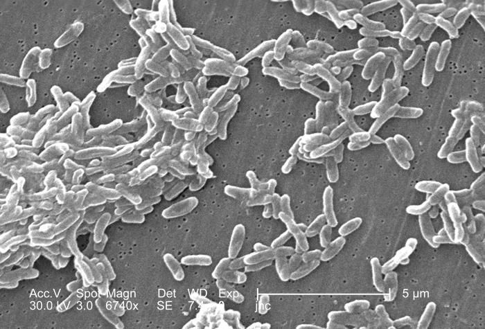
Magnified 6,740x, this scanning electron micrograph (SEM) depicted a grouping of Ralstonia mannitolilytica bacteria, which was harvested from a pure culture.Created: 2006
-
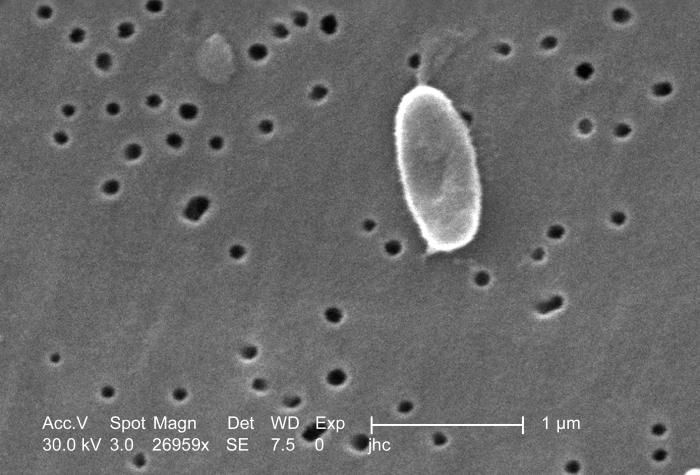
Magnified 26,959x, this scanning electron micrograph (SEM) depicted a highly enlarged view of a Ralstonia mannitolilytica bacterium, which was harvested from a pure culture.Created: 2006
-
Magnified 26,959x, this scanning electron micrograph (SEM) depicted a highly enlarged view of a Ralstonia mannitolilytica bacterium, which was harvested from a pure culture.Created: 2006
-
Magnified 6,740x, this scanning electron micrograph (SEM) depicted three Ralstonia mannitolilytica bacteria, which were harvested from a pure culture.Created: 2006
-
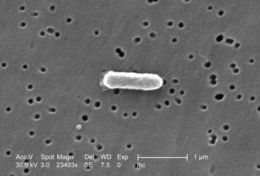
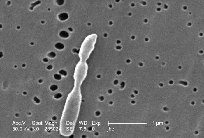
Magnified 23,493x, this scanning electron micrograph (SEM) depicted a highly enlarged view of a Ralstonia mannitolilytica bacterium, which was harvested from a pure culture.Created: 2006
-
Magnified 23,502x, this scanning electron micrograph (SEM) depicted a highly enlarged view of a Ralstonia mannitolilytica bacterium, which was harvested from a pure culture. Note that in this particular instance, the bacterium is in the process of undergoing replication, which when completed, will give rise to three separate bacteria.Created: 2006
-
-
-
-