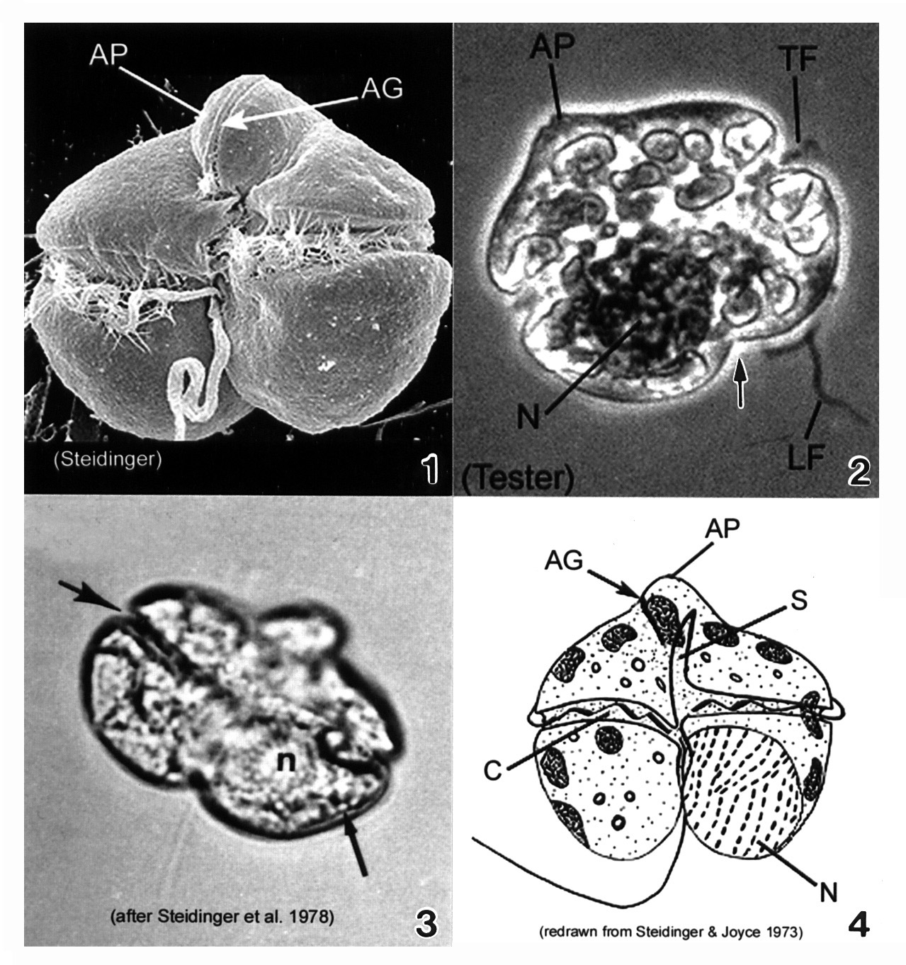
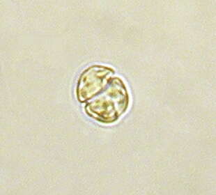
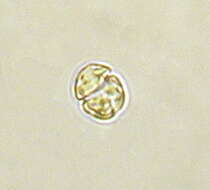
-
Galende, Castile and Len, Spain
-
Ribadelago de Franco, Castille and Leon, Spain
-
Villar del Pedroso, Extremadura, Spain
-
Galende, Castile and Len, Spain
-
San Martin De Castaneda, Castille and Leon, Spain
-
Dehesa de Montejo, Castille and Leon, Spain
-
Galende, Castile and Len, Spain
-
San Martn de Castaeda, Castilla y Len, Espaa
-
Villar del Pedroso, Extremadura, Espaa
-
Galende, Castile and Len, Spain
-
Plate 22. Gymnodinium breve. Fig. 1. SEM: ventral view. Cell small, wider than long, dorso-ventrally flattened. Cell nearly square in outline; prominent apical process (AP) directed ventrally. Apical groove (AG) present on apical process, adjacent to sulcus. Figs. 2-3. LM. Fig. 2. Dorsal view: large nucleus (N) in hypotheca. Transverse (TF) and longitudinal (LF) flagella present. Hypotheca bilobed (arrow). Fig. 3. Ventral view: displaced cingulum (large arrow) and lipid globule (small arrow). Fig. 4. Line drawing. Cingulum (C) displaced, descending. Long sulcus (S) extends to apex of cell.
-
Plate 23. Gymnodinium catenatum. Figs. 1-3. SEM: ventral view. Fig. 1. Cell small, elongate-ovoid with slight dorso-ventral compression. Conical apex; rounded and notched antapex. Cingulum (C) excavated; sulcus (S) long. Distinctive horse-shoe shaped apical groove (AG) encircles apex. Fig. 2. Two cell chain; attachment point visible (arrow). Premedian cingulum displaced 2X its width. Longitudinal (LF) and transverse (TF) flagella visible. Fig. 3. Chain cells with anterior-posterior compression. Terminal cell slightly longer. Thecal surface rugose to smooth (Blackburn et al. 1989). Figs. 4-5. LM. Fig. 4. Chain-formation (Yuki and Yoshimatsu 1987). Fig. 5. Single cell. Conical epitheca with concave to flat apex. Bilobed hypotheca (arrow). Fig. 6. Line drawing. Fig. 7. SEM: cyst with microreticulations. ag=apical groove; c=cingulum
-
Plate 24. Gymnodinium mikimotoi. Figs. 1-4. SEM. Fig. 1. Ventral view: cell small, broadly oval to almost round. Epitheca slightly smaller than hypotheca. Characteristic straight apical groove (AG). Cingulum (C) deep, displaced 2 times its width. Sulcus (S) slightly invades epitheca (arrowheads). Hypotheca notched by widening sulcus (arrow). Fig. 2. Dorsal view: apical groove extends to dorsal side of epitheca creating slight indentation at the apex (arrowhead). Hypotheca bilobed (arrow). Fig. 3. Apical view of apical groove (arrow)(after Fukuyo et al.). Fig. 4. Cell compressed dorso-ventrally (after Fukuyo et al.). Figs. 5-7. LM. Fig. 5. Cingulum displaced 2 times its width (arrows)(from Larsen & Moestrup 1989: fig. 16g). Fig. 6. Large nucleus (N) in left lobe of hypotheca. Fig. 7. Vegetative division. Division plane oblique.
-
Plate 25. Gymnodinium pulchellum. Figs. 1-2. SEM: ventral view. Fig. 1. Cell small and broadly oval. Cingulum wide, displaced 1-1.5 X its width. Deeply excavated sulcus creates lobed hypotheca. Conspicuous undulating apical groove (AG). Fig. 2. Well-developed apical groove: reverse S-shape. Transverse flagellum (TF) housed in cingulum. Sulcus slightly invades epitheca with finger-like projection (arrow). Figs. 3-5. LM: ventral view. Figs. 3-4. Apical groove distinguishable (small arrows). Chloroplasts and pyrenoids present. Lobed hypotheca (large arrow). Fig. 5. Large elliptical nucleus (N) in left central part of cell. Fig. 6. Line drawing. C=cingulum
-
Plate 26. Gymnodinium sanguineum. Figs. 1-3. LM. Cell large, pentagonal, and slightly dorso-ventrally flattened. Cells vary in shape and size. Fig. 1. Ventral view. Epitheca and hypotheca nearly equal in size: epitheca conical, hypotheca bilobed (arrows). Fig. 2. Ventral view. Deep cingulum median, displaced 1-2 times its width. Sulcus deeply notches hypotheca. Apical groove present (arrow). Fig. 3. Cell deeply pigmented; central nucleus (n). Fig. 4. Line drawing. Spindle-shaped chloroplasts radially arranged.
-
Plate 27. Gymnodinium veneficum. Figs. 1-3. Line drawings. Fig. 1. Ventral view: cell small and ovoid. Epitheca slightly pointed, without apical groove. Cingulum deep and displaced 1-2 times its width. Fig. 2. Dorsal view: large central nucleus (N). Two to eight irregular chloroplasts present (C). Fig. 3. Sigmoid sulcus slightly invades epitheca (arrow).
-
Gymnodinium (jim-no-din-ee-um), a so-called typical dinoflagellate. Most dinoflagellates have two flagella and they lie in grooves in the cell surface. The flagella are not evident here, but the grooves are. There is an circumferential groove (the girdle or cingulum) which wraps around the cell, and a longitudinal groove which extends from the point of flagellar insertion towards the back of the cell. This is an autotrophic dinoflagellate with numerous plastids with chlorophylls a and c. Also with an eyespot. Differential interference contrast.
-
Gymnodinium (jim-no-din-ee-um), a so-called typical dinoflagellate. Most dinoflagellates have two flagella and they lie in grooves in the cell surface. The flagella are not evident here, but the grooves are. There is an circumferential groove (the girdle or cingulum) which wraps around the cell, and a longitudinal groove which extends from the point of flagellar insertion towards the back of the cell. This is an autotrophic dinoflagellate with numerous plastids with chlorophylls a and c. The nucleus is the granular structure in the lower (hypocone) part of the cell. Differential interference contrast.
-
Gymnodinium (jim-no-din-ee-um), a so-called typical dinoflagellate. Most dinoflagellates have two flagella and they lie in grooves in the cell surface. The flagella are not evident here, but one groove -the circumferential groove - is. This is an autotrophic dinoflagellate with numerous plastids with chlorophylls a and c. The nucleus is the granular structure in the upper (epicone) part of the cell. Differential interference contrast.
-
Gymnodinium (jim-no-din-ee-um), a so-called typical dinoflagellate. Most dinoflagellates have two flagella and they lie in grooves in the cell surface. The flagella are not evident here, but one groove -the circumferential groove - is., and the second (trailing) flagellum can be seen extending out the back of the cell. This is an autotrophic dinoflagellate with numerous plastids with chlorophylls a and c. Phase contrast.
-
-
Gymnodinium (jim-no-din-ee-um) danicans Campbell 1973. The image shows a cell in ventral view. The red stigma is visible in the sulcal area. The plastids are yellow-brown and multiple.
-
Image showing the off green colour of the chloroplasts of this dinoflagellate, the equatorial groove and the longitudinal (trailing) flagellum.
-
Dinoflagelate with chloroplasts. There are two flagella, one in the groove that runs around the middle of the body and the second lies in the longitudinal groove and extends behind the swimming cell.