Plate 25
Description:
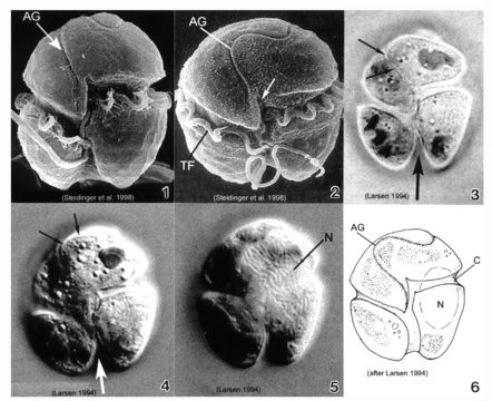
Plate 25. Gymnodinium pulchellum. Figs. 1-2. SEM: ventral view. Fig. 1. Cell small and broadly oval. Cingulum wide, displaced 1-1.5 X its width. Deeply excavated sulcus creates lobed hypotheca. Conspicuous undulating apical groove (AG). Fig. 2. Well-developed apical groove: reverse S-shape. Transverse flagellum (TF) housed in cingulum. Sulcus slightly invades epitheca with finger-like projection (arrow). Figs. 3-5. LM: ventral view. Figs. 3-4. Apical groove distinguishable (small arrows). Chloroplasts and pyrenoids present. Lobed hypotheca (large arrow). Fig. 5. Large elliptical nucleus (N) in left central part of cell. Fig. 6. Line drawing. C=cingulum
Included On The Following Pages:
This image is not featured in any collections.
Source Information
- license
- cc-publicdomain
- bibliographic citation
- Faust, Maria A. and Rose A. Gulledge. Identifying Harmful Marine Dinoflagellates. Smithsonian Contributions from the United States National Herbarium, volume 42: 1-144 (including 48 plates, 1 figure and 1 table).
- original
- original media file
- visit source
- partner site
- NMNH Marine Dinoflagellates
- ID