This specimen grows slowly at 30°C and faster at 37°C, being the second temperature of incubation the conversion point of the pathogen to yeast phase under anaerobic incubation conditions (Liu 2011; Ribes et al. 2000).
C. recurvatus can be differentiated from another sporangiola producing species based on morphological characteristics like the number of cells in the sporangiole, length, and morphology of the sterigmata and production of zygospores (Parker et al. 2011).
Cokeromyces recurvatus was cultured for the first time in 1949 from rabbit feces in Illinois (Parker et al. 2011). Since then, in further investigations it has been isolated from soil and feces of lizards and numerous small rodents including rats, squirrels, and mice (Ribes et al. 2000).
The geographic distribution of Cokeromyces recurvatusisolates from feces covered parts of Mexico and some states in the US such as California, Arizona, Illinois, Michigan, and Florida. While Texas was the state where the isolate from soil was obtained (Ribes et al. 2000).
The Zygomycetes class is mainly characterized by their sexual reproduction that consist on the fusion of coenocytic gametangia forming zygospores with thick walls. There are three types of mating reproduction: heterothallic, the gametangia formed from different mycelia; homothallic, gametangia formed by a single mycelium; and azygosporic, when a single gametangium produce zygospores (Schipper and Stalpers 2003). Since C. recurvatus is homothallic, it requires only one mating type for sexual reproduction, zygospores are produced within a single isolate. These spores are globular, rough-walled, with dark brown-black or sometimes golden brown structures and a diameter measuring 33.5 to 54.5 µm. Studies have shown that higher temperatures (35-37°C), promote an abundant production of zygoespores (Liu 2011; Ribes et al. 2000).
This fungus present a dimorphic characteristic that allows the specimen to exist in a filamentous and yeast form with similar morphology to the yeast phase of Paracoccidioides braziliensis. Dimorphism of C. recurvatus depends on three factors: degree of anaerobiosis, temperature of incubation and the culture medium. When it is expose to room temperature, the specimen grows as a filamentous fungus showing a visible growth within 1 week and the growth present colonies with a tan-gray color and concentric zones or rings. The gray color portion on the colonies represents areas containing large numbers of sporangioles and the tan or lighter portions are the vegetative mycelium. With age, colonies develop wrinkles with radial folds. Studies have shown the mycelium growth rate is 15 to 20 mm in 5 days while in other results it was observed 30 mm in 3 days. This dimorphic form makes C. recurvatus different from other Zygomycetes species because the mycelium is usually lower and the colonial growth is slower and less aggressive (Parker et al. 2011; Ribes et al. 2000).
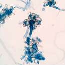
The filamentous form of this fungus has microscopic morphology with coenocytic ribbon like hyphae, which means they are made up of a multinucleate and continuous mass of protoplasm enclosed by a cell wall. Hyphae has a diameter of 5 to 10 µm, a regular characteristic within the Zygomycetes. The sporangiospores are mostly unbranched and tall, 300 to 500 µm long, with a terminal vesicle of 13 to 31 µm in diameter. From the vesicle the sporangiola are formed, which have a diameter of 8 to 11 µm, supported on thin, recurving, and pigmented stalks. Each sporangiolum is composed of 12 to 20 oval that would produce sporangiospores with smooth walls (Ribes et al. 2000).
Cokeromyces recurvatus can be forced into a yeast phase. Studies indicate that the conversion to yeast phase would more likely occur under conditions that encourage anaerobic fermentation, high concentrations of carbon dioxide, high temperatures, pH range between 5.8 – 6.5, glucose availability, and inhibitors of respiration or mitochondrial protein synthesis (Parker et al. 2011). The yeast cells have thick walls with a diameter of 30 to 90 µm and the large yeast cells are usually encircled by smaller yeast buds (Ribes et al. 2000).
The Zygomycetes class only produce hyphae with no regular septation, allowing the nuclei to move freely in the cytoplasm. Septa are produced only near reproductive structures with the purpose to separate old or damaged parts of the mycelium(Schipper and Stalpers 2003).
Microscopic characteristics ofCokeromyces recurvatus can be found on the fallowing link:http://cmr.asm.org/content/13/2/236/F17.expansion.html
Cokeromyces recurvatus is a sporangiola-forming species of the class Zygomycetes within the order Mucorales (Schipper and Stalpers 2003). It was originally designated to the family Choanephoraceae but the genus Cokeromyces is now grouped within the family Thamnidiaceae (Liu 2011). Cokeromyces recurvatus was described by Shanor et al. (1950) from Illinois and Gilman et al. (1957) from Iowa. It has been isolated only in North America: Unites States and Mexico (Benny 2015). This fungus present a dimorphic characteristic, allowing the specimen to exist in two forms: filamentous, that help to differentiate C. recurvatus from other Zygomycetes species, and a yeast form (Parker et al. 2011; Ribes et al. 2000). Since C. recurvatus is homothallic, it requires only one mating type for sexual reproduction and zygospores are produced within a single isolate (Liu 2011; Ribes et al. 2000). Cokeromyces recurvatus is a rare cause of gastrointestinal and urogenital infections, and also of cystisis, peritonitis, and myositis in humans (Liu 2011).
The Zygomycetes class contains two orders of medical importance; Mucorales and Entomophthorales, producing different clinical manifestation of human infections. Pathogenic fungi belonging to these orders can cause Zygomycosis, which is the term used for fungal infection being originated by a fungus within the class of zygomycetes. The order Entomophthorales is the group responsible of many diseases called entomophthoramycoses that are more inactive and chronically progressive. While Mucorales produce diseases called mucormycosis, which was described before as phycomycosis. These diseases are fatal and have a relative rapid progression. Infections caused by the group of fungi under the order Mucorales are the majority of human cases of zygomycoses (Chayakulkeeree et al 2006). Mucormycosys present 5 mayor clinical form: cutaneous, gastrointestinal, disseminated, rhinocerebral and pulmonary diseases, the last 2 are the most common (Moore and Richardson 2014).
Gastrointestinal diseases caused or related to zygomycosis is uncommon in humans, the risk factors associated with this disease present gastrointestinal ulceration, protein-calorie malnutrition, and diarrhea (Parker et al. 2011). Cokeromyces recurvatus is a rare cause of gastrointestinal and urogenital infections, and also of cystisis, peritonitis, and myositis in humans. This pathogen has a higher probability of infection to humans when the person is immunocompetent and immunosuppressed caused by ulcer, diabetes mellitus, myeloma, and cancer condition, including chronic alcoholism (Liu 2011). This pathogen can function as opportunistic and make a contribution to the pathogenesis of intestinal inflammation (Parker et al. 2011).
Transmission
Currently the transmission mode to humans is unknown but its presence is assumed for some cases when the person already have a symptomatic disease (Ribes et al. 2000).
Host Characteristic
C. recurvatus has been reported in 8 cases of human diseases. One case present in a healthy host while the remaining patients had factors for immune system compromise risk or dysfunction like diabetes mellitus, pregnancy, alcoholism, diverticulitis, cystisis, and treatment with immunosuppressants (Nielsen et al. 2005).
Virulence Factors
The pathogenic potential of Cokeromyces recurvatus is unclear (Ribes et al. 2000). Studies have suggested that the pathogenicity depends on 1 or more extracellular mycotoxins and this is because it is not frequently observed to invade tissue but in some patients it appears to cause disease (Nielsen et al. 2005).
Diagnosis
The first human case of a C. recurvatus infection was reported by Rippon and Dolan, they isolated the pathogen from a vaginal sample (Ramani 2000). This specimen has similar sizes and morphology characteristics to the yeast form of Paracoccidoides brasiliensis and spherules of Coccidioides immitis that can lead to a misdiagnosis (Ryan 2009).
Treatment
A specific antifungal therapy is currently unknown for the treatment of infections caused by this fungus. However, amphotericin B which is the drug used to treat other agents of zygomycosis like species of Rhizopus, could be used for the treatment of Cokeromyces recurvatus (Munipalli 1996). Other studies have shown that surgical intervention accompanied with amphotericin B have successfully eradicated the fungus (Ramani 2000).
Cokeromyces recurvatus is a sporangiola-forming species of the class Zygomycetes within the order Mucorales. The Thrichomycetes, which are obligate parasites on arthropods, and the Zygomycetes are the two classes belonging to the phylum Zygomycota (Schipper and Stalpers 2003). The class of Zygomycetes is considered a natural group because of the two types of reproduction: sexual and asexual, the first is based on the formation of zygospore by the fusion of gametangia and the second, is based on the production of aplanospores. This class is represented by three orders: the Mucorales, Entomophthorales and Zoopagales (Hesseltine and Ellis 1973). The order Mucorales is composed by 13 families and many of them do not form zygospores but because of their similar asexual characteristics, they can be easily stablished in this order (Schipper and Stalpers 2003). This group is typically a saprobic fungi group and its members are the most commonly found when isolating microorganism from decaying plant material, dung, air, or soil (Hesseltine and Ellis 1973).C. recurvatus was originally designated to the family Choanephoraceae but the genus Cokeromyces is now grouped within the family Thamnidiaceae that consist of 12 genera: Backusella, Cokeromyces, Dicranophora, Ellisomyces, Fennellomyces, Helicostylum, kirkomyces, Phascolomyces, Pirella, Thamnidium, Thamnostylum, and Zychaea (Liu 2011). The common characteristic of all the species belonging to this family is the forming of the sporangiola which are distinct from a large Mucor-like sporangia found in some genera (Hesseltine and Ellis 1973).
Cokeromyces recurvatus was described by Shanor et al. (1950) from Illinois and Gilman et al. (1957) from Iowa. This pathogen has been isolated only in North America, being Unites States and Mexico the two countries where it can be found. In 1976, the specimen was described and illustrated by Benny and Benjamin who studied isolates of C. recurvatus that were isolated in the countries mentioned before. Within the genus Cokeromyces there is an identified species (Cokeromyces recurvatus) and one species that has not been identified yet (Cokeromyces sp. SW078). In the same genus the species Cokeromyces poitrasii was reclassified to Benjaminiella poitrasii (Benny 2015; Nielsen et al. 2005).
Cokeromycesproduces relatively short sporangiophores that arise directly from the substrate and the apex terminates in a single vesicle that bears several recurved to twisted and contorted pedicels. Sporangia are formed at the apex of each pedicel that are globose, columellate, multispored, and they have a persistent wall. Zygospores with rough, dark walls and opposed suspensors are formed just above the surface of the substrate; the fungus in homothallic.
Type species:C. recurvatus
Species ofCokeromyces:
C. recurvatusPoitras, 1950 (in Shanor, Poitras, and Benjamin, Mycologia 42:272).
Cokeromyces recurvatuswas described by Shanor et al. (1950) from Illinois and Gilman et al. (1957) isolated it from Iowa. Benny and Benjamin (1976) studied specimens ofC. recurvatusfrom Mexico (Baja California) and the U.S.A. (Arizona, California, Florida, Illinois, Michigan, Texas); all cultures were isolated from dung except the one made in Texas (soil).Cokeromyceswas redescribed and illustrated by Benny and Benjamin, 1976). The colony ofC. recurvatusis low growing (less than 1 mm high) and dark brown in color.Cokeromycesis the host of choice for several mycoparasites in the Dimargaritales and many species ofPiptocephalis. Some species ofSyncephalis, for exampleS. depressamay also be grown onCokeromyces recurvatus.
Benny et al. (1985) discussed yeast formation inC. recurvatus. Ultrastructural studies onCokeromyceshave been published by Jeffries and Young (1983a, 1983b). O’Donnell (1979) publishedSEMphotographs ofC. recurvatus.
Poitras (1957) found that zygospores and sporangia were formed when grown under 24 hr of fluorescent light but under alternating fluorescent light and dark the colony formed rings because sporangia were not produced without light. Direct sunlight inhibited sporulation and placing the cultures close to a window receiving incidental sunlight induced sporangial formation but inhibited zygospore production. Incubation temperature (Poitras, 1957) was thought to also be important for reproduction ofC. recurvatus. The best growth and sporulation ofC. recurvatuswas obtained when grown on Emerson’s YpSs agar (Difco) andMEYEagars at 26 C (Benny and Benjamin, 1976). Growth rate was about the same at 26 C and 31 C but there was little growth at 36 C (G. Benny, unpublished data, 1971).
Cokeromyces recurvatusalso is known be a rare cause of mucormycosis in humans (Axelrod et al., 1987; Alvarez et al., 1995; Munipalli et al., 1996; Tsai et al., 1997). There also have been papers published on the isolation, identification, and storage ofC. recurvatusin the clinical laboratory (McGough et al., 1990; Pasarelli and McGinnis, 1992; Kemna et al., 1994).
Cokeromyces recurvatus is a pathogenic fungus.[3][4] Described as a new species in 1950, it was isolated from rabbit dung collected in Illinois.[2]
The genus name of Anzia is in honour of William Chambers Coker (1872 – 1953), was an American botanist and mycologist.[5]
The genus was circumscribed by Leland Shanor in Mycologia Vol.42 (Issue 2) on page 272 in 1950.
It appears similar to Coccidioides immitis.[6]
Cokeromyces recurvatus is a pathogenic fungus. Described as a new species in 1950, it was isolated from rabbit dung collected in Illinois.
The genus name of Anzia is in honour of William Chambers Coker (1872 – 1953), was an American botanist and mycologist.
The genus was circumscribed by Leland Shanor in Mycologia Vol.42 (Issue 2) on page 272 in 1950.
It appears similar to Coccidioides immitis.