Under a magnification of 201X, this scanning electron micrographic (SEM) image depicted a dorsal view of an unidentified engorged female tick, which had been extracted from the skin of a pet cat while in the process of obtaining its blood meal. Note the presence of some of the cats fur, along with some of its skin tissue in which the ticks gnathosoma were still embedded. See PHIL 9972 and 9973 for additional, less magnified views of this scenario. It is from the basis capituli that the two spread pedipalps, and hidden skin-piercing hypostome and chelicerae emanate. On the dorsal surface of the basis capituli youll see two depressed areas known as the porose areas, through which secretions produced by dermal glands are released.Created: 2006
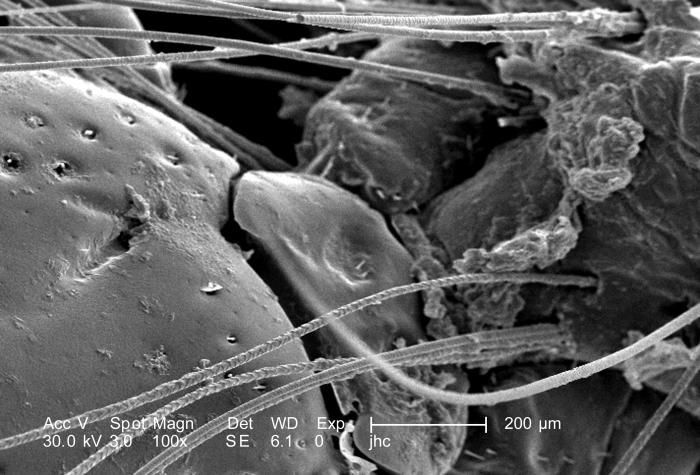
Under a low magnification of 100X, this scanning electron micrographic (SEM) image depicted a dorsal view of an unidentified engorged female tick, which had been extracted from the skin of a pet cat while in the process of obtaining its blood meal. Note the presence of some of the cats fur, along with some of its skin tissue in which the ticks gnathosoma were still embedded. See PHIL 9972 and 9973 for additional, less magnified views of this scenario. It is from the basis capituli that the two spread pedipalps, and hidden skin-piercing hypostome and chelicerae emanate. On the dorsal surface of the basis capituli youll see two depressed areas known as the porose areas, through which secretions produced by dermal glands are released.Created: 2006
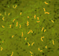
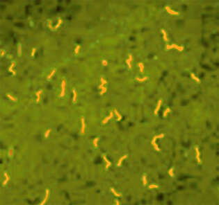
Description: Deutsch: Borrelia duttoni in der Fluoreszenzfärbung mit Acridin-Orange. Date: 25 June 2006 (original upload date). Source: Referenzaufnahme des Benutzers Gleiberg. Author: Benutzers Gleiberg.
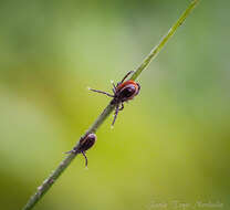
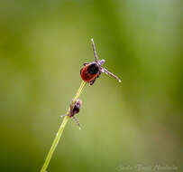
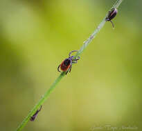
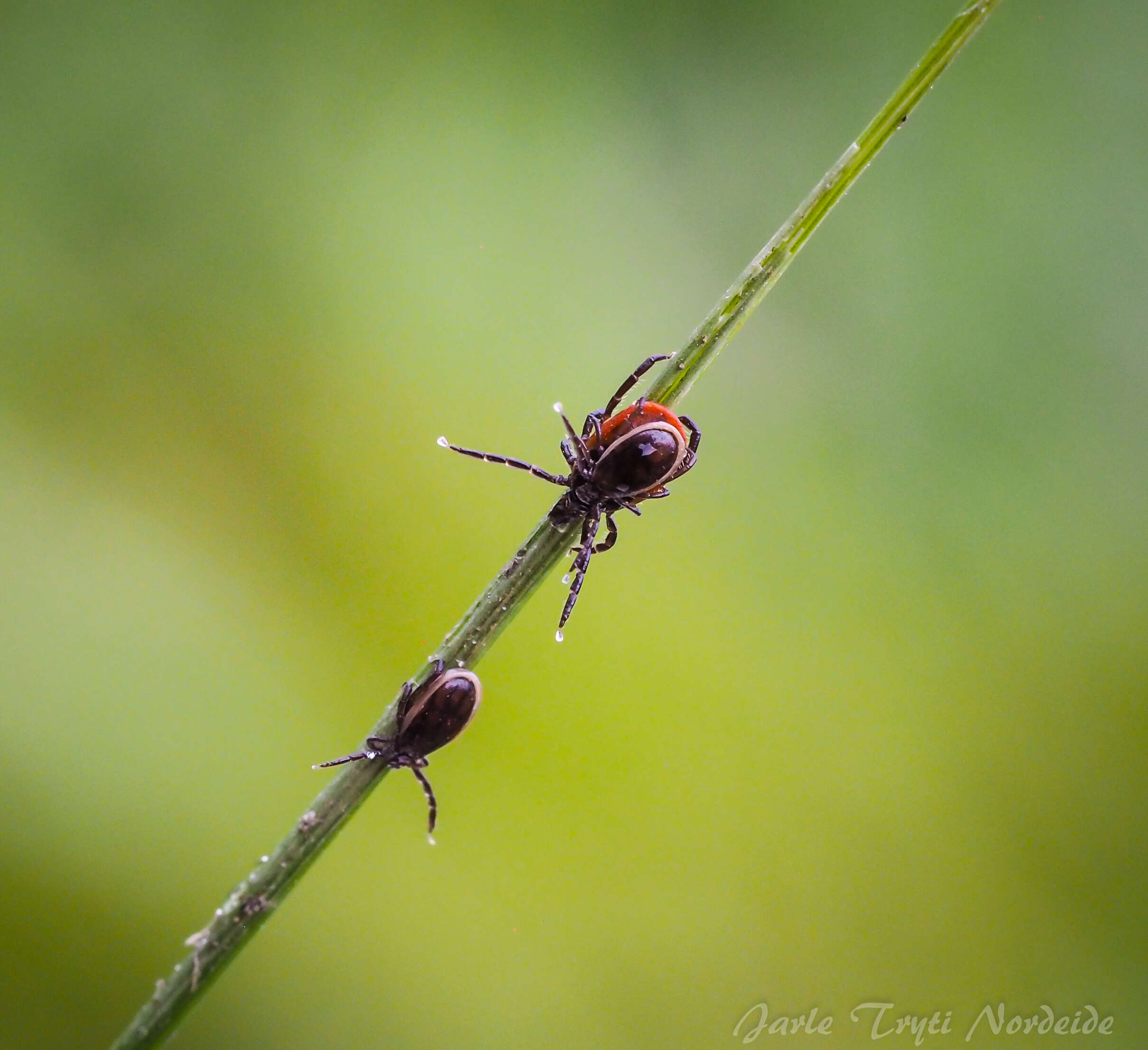
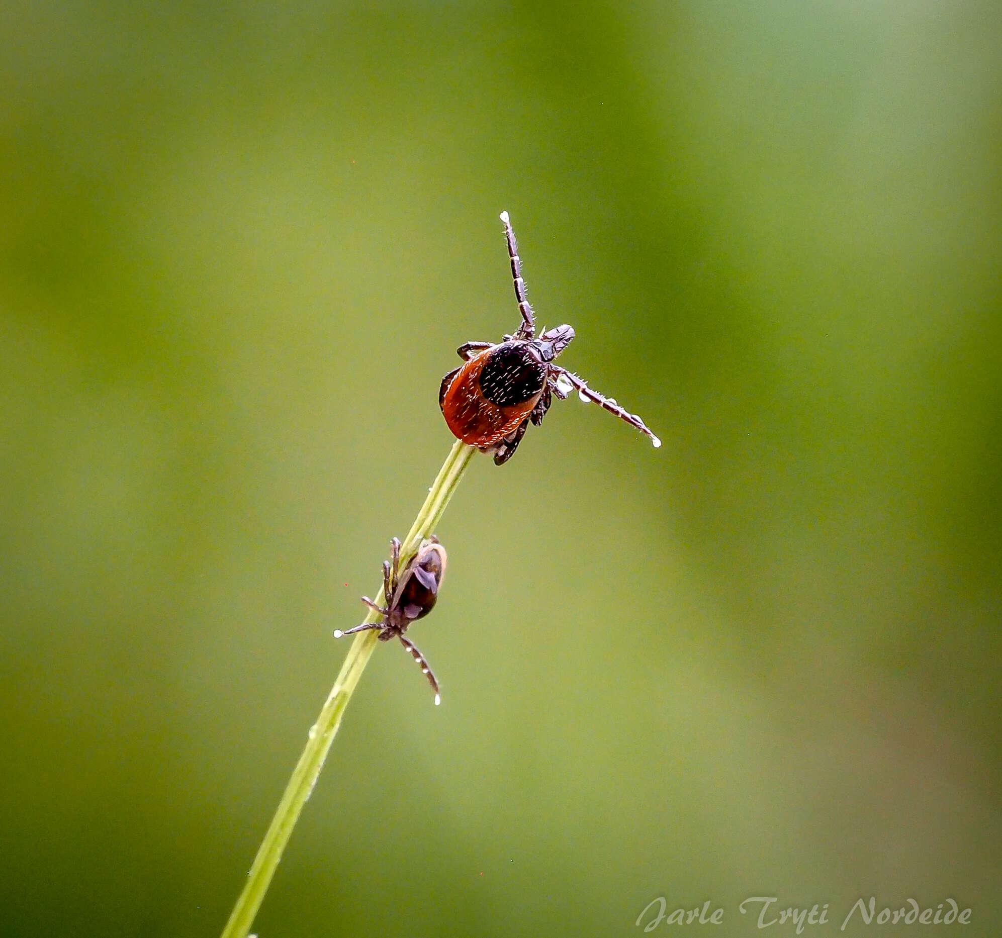
Description: English: The castor bean tick, Ixodes ricinus. A reddish female with a black male at her back, plus another male below. Date: 5 July 2020, 17:11:01. Source: Own work. Author: Jtrytin (Jarle Tryti Nordeide).