-
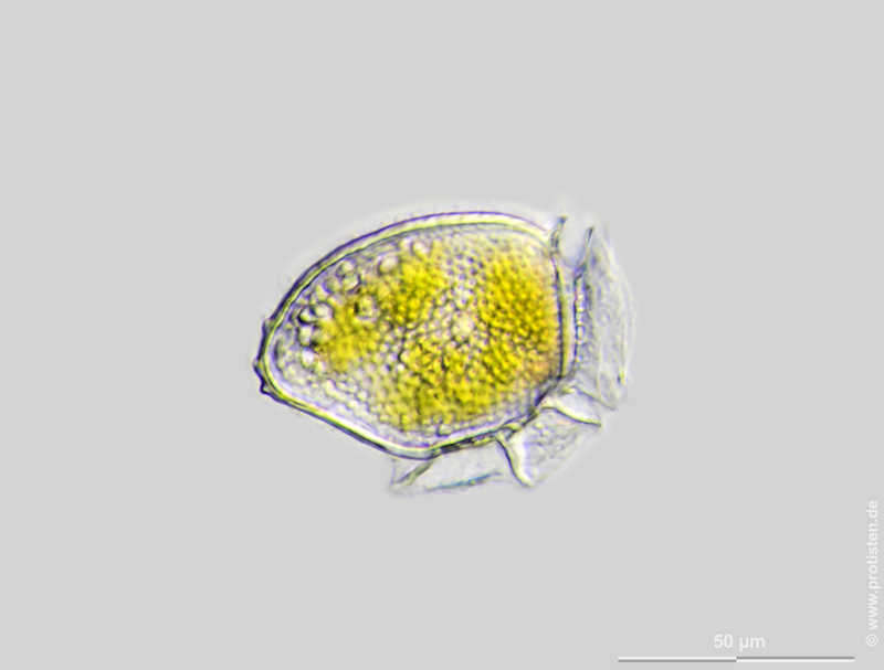
Dinophysis acuta Scale bar indicates 50 µm. The specimen was gathered in the Kieler Förde (Baltic Sea). Sampling date 2/2022. The image was built up using several photomicrographic frames with manual stacking technique. Images were taken using Zeiss Axioplan with Olympus OM-D M5 MKII. Image under Creative Commons License V 3.0 (CC BY-NC-SA). Place name: Baltic Sea, Kieler Förde, Kiel Fjord (Germany) Latitude: 54.3894126 Longitude: 10.1749055 Multiebenen-Abbildung, manuell gestapelt. Der Messbalken markiert eine Länge von 50 µm. Probe aus der Kieler Förde. Datum der Aufsammlung: 2/2022. Mikrotechnik: Zeiss Axioplan, Kamera: Olympus OM-D M5 MKII. Creative Commons License V 3.0 (CC BY-NC-SA). For permission to use of (high-resolution) images please contact postmaster@protisten.de.
-
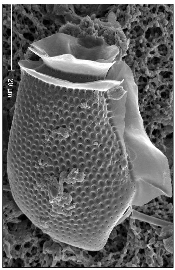
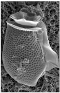
Fig 4 Eletron micrograph of a D. acuta cell showing details of pore structure and sulcal lists
-
Plate 12. Dinophysis acuta. Fig. 1. SEM: lateral view. Cell oblong and robust; theca heavily areolated. Well developed cingular lists (CL) and left sulcal list (LSL). Pointed antapex. Figs. 2-3. LM: lateral view (from Larsen & Moestrup 1992: fig.s 2a,d; scale bars=20 _). Fig. 2. Large areolae, each with a pore (arrows). Fig. 3. Widest point below mid-section (dashed line) aligned with third sulcal rib (arrow). Fig. 4. Line drawing.
-
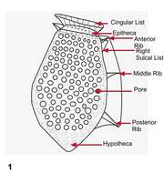
Fig 1: Dinophysis acuta Schematic drawing of a cell in lateral view
-
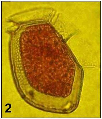
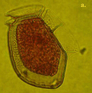
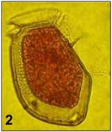
Fig 2: Dinophysis acuta Lugol's preserved cell showing shrinkage of cell contents
-
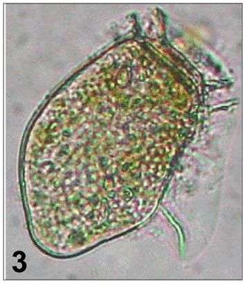
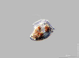
Fig 3: Dinophysis acuta Live cell in lateral view
-
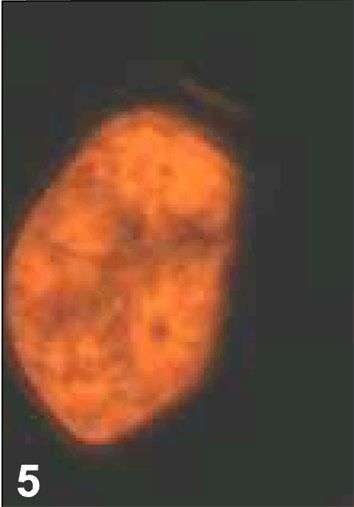
Fig 5 Epifluorescence image under blue light
-
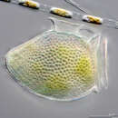
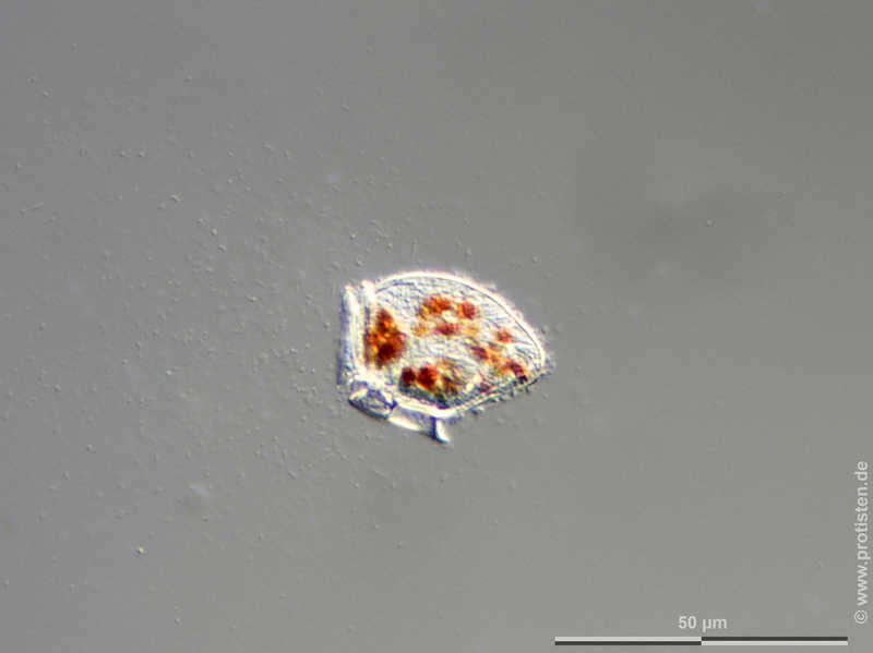
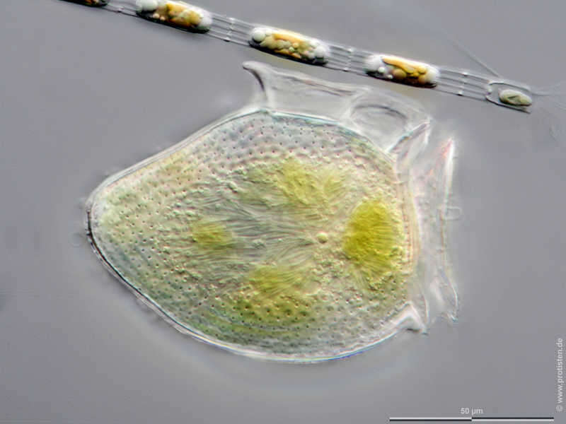
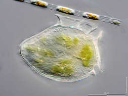
Dinophysis acuta The specimen was gathered in the Kieler Förde (German Baltic Sea). Scale bar indicates 50 µm. The image was built up using several photomicrographic frames with manual stacking technique. Images were taken using Zeiss Axioplan with MFT camera Olympus OM-D E-M5 II.Image under Creative Commons License V 3.0 (CC BY-NC-SA). Place name: Baltic Sea, Kieler Förde, Kiel Fjord (Germany) Latitude: 54.3894126 Longitude: 10.1749055 Multiebenen-Abbildung, manuell gestapelt. Der Messbalken markiert eine Länge von 50 µm. Probe aus der Kieler Förde. Mikrotechnik: Zeiss Axioplan, Kamera: Olympus OM-D E-M5 II. Creative Commons License V 3.0 (CC BY-NC-SA). For permission to use of (high-resolution) images please contact postmaster@protisten.de.
-
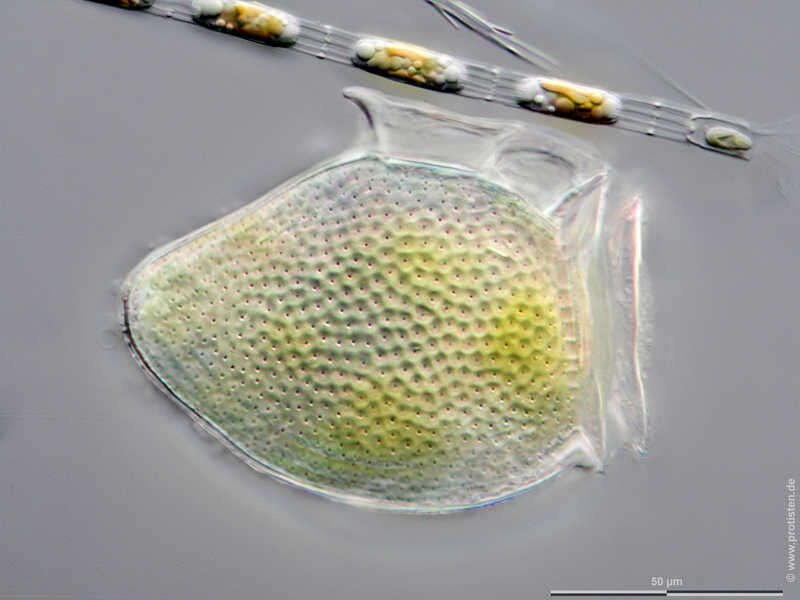
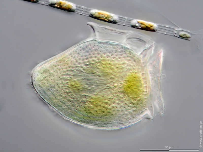
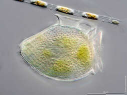
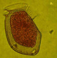
Dinophysis acuta Scale bar indicates 50 µm. The specimen was gathered in the Kieler Förde (Baltic Sea). Sampling date 2/2022. The image was built up using several photomicrographic frames with manual stacking technique. Images were taken using Zeiss Axioplan with Olympus OM-D M5 MKII. Image under Creative Commons License V 3.0 (CC BY-NC-SA). Place name: Baltic Sea, Kieler Förde, Kiel Fjord (Germany) Latitude: 54.3894126 Longitude: 10.1749055 Multiebenen-Abbildung, manuell gestapelt. Der Messbalken markiert eine Länge von 50 µm. Probe aus der Kieler Förde. Datum der Aufsammlung: 2/2022. Mikrotechnik: Zeiss Axioplan, Kamera: Olympus OM-D M5 MKII. Creative Commons License V 3.0 (CC BY-NC-SA). For permission to use of (high-resolution) images please contact postmaster@protisten.de.
-
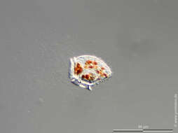
It is often confused with D. norvegica. However, the widest part of the cell lies further posteriorly than in D. norvegica, with a blunt antapical tip. Length: 54-94 microns.