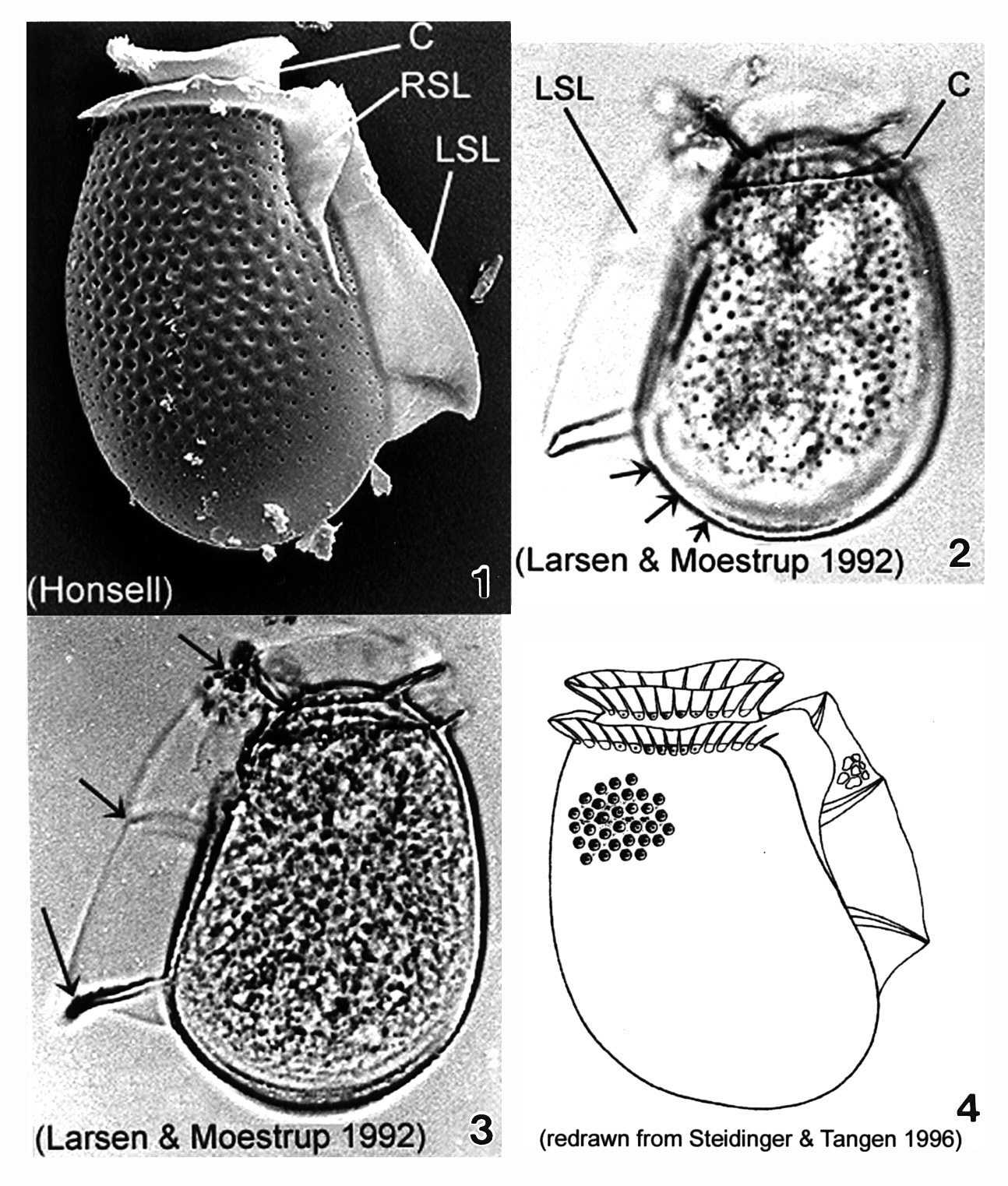
Plate 14. Dinophysis fortii. Fig. 1. SEM: lateral view. Left sulcul list (LSL) long and well-developed. Right sulcal list (RSL) present. Cingulum (C) obscures low and small epitheca. Thecal surface covered with areolae. Figs. 2-3. LM: lateral view. Fig. 2. Cell subovate with a wide round posterior bottom (dorsal bulge)(arrows). Fig. 3. LSL supported by three strong ribs (arrows). Smoothly convex dorsal margin. Fig. 4. Line drawing.