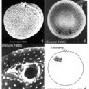
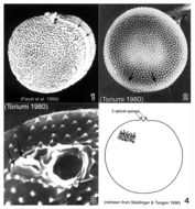
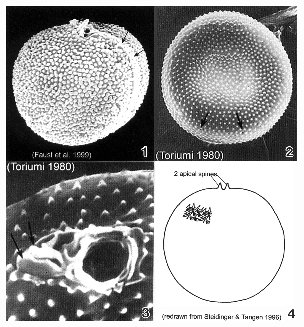
Plate 38. Prorocentrum balticum. Figs. 1-3. SEM. Fig. 1. Valve view: cell round to spherical, covered with many tiny spines. Apical spine apparent. Intercalary band broad, transversely striated (arrows). Fig. 2. Surface with scattered rimmed pores (arrows). Fig. 3. Periflagellar region: two different sized pores and two small apical projections (arrows). Fig. 4. Line drawing.