Ecology
provided by NMNH Marine Dinoflagellates
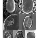
P. lima is a benthic and epiphytic species that can be tycoplanktonic. Cultured cells readily adhere to the culturing vessel via mucous strands and rarely swim freely (Fukuyo 1981; Steidinger & Tangen 1996). This species produces a pale colored resting cyst as part of its life cycle. Cysts are large (70-75 µm diameter) and round with a smooth triple-layered wall (Faust 1993c).
- bibliographic citation
- Faust, Maria A. and Rose A. Gulledge. Identifying Harmful Marine Dinoflagellates. Smithsonian Contributions from the United States National Herbarium, volume 42: 1-144 (including 48 plates, 1 figure and 1 table).
Habitat and Locality
provided by NMNH Marine Dinoflagellates
Prorocentrum lima is a neritic, estuarine species with world-wide distribution (Steidinger & Tangen 1996). Cells can be found in temperate (Lebour 1925; Schiller 1933; Carter 1938) as well as tropical oceans (Fukuyo 1981; Steidinger 1983; Carlson 1984; Faust 1990b). This species occurs in sand (Lebour 1925; Drebes 1974; Dodge 1985), attached to the surface of red and brown algae and benthic debris (Fukuyo 1981; Steidinger 1983; Carlson 1984), associated with coral reefs (Yasumoto et al. 1980; Fukuyo 1981; Bomber et al. 1985; Carlson & Tindall 1985), or can be found attached to floating detritus in mangrove habitats (Faust 1991).
- bibliographic citation
- Faust, Maria A. and Rose A. Gulledge. Identifying Harmful Marine Dinoflagellates. Smithsonian Contributions from the United States National Herbarium, volume 42: 1-144 (including 48 plates, 1 figure and 1 table).
Morphology and Structure
provided by NMNH Marine Dinoflagellates
Prorocentrum lima is a photosynthetic species containing two chloroplasts, a central pyrenoid and a large posterior nucleus (Figs. 5, 6) (Dodge 1975).
- bibliographic citation
- Faust, Maria A. and Rose A. Gulledge. Identifying Harmful Marine Dinoflagellates. Smithsonian Contributions from the United States National Herbarium, volume 42: 1-144 (including 48 plates, 1 figure and 1 table).
Nomenclatural Types
provided by NMNH Marine Dinoflagellates
Holotype: Prorocentrum lima (Ehrenberg) Dodge, 1975: 109, figs. 1 E, F, plate 1B, C
Type Locality: unknown
- bibliographic citation
- Faust, Maria A. and Rose A. Gulledge. Identifying Harmful Marine Dinoflagellates. Smithsonian Contributions from the United States National Herbarium, volume 42: 1-144 (including 48 plates, 1 figure and 1 table).
Reproduction
provided by NMNH Marine Dinoflagellates
P. lima reproduces asexually by binary fission. This species also exhibits an alternate form of asexual reproduction in which a chain of cell pairs is enclosed within a thin-walled cyst. In this mode multiple vegetative divisions occur within a hyaline envelope (a division cyst) which may contain a chain of 4 to 32 cells (Faust 1993d). Sexual reproduction has also been documented: isogamous gametes form, conjugation takes place, and a large hypnozygote (resting cyst) is produced (Fig. 7)(Faust 1993c).
- bibliographic citation
- Faust, Maria A. and Rose A. Gulledge. Identifying Harmful Marine Dinoflagellates. Smithsonian Contributions from the United States National Herbarium, volume 42: 1-144 (including 48 plates, 1 figure and 1 table).
Species Comparison
provided by NMNH Marine Dinoflagellates
P. lima is difficult to identify due to its similar morphology to several other Prorocentrum species with a triangular periflagellar area and an oval or ovoid shape (e.g. P. foraminosum, P. concavum and P. hoffmannianum). P. lima can be distinguished by its size, shape, narrow periflagellar area and the presence of valve and marginal pores. P. concavum, however, is larger, broader, has more valve pores and does not have marginal pores. P. foraminosum and P. hoffmannianum are also similar in shape to P. lima, though both are larger species with very different valve pore numbers and arrangements. P. hoffmannianum, moreover, is much broader and its valve surface is deeply areolated (Steidinger 1983; Steidinger & Tangen 1985; 1996; Fukuyo 1981; Faust 1990b; 1991; 1993b). Steidinger (1983) recognized that the marginal pores of P. lima can be used to differentiate this species at the light microscope level from completely areolated species such as P. concavum or P. compressum which are similar in shape.
- bibliographic citation
- Faust, Maria A. and Rose A. Gulledge. Identifying Harmful Marine Dinoflagellates. Smithsonian Contributions from the United States National Herbarium, volume 42: 1-144 (including 48 plates, 1 figure and 1 table).
Species Overview
provided by NMNH Marine Dinoflagellates
P. lima is an armoured, marine, benthic dinoflagellate species with world-wide distribution.
- bibliographic citation
- Faust, Maria A. and Rose A. Gulledge. Identifying Harmful Marine Dinoflagellates. Smithsonian Contributions from the United States National Herbarium, volume 42: 1-144 (including 48 plates, 1 figure and 1 table).
Synonyms
provided by NMNH Marine Dinoflagellates
Exuviaella marina Cienkowski, 1881
Exuviaella lima (Ehrenberg) Bütschli, 1885
Exuviaella marina var. lima (Ehrenberg) Schiller 1933
Basionym: Cryptomonas lima Ehrenberg, 1860
- bibliographic citation
- Faust, Maria A. and Rose A. Gulledge. Identifying Harmful Marine Dinoflagellates. Smithsonian Contributions from the United States National Herbarium, volume 42: 1-144 (including 48 plates, 1 figure and 1 table).
Taxonomic Description
provided by NMNH Marine Dinoflagellates
P. lima is a bivalvate species often observed in valve view. Cells are oblong to ovate, small to medium-sized, broadest in the mid-region, and narrow toward the anterior end (Figs. 1, 2, 4-6). Cell size ranges between 32-50 µm in length and 20-28 µm in width. Thecal valves are thick and smooth with scattered surface pores (Figs. 1-4). Each valve contains about 50-80 small round marginal pores evenly spaced around the perifery of the valve (0.6 µm in diameter) (Figs. 1, 3), and about 60-100 larger round to oblong unevenly distributed valve pores with trichocysts (0.48 µm in diameter) (Figs. 1,2,4). All pores have smooth edges (Figs. 3,4). The center is devoid of pores (Figs. 1, 2, 4). Marginal pores are a useful diagnostic feature of this species distinguishing it from other Prorocentrum species. Occasionally P. lima can be found without marginal pores or with partially filled pores. In older cells, the thecal surface can become vermiculate. The intercalary band appears as a thick, smooth, and well-defined margin at the periphery of the valve giving the appearance of a flared ridge (Figs. 1, 2, 4-6) (von Stosch 1980; Dodge 1975; Faust 1990b; Faust 1991; Steidinger & Tangen 1996). The periflagellar area is a shallow V-shaped depression on the right valve (Fig. 3) made up of eight platelets and two pores: a larger flagellar pore and a smaller auxiliary pore (Figs. 1, 3-5). A protruding periflagellar collar surrounds the auxiliary pore (Fig. 3). Both valves are anteriorly indented; the left valve margin has a flattened apical ridge that borders the periflagellar area (Figs. 1, 2, 6) (Faust 1991; Steidinger & Tangen 1996).
- bibliographic citation
- Faust, Maria A. and Rose A. Gulledge. Identifying Harmful Marine Dinoflagellates. Smithsonian Contributions from the United States National Herbarium, volume 42: 1-144 (including 48 plates, 1 figure and 1 table).
Toxicity
provided by NMNH Marine Dinoflagellates
Prorocentrum lima is a toxic dinoflagellate species known to produce a number of toxic substances: fast-acting toxin (FAT)(Tindall et al. 1989); prorocentrolide (Torigoe et al. 1988); and diarrhetic shellfish poison (DSP) toxins (Yasumoto et al. 1987): okadaic acid (OA)(Murakami et al. 1982; Lee et al. 1989; Marr et al. 1992); Dinophysistoxin-1 (DTX1) (Marr et al. 1992); Dinophysistoxin-2 (DTX2) (Hu et al. 1993); and Dinophysistoxin-4 (DTX4) (Hu et al. 1995).
- bibliographic citation
- Faust, Maria A. and Rose A. Gulledge. Identifying Harmful Marine Dinoflagellates. Smithsonian Contributions from the United States National Herbarium, volume 42: 1-144 (including 48 plates, 1 figure and 1 table).