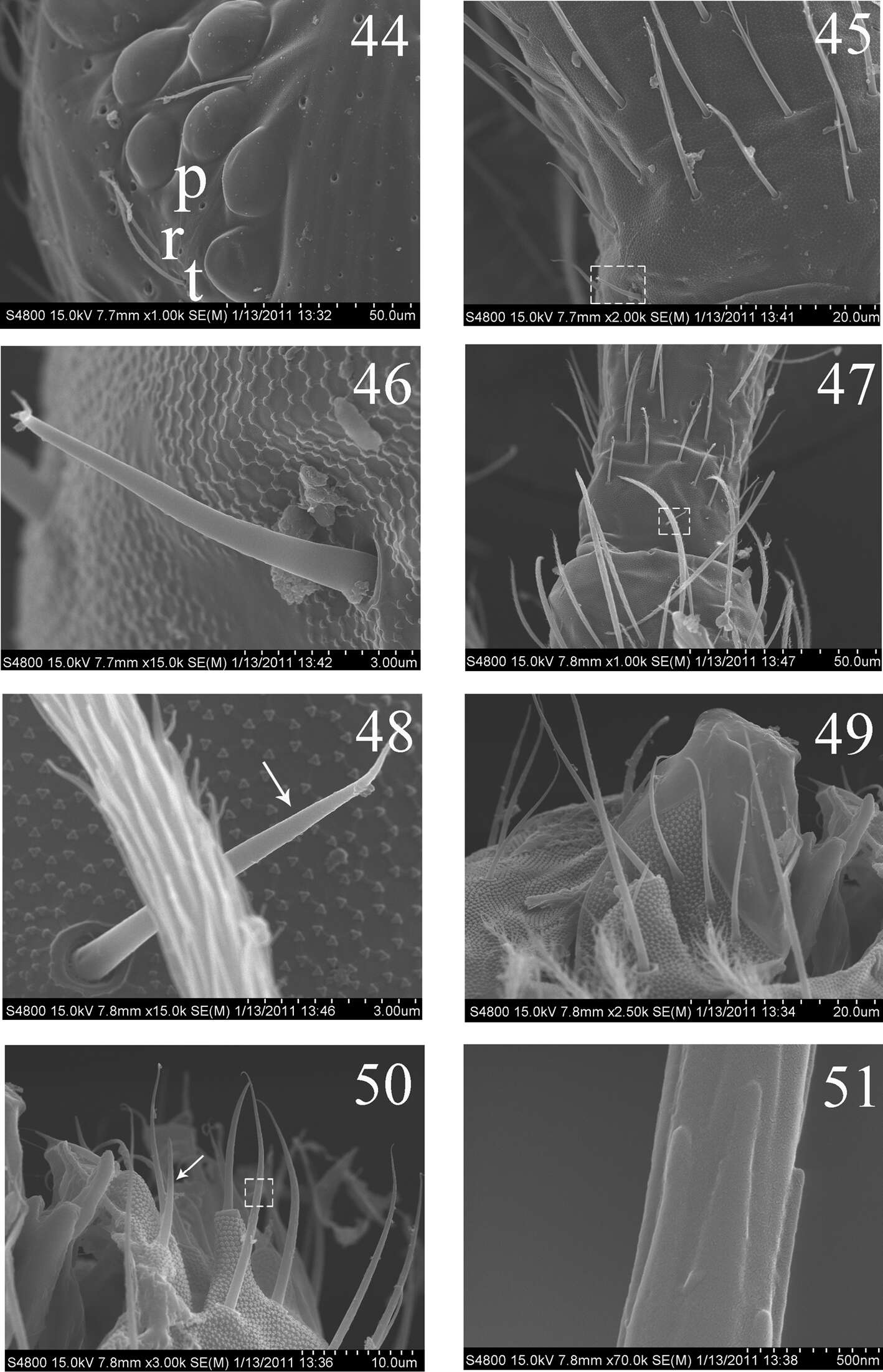
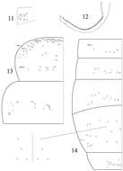
Figures 44–51.SEM photos of adult Homidia jordanai sp. n. 44 eye patches 45 base of Ant. I, dorsal side 46 spiny setae on base of Ant. I, dorsal side 47 joint of Ant. I and Ant. II 48 spiny seta on base of Ant. II 49 maxillary outer lobe 50 labial palp 51 micro-architecture of proximal setae.
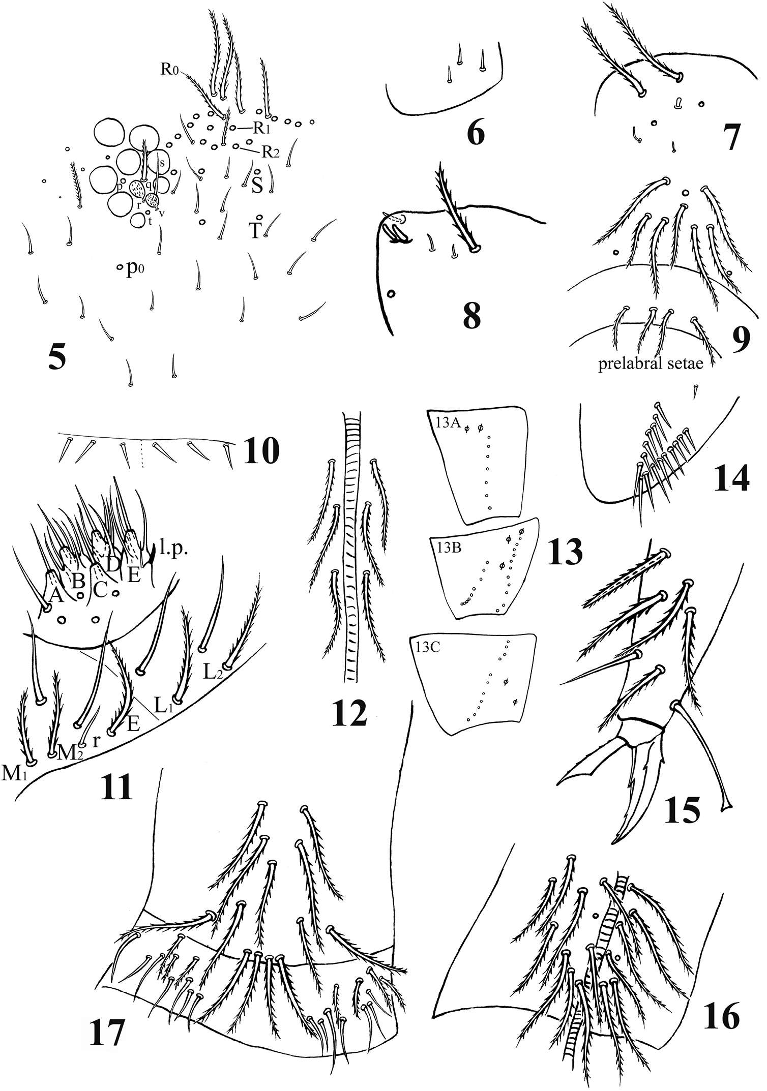
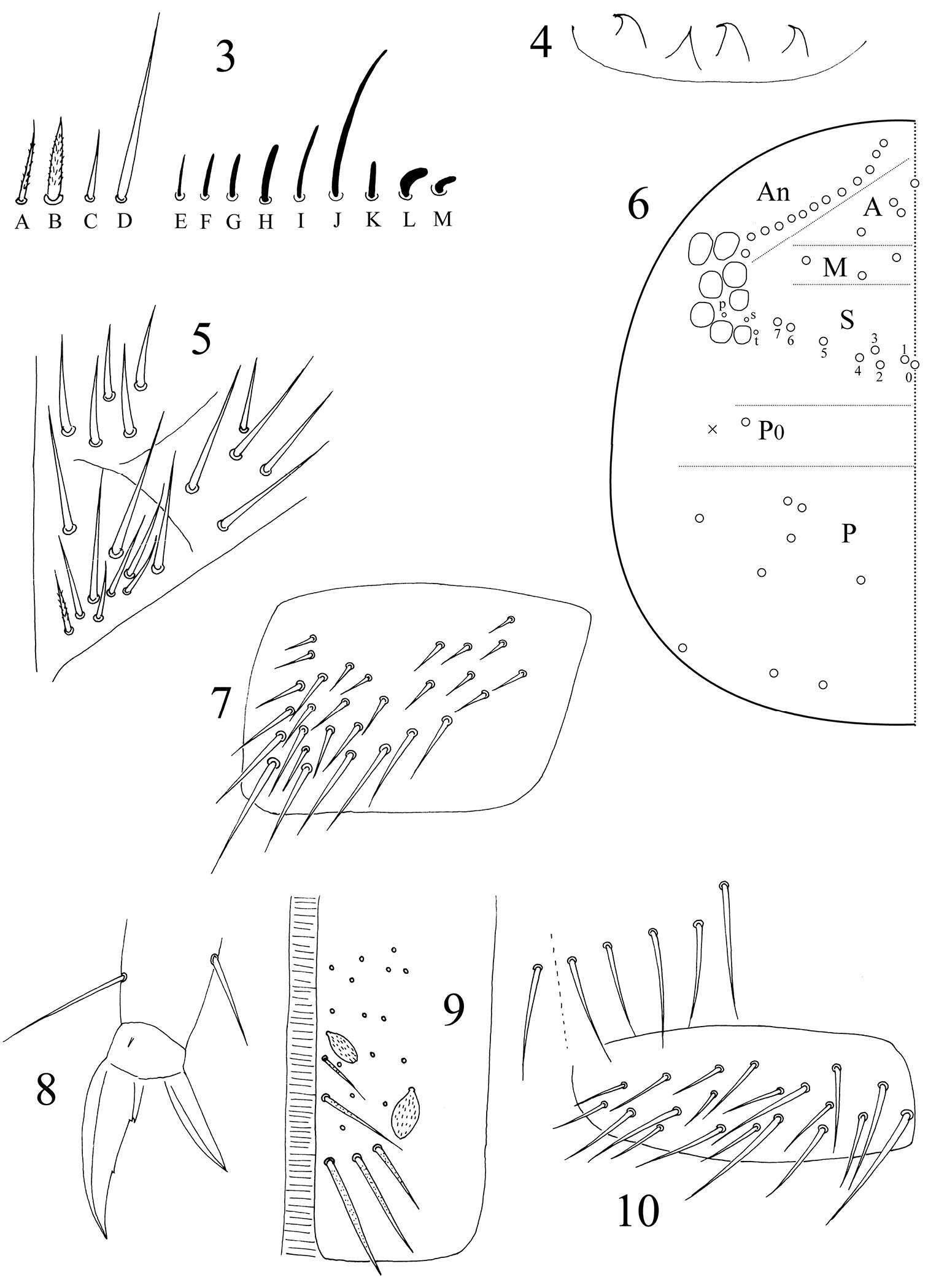
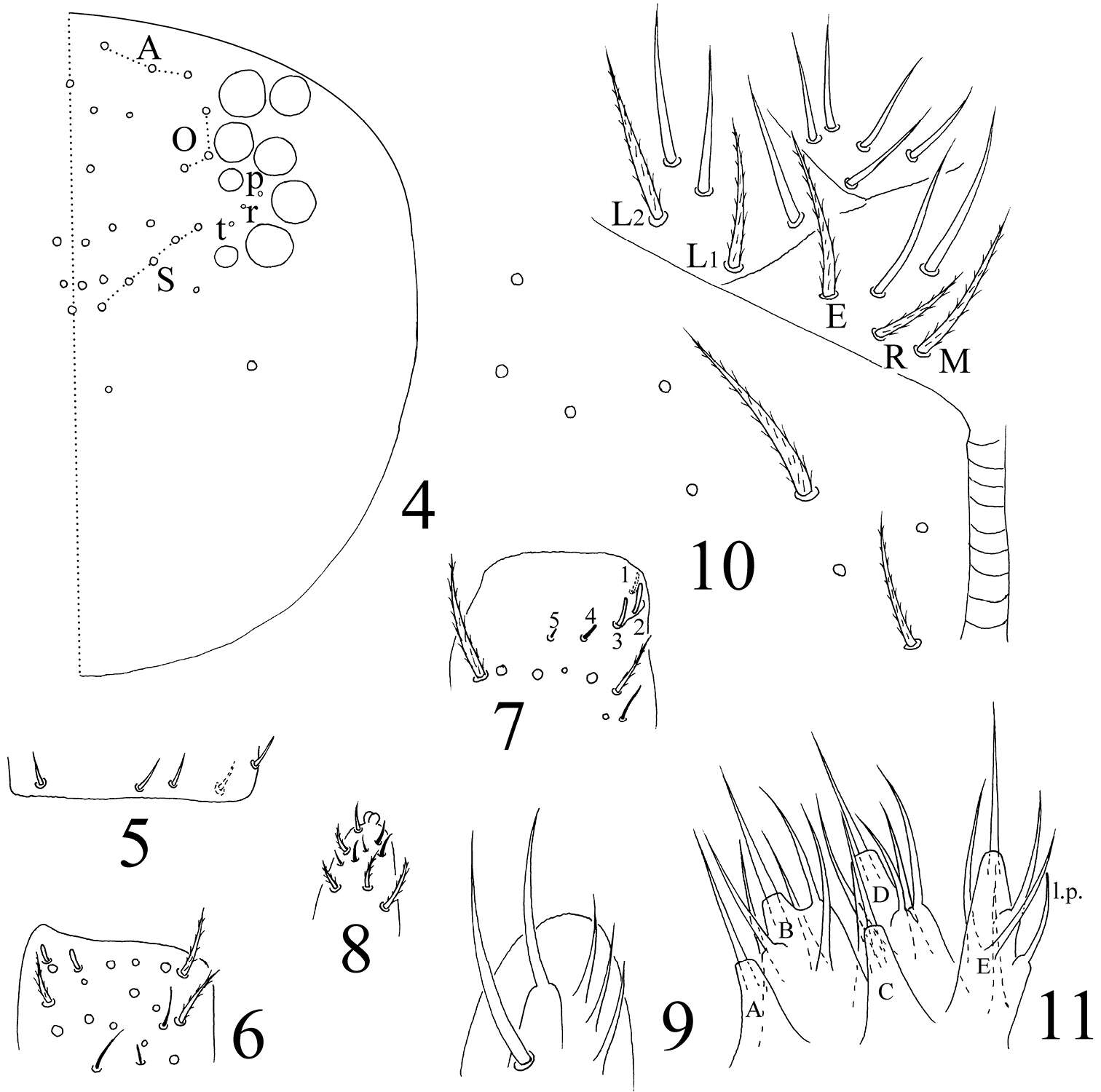
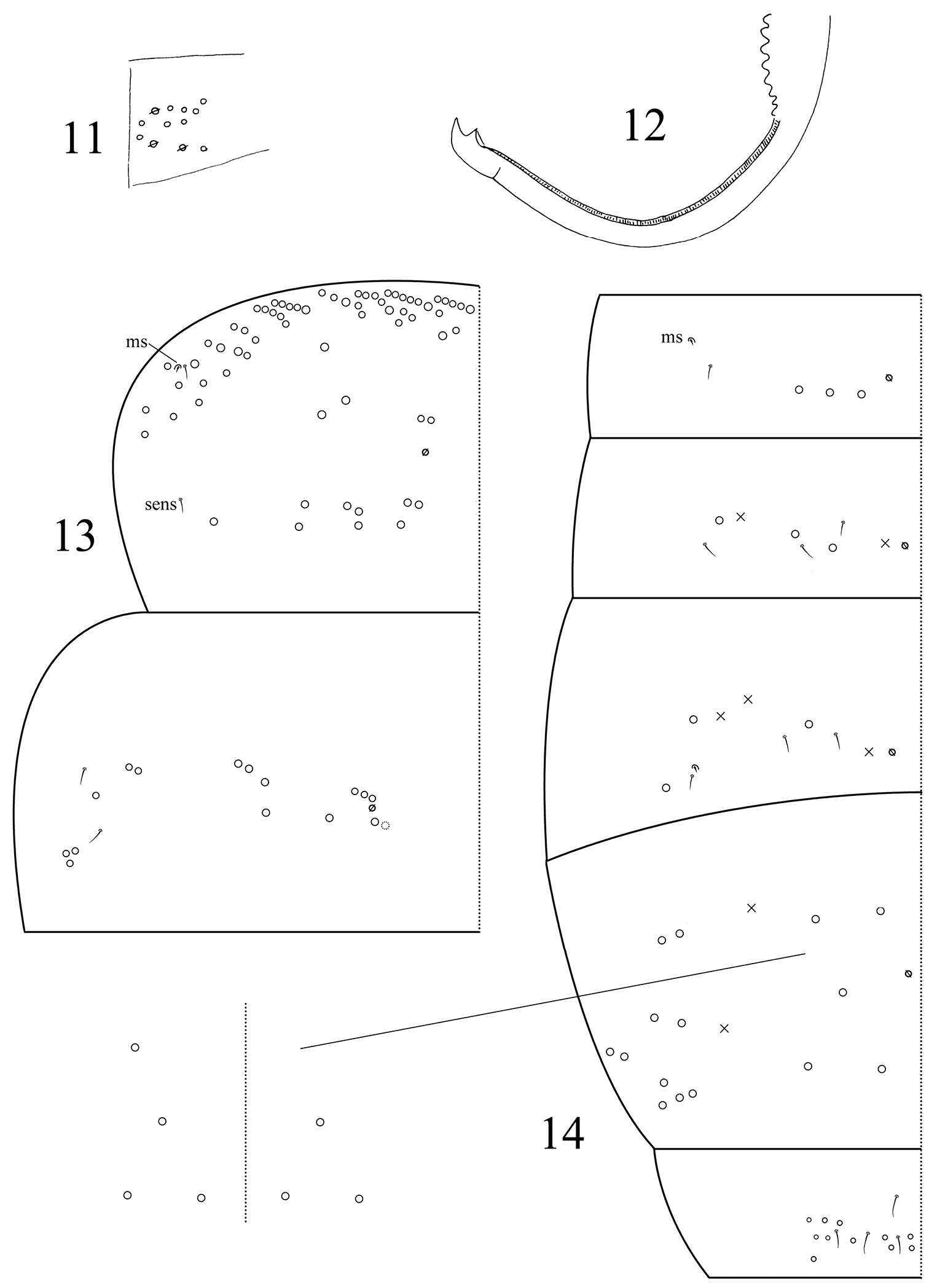
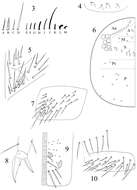
Figures 5–17.Acrocrytus zhujiensis sp. n. 5 head cheatotaxy 6 basal Ant. I 7 three kinds S-chaetae on Ant. II 8 Ant. III organ 9 clypeal chaetae 10 cervical chaetae 11 labial base and labial palp 12 cephalic groove 13 coxal macrochaetae (13A fore legs 13B mid legs 13C hind legs) 14 trochanteral organ 15 hind claw 16 anterior side of ventral tube 17 posterior side and lateral flap of ventral tube.
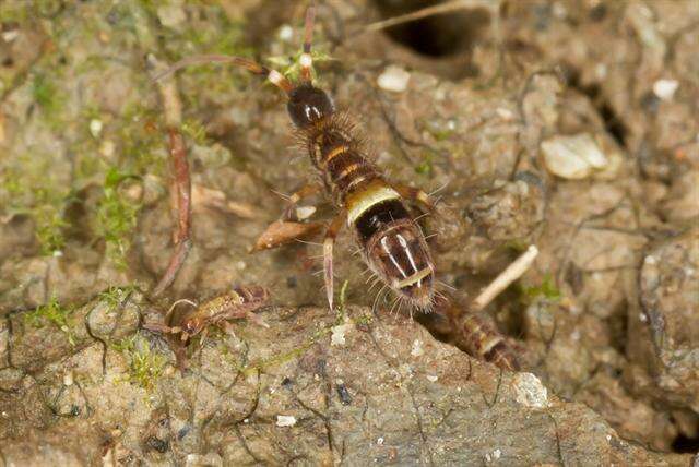
Figures 52–59.SEM photos of adult Homidia jordanai sp. n. 52 distal part of Ant. II 53 s on distal part of Ant. II 54 Ant. III organ 55 internal two s of Ant. III organ 56 external s of Ant. III organ 57 Ant. IV 58–59 s on Ant. IV.
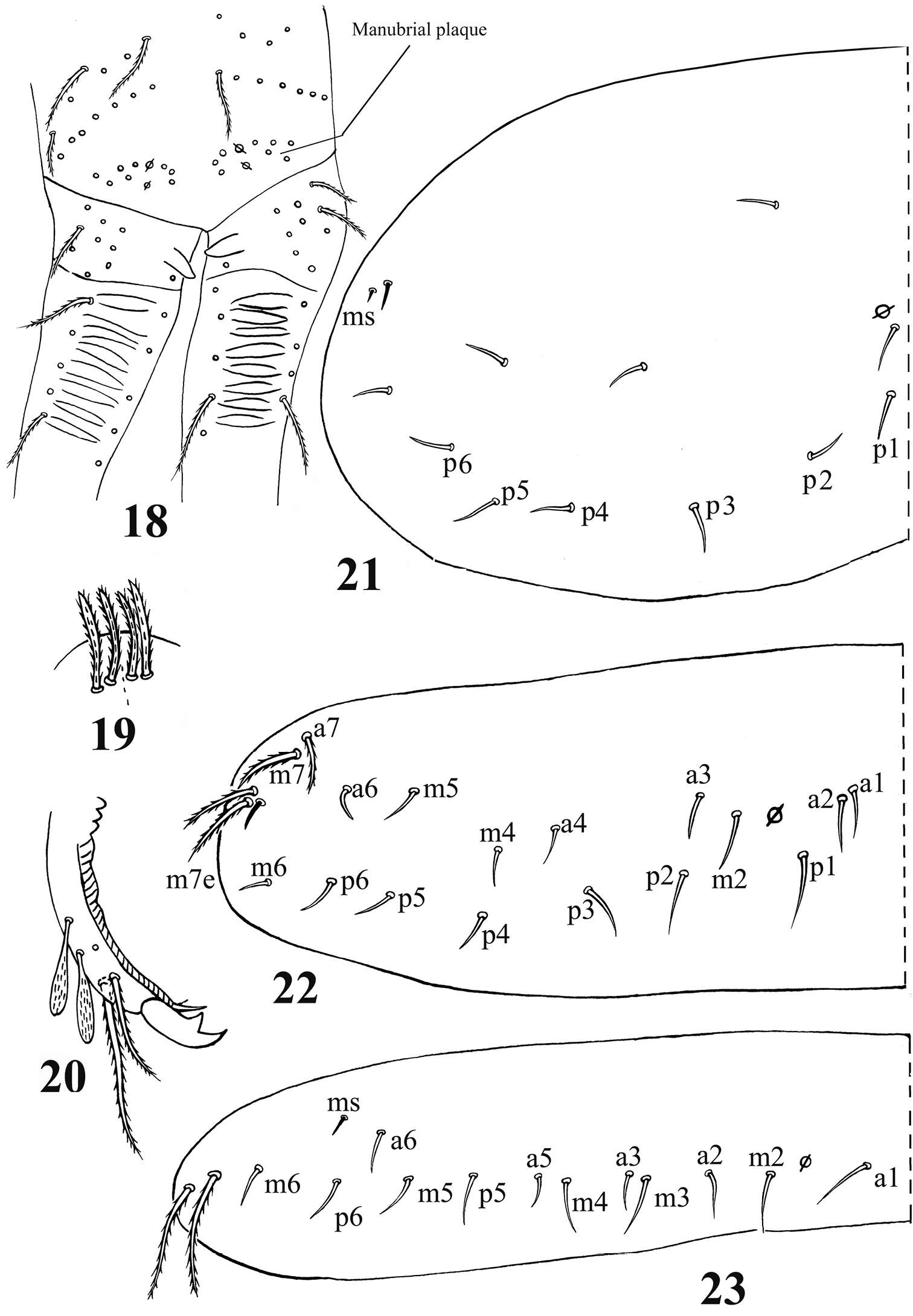
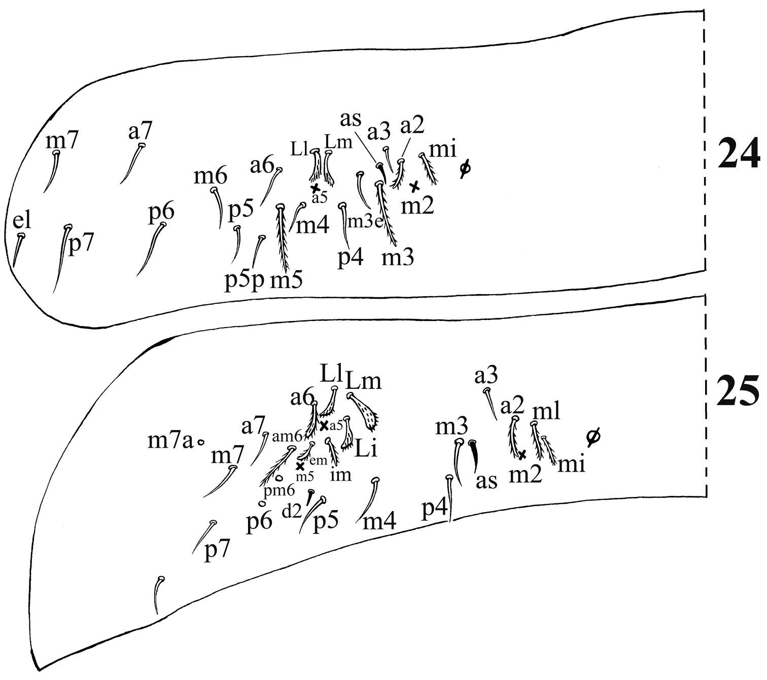
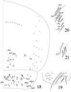
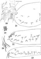
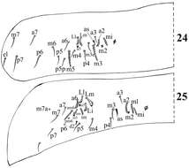
Figures 18–23.Acrocrytus zhujiensissp. n. 18 distal manubrium and basal dens 19 distal part of ventral manubrium 20 mucro 21–23 dorsal chaetotaxy 21 Th. II 22 Th. III 23. Abd. I.
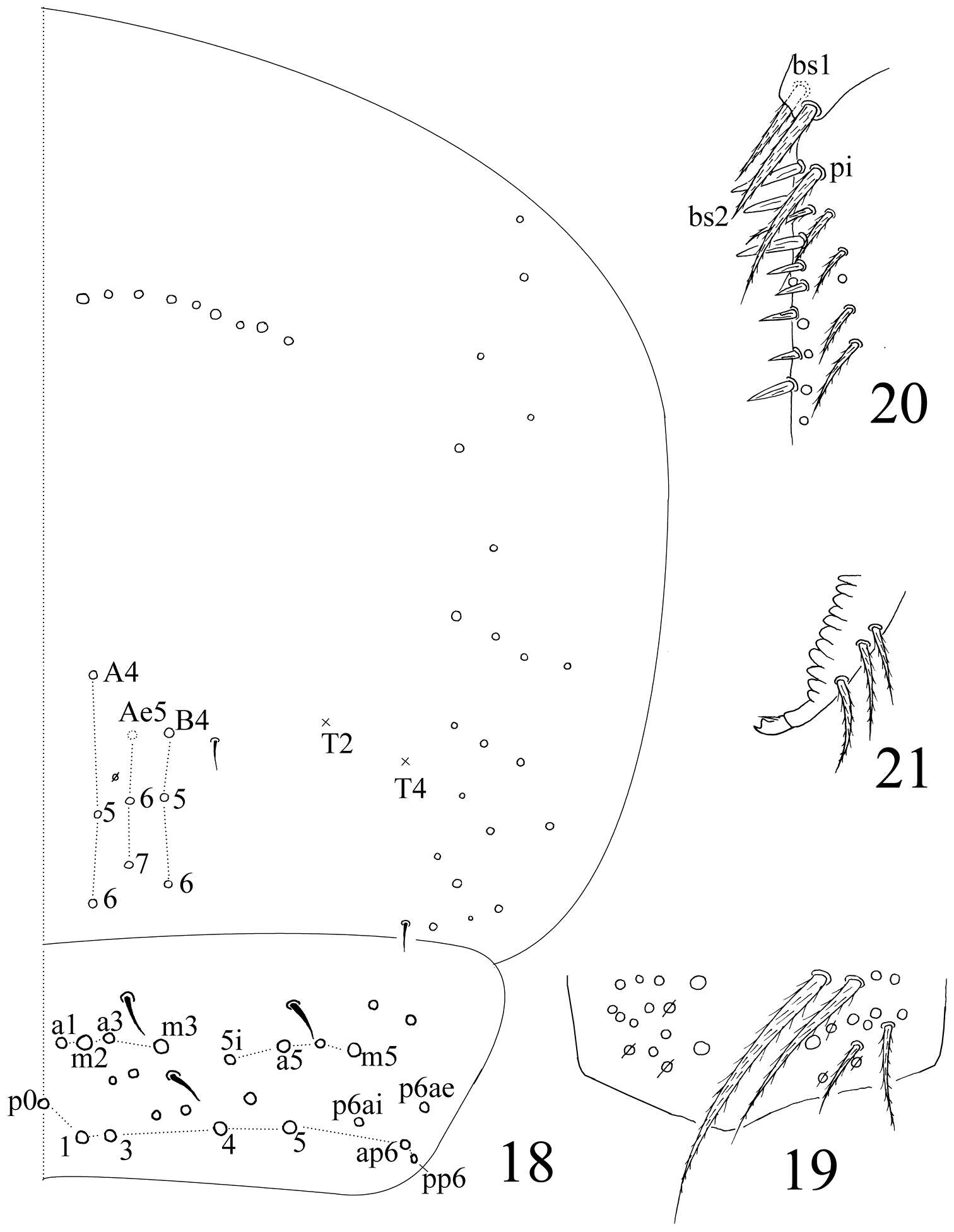
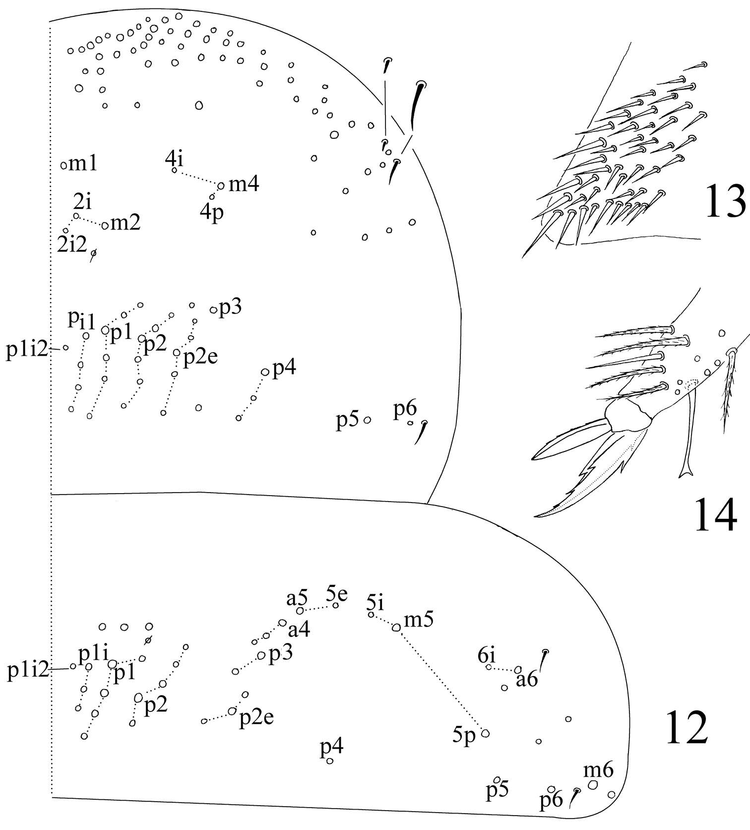
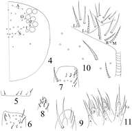
Figures 4–10.Homidia taibaiensis sp. n. 4 dorsal cephalic chaetotaxy 5 basal spiny chaetae of Ant. II 6 distal Ant. II 7 Ant. III organ 8 apical bulb of Ant. IV 9 maxillary outer lobe 10 labial base 11 labial palp.
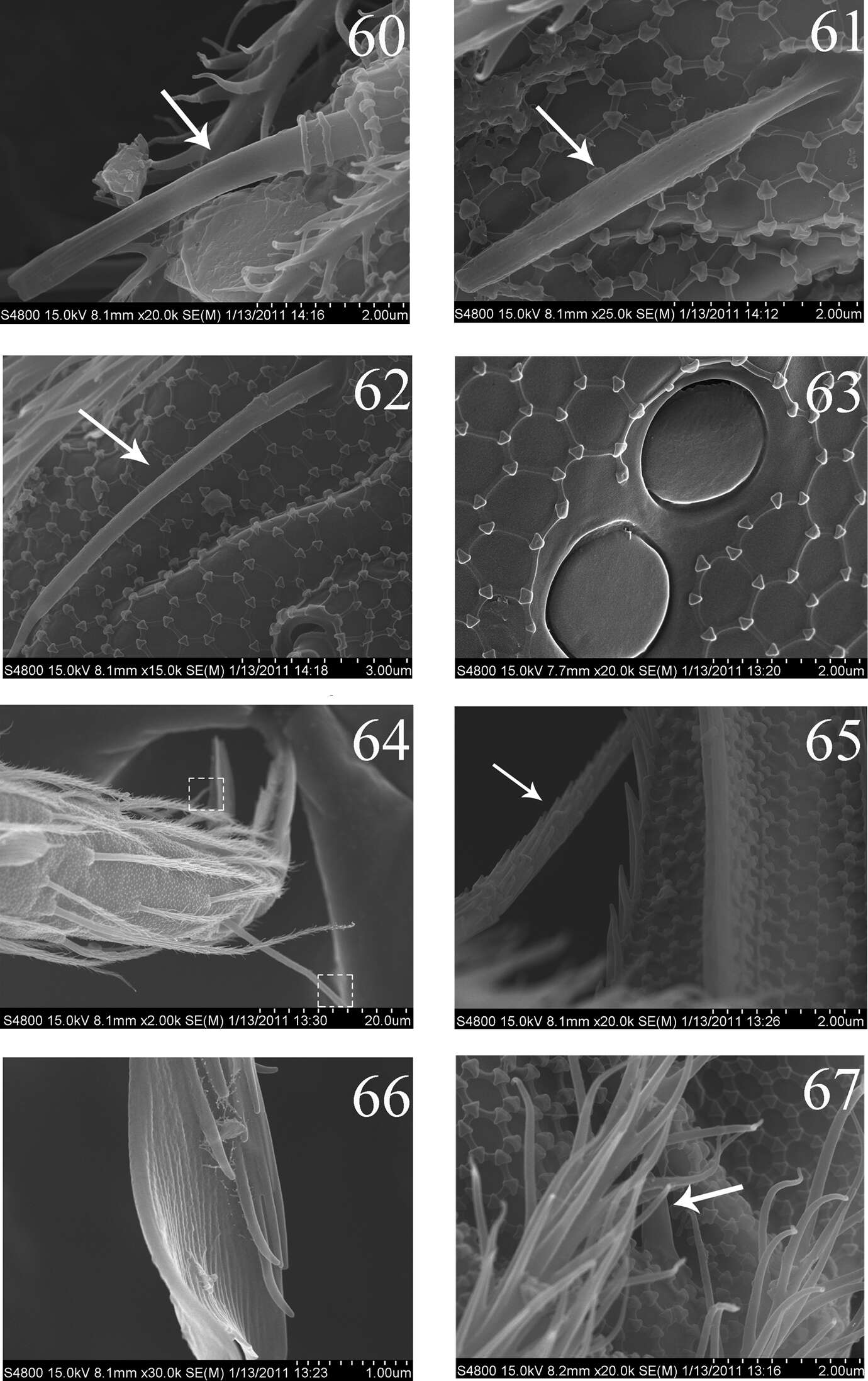
Figures 60–67.SEM photos of adult Homidia jordanai sp. n. 60–62 s of Ant. IV 63 pseudopores of coxa 64 Tita and claw of hind leg 65 supraempodial seta and outer edge of unguiculus 66 distal part of tenent hair 67 spiny seta on pretarsus.