-
At a magnification of 1000X, twice that of PHIL 10557, this scanning electron micrograph (SEM) revealed some of the minute exoskeletal details found at the proboscis tip of an unidentified mosquito found deceased in the suburbs of Decatur, Georgia. The proboscis is the organ used by this, as well as other like insects, to feed upon the blood of a warm-blooded host, including human beings. What you see here, is the sheath that encases a pair of needle-sharp "stylets", which together are known as the "fascicle". The larger of the two stylets, known as the "labrum", when viewed in cross-section, takes on the shape of a "V", and acts as a gutter, which directs the ingested host blood towards the insect's mouth. The hair-like structures are known as "setae", and are really extensions of the insect's exoskeletal, chitinous covering. These setae act as sensory organs, transmitting impulses indicating changes in the organism's environment.Created: 2008
-
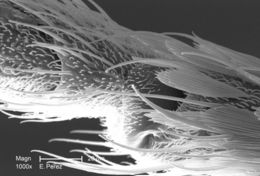
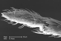
Magnified 500X, this scanning electron micrograph (SEM) revealed some of the minute exoskeletal details found at the proboscis tip of an unidentified mosquito found deceased in the suburbs of Decatur, Georgia. The proboscis is the organ used by this, as well as other like insects, to feed upon the blood of a warm-blooded host, including human beings. What you see here, is the sheath that encases a pair of needle-sharp "stylets", which together are known as the "fascicle". The larger of the two stylets, known as the "labrum", when viewed in cross-section, takes on the shape of a "V", and acts as a gutter, which directs the ingested host blood towards the insect's mouth. The hair-like structures are known as "setae", and are really extensions of the insect's exoskeletal, chitinous covering. These setae act as sensory organs, transmitting impulses indicating changes in the organism's environment.Created: 2008
-
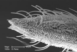
Magnified 1500X, this scanning electron micrograph (SEM) revealed some of the minute exoskeletal details found at the proboscis tip of an unidentified mosquito found deceased in the suburbs of Decatur, Georgia. The proboscis is the organ used by this, as well as other like insects, to feed upon the blood of a warm-blooded host, including human beings. What you see here, is the sheath that encases a pair of needle-sharp "stylets", which together are known as the "fascicle". The larger of the two stylets, known as the "labrum", when viewed in cross-section, takes on the shape of a "V", and acts as a gutter, directing the ingested host blood towards the insect's mouth. The hair-like structures are known as "setae", and are really extensions of the insect's exoskeletal, chitinous covering. These setae act as sensory organs, transmitting impulses indicating changes in the organism's environment.Created: 2008
-
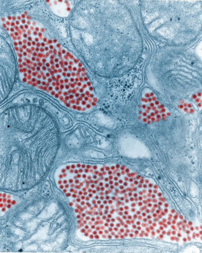
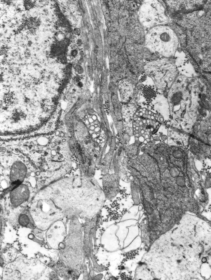
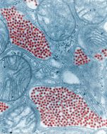
This colorized transmission electron micrograph (TEM) depicts a salivary gland that had been extracted from a mosquito, which was infected by the Eastern equine encephalitis (EEE) virus, which has been colorized red; magnified 83,900x.Created: 1968
-
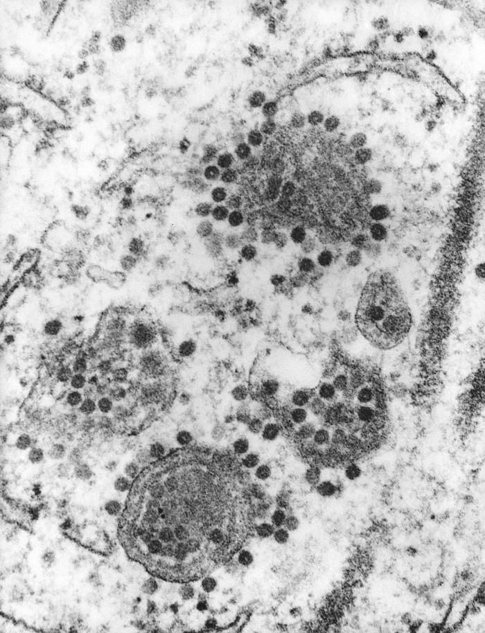
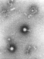
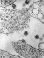
This 1975 transmission electron micrograph (TEM) revealed the presence of a number of Eastern Equine Encephalitis (EEE) virus virions that happened to be in a specimen of central nervous system tissue. EEE is an zoonotic arbovirus, which means that its spead to human beings through the bite of an infected mosquito. EEE virus (EEEV) occurs in the eastern half of the United States where it causes disease in humans, horses, and some bird species. Because of the high mortality rate, EEE is regarded as one of the most serious mosquito-borne diseases in the United States. EEE is a Togaviridae virus family member, and the genus Alphavirus.Created: 1975
-
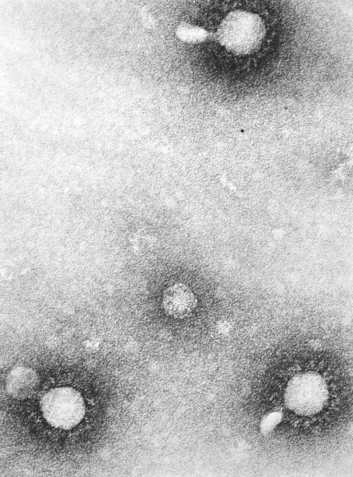
This negatively-stained 1975 transmission electron micrograph (TEM) revealed the presence of a number of Eastern equine encephalitis (EEE) virus virions in this tissue specimen. VEE is a Togaviridae family member, and a member of the genus Alphavirus.Eastern equine encephalitis (EEE) is a mosquito-borne viral disease. EEE virus (EEEV) occurs in the eastern half of the United States where it causes disease in humans, horses, and some bird species. Because of the high mortality rate, EEE is regarded as one of the most serious mosquito-borne diseases in the United States.Created: 1975
-
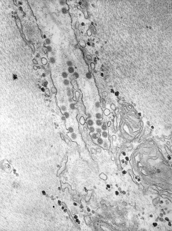
This negatively-stained 1975 transmission electron micrograph (TEM) revealed the presence of a number of Venezuelan equine encephalitis (VEE) virus virions in this tissue specimen, which had additionaly been fixed using phosphotungstic acid (PTA). This chemical is very electron-dense, and due to proposed electrostatic forces of attraction, clings to the capsid surface of each viral particle or virion, thereby, highlighting the presence of such pathogens. VEE is a Togaviridae family member, and a member of the genus Alphavirus.Created: 1975
-
This 1975 transmission electron micrograph (TEM) revealed the presence of a number of Eastern Equine Encephalitis (EEE) virus virions that happened to be in a specimen of central nervous system tissue. EEE is a zoonotic arbovirus, which means that its spead to human beings through the bite of an infected mosquito. EEE virus (EEEV) occurs in the eastern half of the United States where it causes disease in humans, horses, and some bird species. Because of the high mortality rate, EEE is regarded as one of the most serious mosquito-borne diseases in the United States. EEE is a Togaviridae virus family member, and the genus Alphavirus.Created: 1975
-
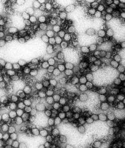
This negatively-stained transmission electron micrograph (TEM) revealed the presence of numerous Semliki Forest virus virions, which were present in a muscle tissue specimen. Named for the region in which they were isolate from mosquitoes, the Semliki Forest, Uganda, this virus is a Togaviridae family member, and the genus, Alphavirus.Created: 1975
-
This 1975 transmission electron micrograph (TEM) revealed the presence of a number of Eastern Equine Encephalitis (EEE) virus virions in this specimen of central nervous system tissue. EEE is an zoonotic arbovirus, which means that its spead to human beings through the bite of an infected arthropod, which in this case, is a mosquito. EEE virus (EEEV) occurs in the eastern half of the United States where it causes disease in humans, horses, and some bird species. Because of the high mortality rate, EEE is regarded as one of the most serious mosquito-borne diseases in the United States. EEE is a Togaviridae virus family member, and the genus Alphavirus.Created: 1975
-
This 1975 transmission electron micrograph (TEM) revealed the presence of a number of Eastern Equine Encephalitis (EEE) virus virions in this tissue specimen. EEE is an zoonotic arbovirus, which means that its spead to human beings through the bite of an infected arthropod, which in this case, is a mosquito. EEE virus (EEEV) occurs in the eastern half of the United States where it causes disease in humans, horses, and some bird species. Because of the high mortality rate, EEE is regarded as one of the most serious mosquito-borne diseases in the United States. EEE is a Togaviridae virus family member, and the genus Alphavirus.Created: 1975
-
Description: English: Everglades virus. Date: 7 February 2017. Source: Own work. Author:
Vishnuteja7991.
-
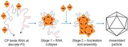
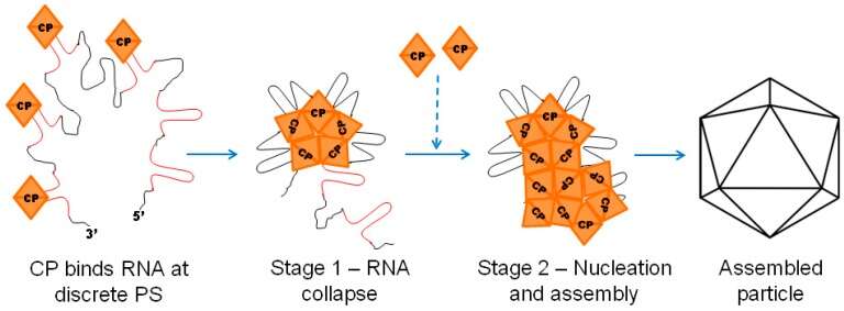
Description: English: Illustration of the two stage viral capsid assembly for an alpha virus. The red sections are packaging signals on the RNA genome. These serve to correctly assemble the capsid proteins. Date: 10 March 2018. Source:
https://www.ncbi.nlm.nih.gov/pmc/articles/PMC5869531/. Author: Adriano Mendes and Richard J. Kuhn.
-
Description: English: cryo-electron microscopy of Sindbis-liposome complex. Date: 3 December 2013, 18:18:21. Source: Own work. Author:
A2-33.
-
: All images in this article were
uploaded in the
JPEG format even though it consists of non-photographic data. This information could be stored more efficiently or accurately in the
PNG or
SVG format. If possible, please upload a PNG or SVG version of this image without
compression artifacts, derived from a non-JPEG source (or with existing artifacts
removed). After doing so, please tag the JPEG version with {{Superseded|NewImage.ext}} and remove this tag. This tag should not be applied to photographs or scans. For more information, see {{
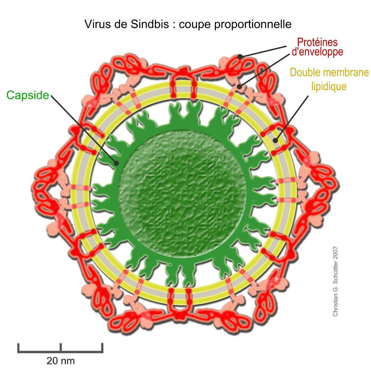
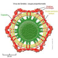
BadJPEG}}.:. Description: English: Proportional model of the Sindbis Virus according to the electron microscope views and thickness measurements by W. Zhang et al. J. Virology (2002) 76: 11645–11658 (Fig. 1)
PMID 12388725 Deutsch: Maßstabgetreues Modell des de:Sindbis-Virus nach den Cryo-Elektronenmikroskopischen Aufnahmen und Dichtemessungen von W. Zhang et al. J. Virology (2002) 76: 11645–11658 (Fig. 1)
PMID 12388725 Français : Section proportionnelle d'un virus de Sindbis selon les vues et les mesures d'épaisseur au microscope électronique de W. Zhang et al. J. Virology (2002) 76: 11645–11658 (Fig. 1)
PMID 12388725. Date: 5 January 2012. Source: This file was derived from:
Sindbis-Virus Struktur.png. Author: Gleiberg. Translated to french by user:Trassiorf on dec. 5 2012. Previous history : The original description page is/was here. All following user names refer to de.wikipedia. Uploaded on14:29, November 3, 2010 by user:Leyo 2007-09-01 00:25 Gleiberg 1772×1772× (753521 bytes) {{Information |Beschreibung = Maßstabgetreues Modell des
Sindbis-Virus nach den Cryo-Elektronenmikroskopischen Aufnahmen und Dichtemessungen von W. Zhang et al. J. Virology (2002) 76: 11645-11658 (Fig. 1)
PMID 12388725 |Quelle = selbst gezeichnet Licensing[
edit] : This file is licensed under the
Creative Commons Attribution-Share Alike 2.0 Germany license.:. https://creativecommons.org/licenses/by-sa/2.0/de/deed.en CC BY-SA 2.0 de Creative Commons Attribution-Share Alike 2.0 de truetrue.
-
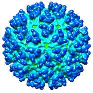
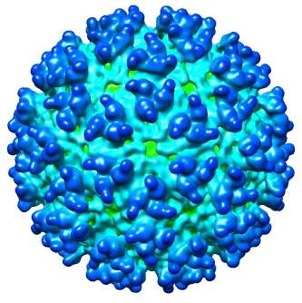
Description: English: CryoEM model of western equine encephalitis virus, 12Å resolution. EMDB entry EMD-5210. Date: 3 December 2013, 20:25:50. Source: Own work. Author:
A2-33.
-
Description: English: This diagram illustrates the methods by which the arbovirus, Western equine encephalitis virus (WEEV) reproduces and amplifies itself in both avian populations, and rodent populations, and is subsequently transmitted to dead end hosts including humans and horses by the Culex tarsalis mosquito. Western equine encephalitis virus, is a member of the family Togaviridae, and the genus Alphavirus, is closely related to Eastern and Venezuelan equine encephalitis viruses. Date: 1976. Source:
https://phil.cdc.gov/Details.aspx?pid=14996. Author: CDC/ Dr. Thomas Monath.
-
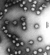
Description: English: Semliki forest virus from purified cellculture supernatant after gel-filtration and ultracentrifugation, magnification 90,000x, ELMI C10 Zeiss. Date: 7 August 2006 (original upload date) (Original text: 2004). Source: Own work (Original text: selfmade). Author:
Gleiberg. Permission(
Reusing this file): Ich, als der Urheber, veröffentliche das Bild unter CC-by-sa. : This file has been superseded by
File:Semliki-Forest-Virus.png. It is recommended to use the other file. Please note that deleting superseded images
requires consent. Reason to use the other file: "A PNG version of this file is now available." :
.
-
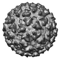
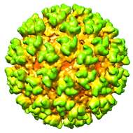
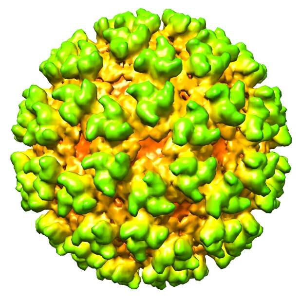
Description: English: Cryo-electron microscopy reconstruction of Semliki Forest virus at 9A resolution. From EMD-1015. Date: 8 December 2013, 10:55:24. Source: Own work. Author:
A2-33.
-
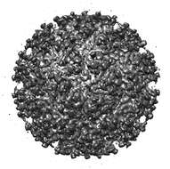
Description: English: CryoEM reconstruction of the Everglades Virus. EMD-5563. Date: 4 December 2013, 17:28:41. Source: Own work. Author:
A2-33.


