-
Asker, Akershus, Norge
-
Jadranka Rota, Scott E. Miller
Zookeys
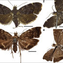
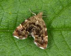
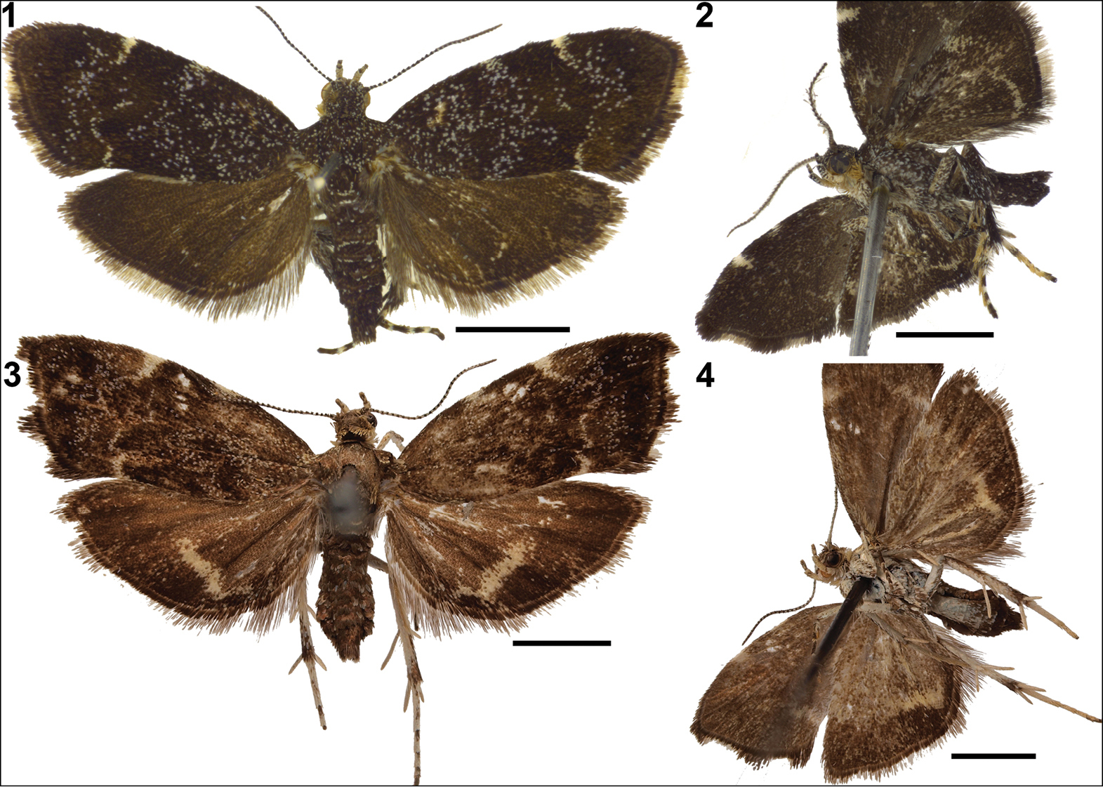
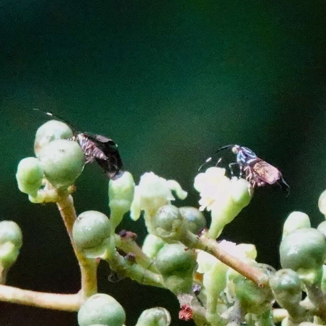
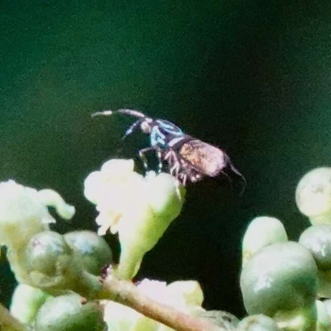
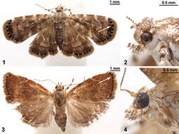
Figures 1–4.Niveas kone: 1 Habitus 2 Head. Niveas agassizi: 3 Habitus 4 Head. (In Figs 1 and 3 arrows point at the terminal black band enclosing white spots.)
-
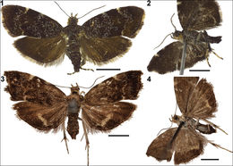
Figures 1–4.Adults in dorsal and lateral view. 1–2. Anthophila threnodes; 3–4. Anthophila fabriciana (scale bar = 2 mm).
-
Asker, Akershus, Norge
-
Jadranka Rota, Scott E. Miller
Zookeys
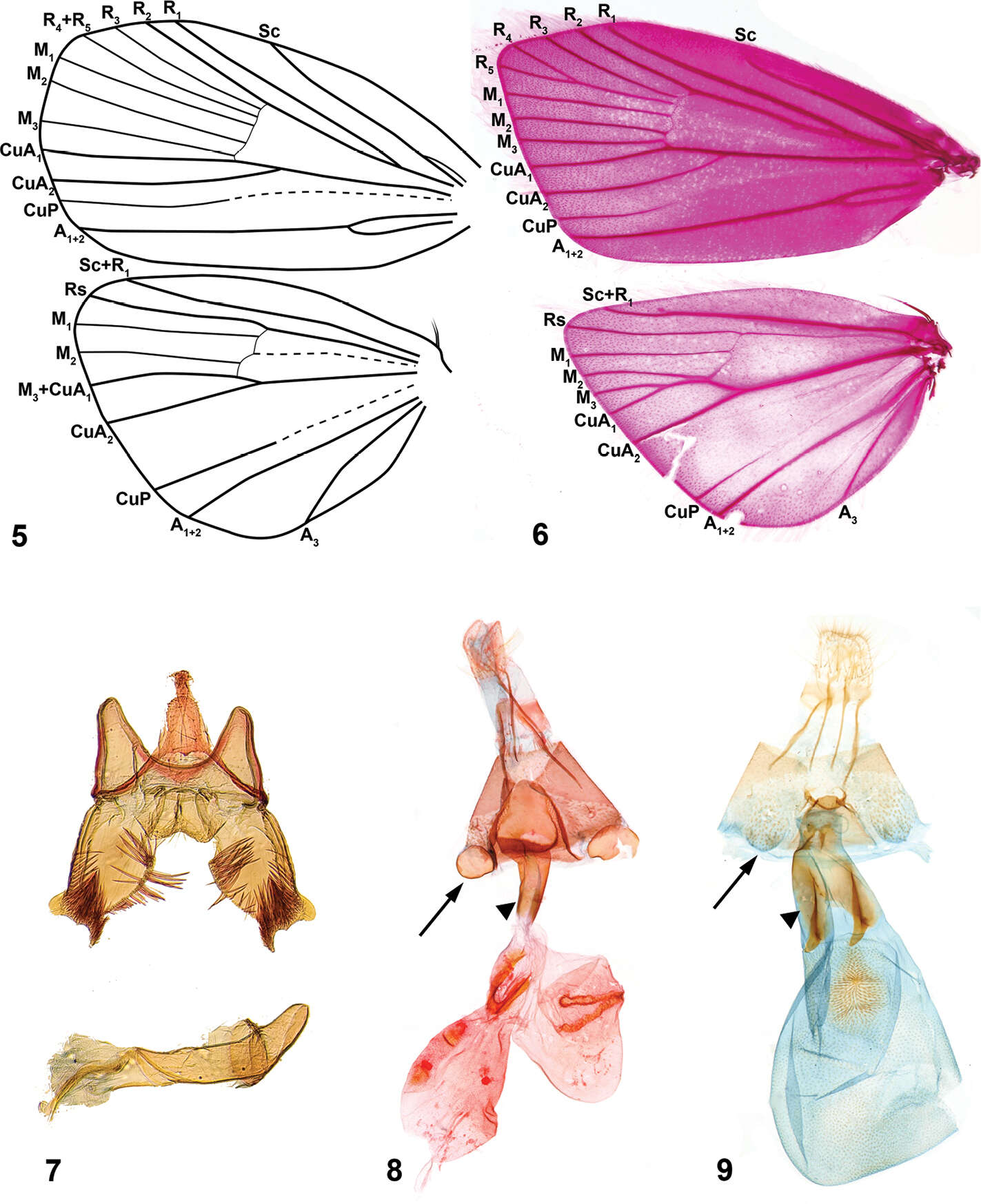
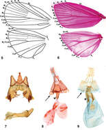
Figures 5–9.Niveas kone: 5 Wing venation 7 Male genitalia 8 Female genitalia. Niveas agassizi: 6 Wing venation 9 Female genitalia. (In Figs 8 and 9 arrows point at the A7 sternite sclerotizations, and triangles point at the lateral sclerotizations on the ductus bursae.)
-
Asker, Akershus, Norge
-
Jadranka Rota, Scott E. Miller
Zookeys
Figures 1–4.Niveas kone: 1 Habitus 2 Head. Niveas agassizi: 3 Habitus 4 Head. (In Figs 1 and 3 arrows point at the terminal black band enclosing white spots.)
-
Asker, Akershus, Norge
-
Jadranka Rota, Scott E. Miller
Zookeys
Figures 5–9.Niveas kone: 5 Wing venation 7 Male genitalia 8 Female genitalia. Niveas agassizi: 6 Wing venation 9 Female genitalia. (In Figs 8 and 9 arrows point at the A7 sternite sclerotizations, and triangles point at the lateral sclerotizations on the ductus bursae.)
-
Jadranka Rota, Scott E. Miller
Zookeys
Figures 1–4.Niveas kone: 1 Habitus 2 Head. Niveas agassizi: 3 Habitus 4 Head. (In Figs 1 and 3 arrows point at the terminal black band enclosing white spots.)
-
Jadranka Rota, Scott E. Miller
Zookeys
Figures 5–9.Niveas kone: 5 Wing venation 7 Male genitalia 8 Female genitalia. Niveas agassizi: 6 Wing venation 9 Female genitalia. (In Figs 8 and 9 arrows point at the A7 sternite sclerotizations, and triangles point at the lateral sclerotizations on the ductus bursae.)
-
Jadranka Rota, Scott E. Miller
Zookeys
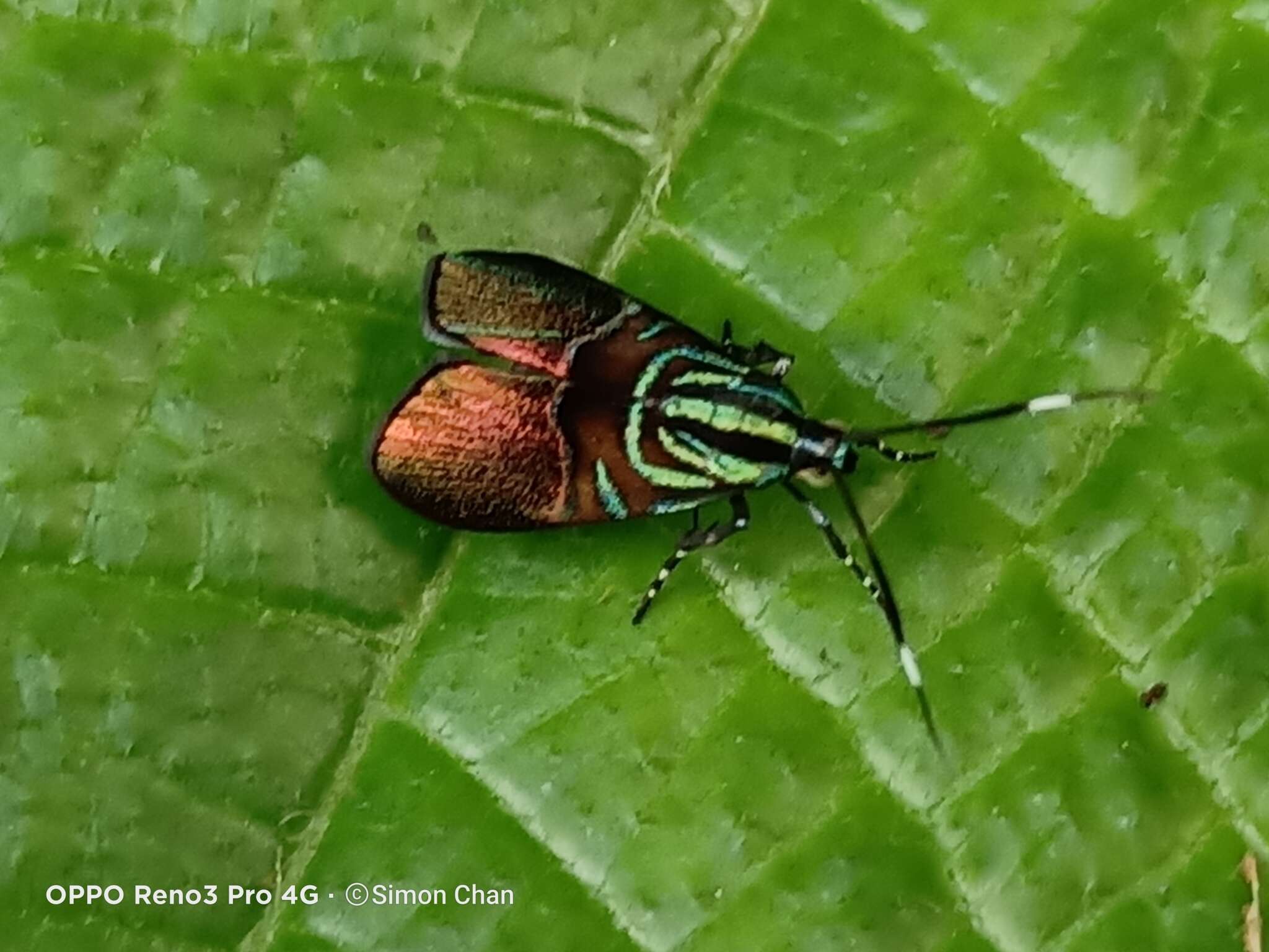
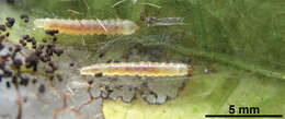
Figure 12.A photograph of the Niveas kone larvae made in the field.
-
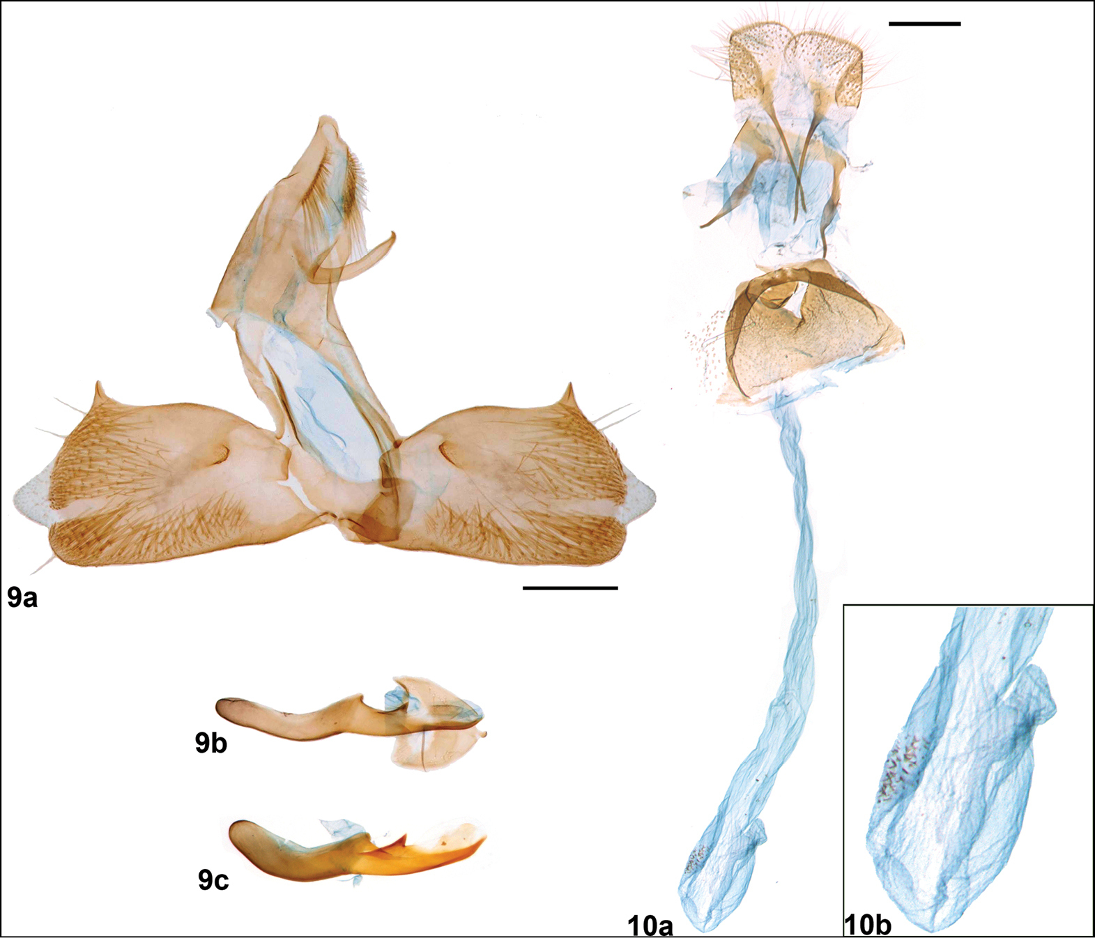
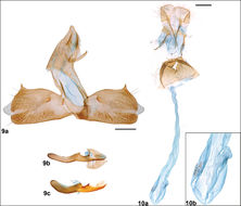
Figures 9–10.Anthophila threnodes: male genitalia (9a), phallus (9b, c) (9a and 9b from slide Karsholt 5236, ZMUC; 9c from slide JR2013-04, ZMUC), female genitalia (10a), inset showing magnified corpus bursae (10b) (scale bar = 0.2 mm).
-
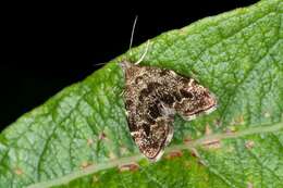
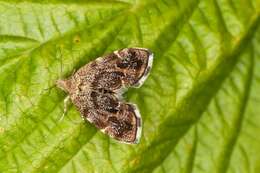
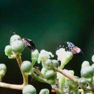
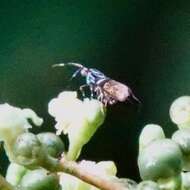
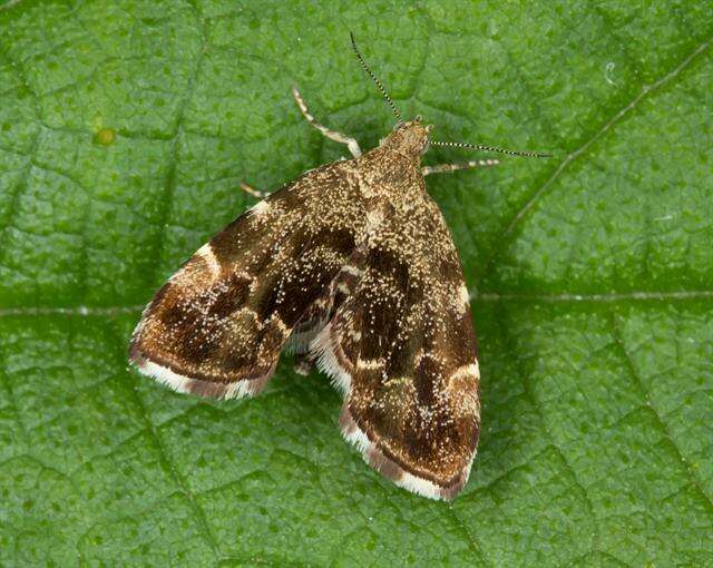
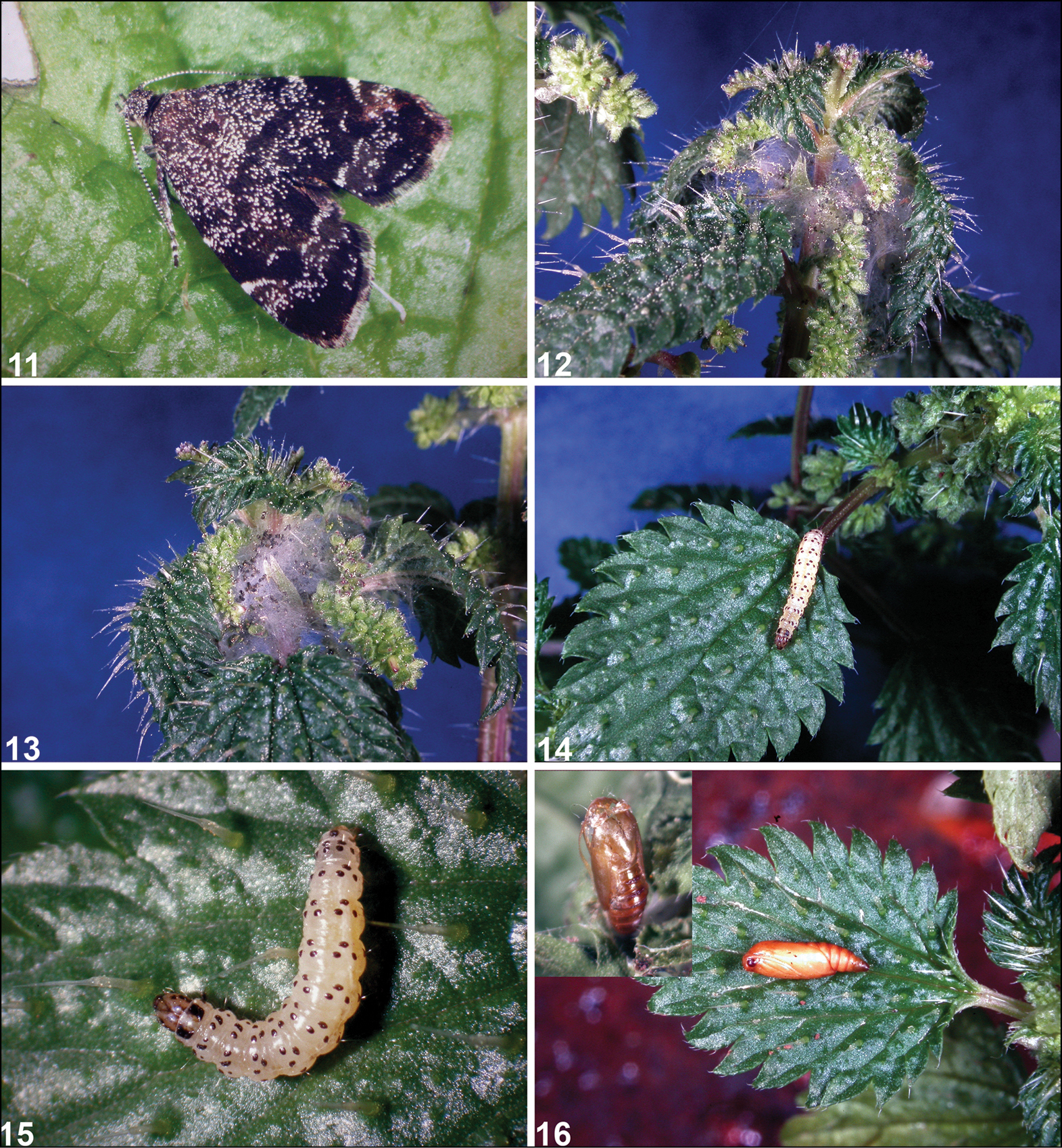
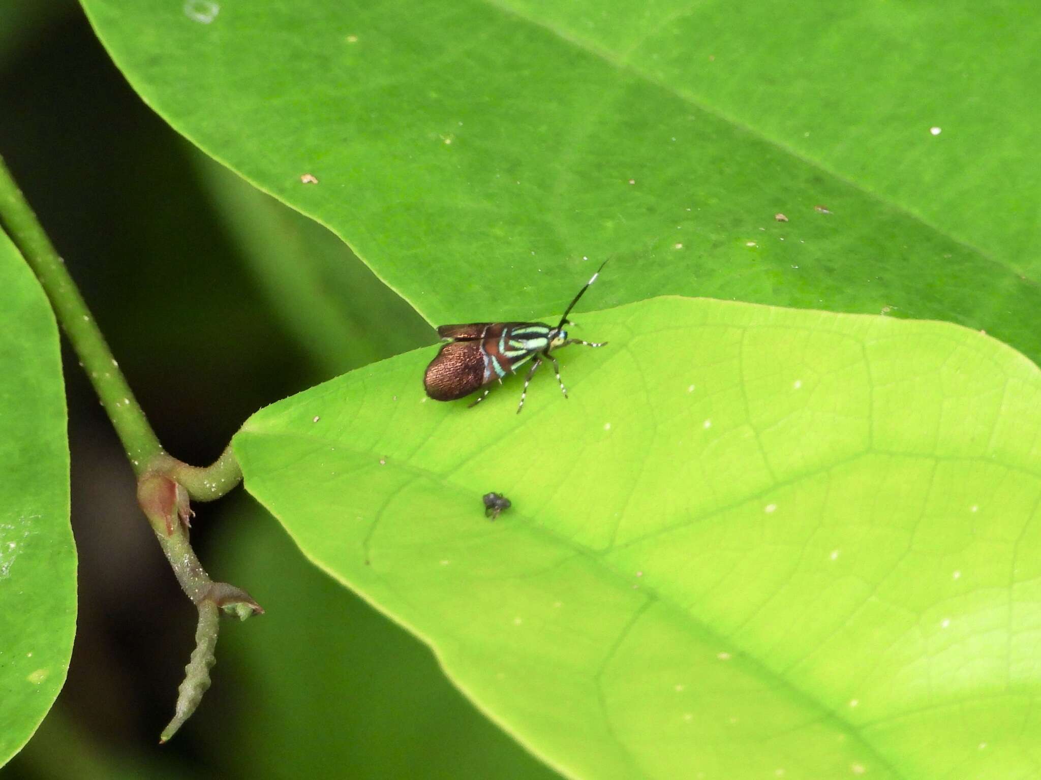
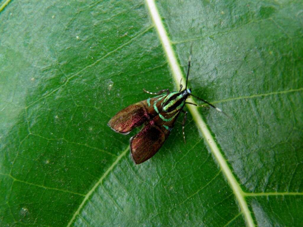
Figures 11–16.Anthophila threnodes: 11. Adult on its host plant; 12–13. Larval webbing tying young leaves; 14–15. Larva; 16. Pupa and an empty pupal shell in the inset.
-
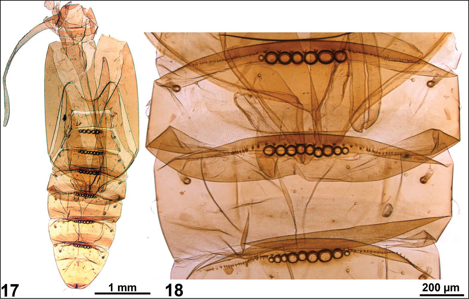
Figures 17–18.Anthophila threnodes pupa (17) with the close-up of dorsal spines and lacunae (18).
-
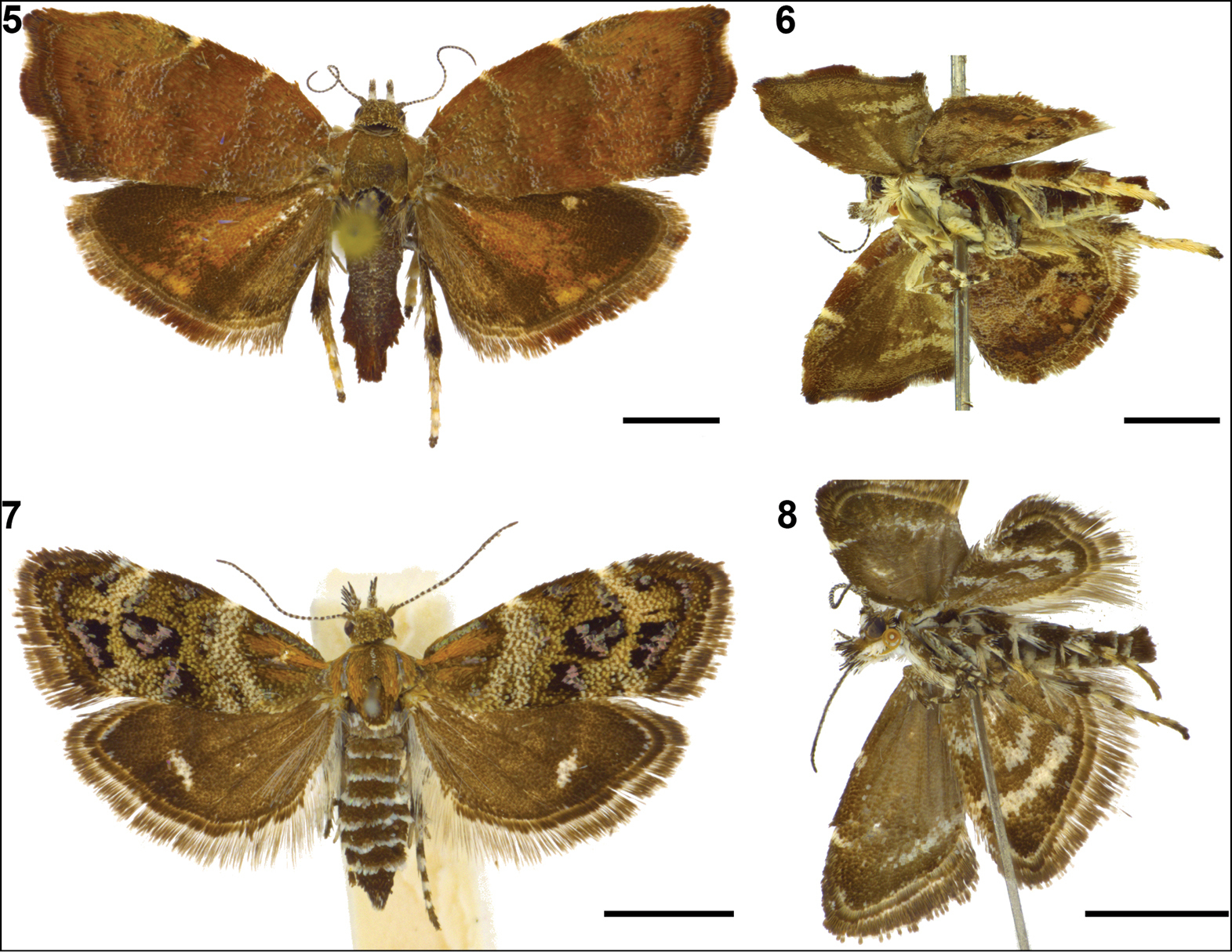
Figures 1–4.Adults in dorsal and lateral view. 1–2. Anthophila threnodes; 3–4. Anthophila fabriciana (scale bar = 2 mm).
-
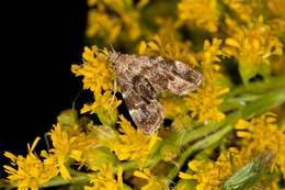
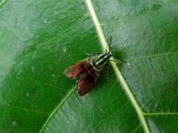
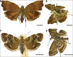
Figures 5–8.Adults in dorsal and lateral view. 5–6. Choreutis nemorana; 7–8. Tebenna micalis (scale bar = 2 mm).
-
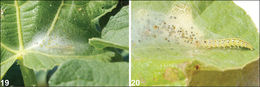
Figures 19–20.Choreutis nemorana: larva under its webbing on the host plant Ficus carica (19) and larva exiting its web-shelter after being disturbed (20).
-
Figures 5–8.Adults in dorsal and lateral view. 5–6. Choreutis nemorana; 7–8. Tebenna micalis (scale bar = 2 mm).
-
-
-
-
-