-
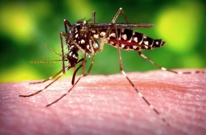
This 2006 image depicted a female Aedes aegypti mosquito as she was completing the activity of obtaining a blood-meal from a human host through her fascicle, which shed begun to resheath in her labium. Both structures are part of her feeding organ known as the proboscis. In this case, what would normally be an unsuspecting host was actually the CDCs biomedical photographers own hand, which hed offered to the hungry mosquito so that shed alight, and be photographed while feeding. After it filled with blood, the abdomen became distended, stretching the exterior exoskeletal surface, thereby, causing it to become transparent, allowing the collecting blood to become visible as an enlarging intra-abdominal red mass.Created: 2006
-
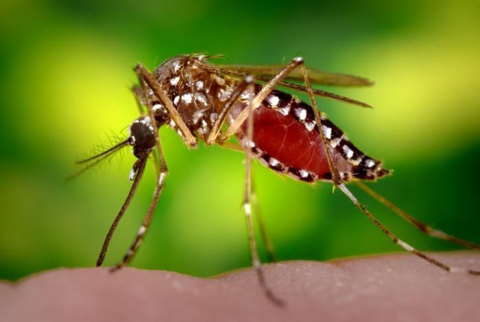
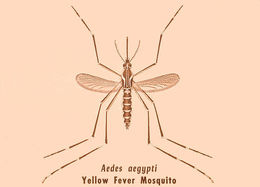
Illustration of Aedes aegypti adult mosquito, vector of yellow fever.Created:
-
This Aedes aegypti female was from a strain of mosquitoes named LVP-IB12, an acronym representing the fact that these mosquitoes were derived from the Liverpool strain (LVP), and that they were inbred 12 times (IB12), in order to create a more homogeneous genotype. Also, of great importance is the additional fact that this specie is being used in the A. aegypti genome sequencing project. Though the mosquitos geographical origin is not known, it is believed to be somewhere in Africa.Created: 2007
-
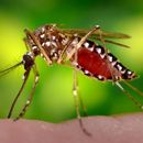
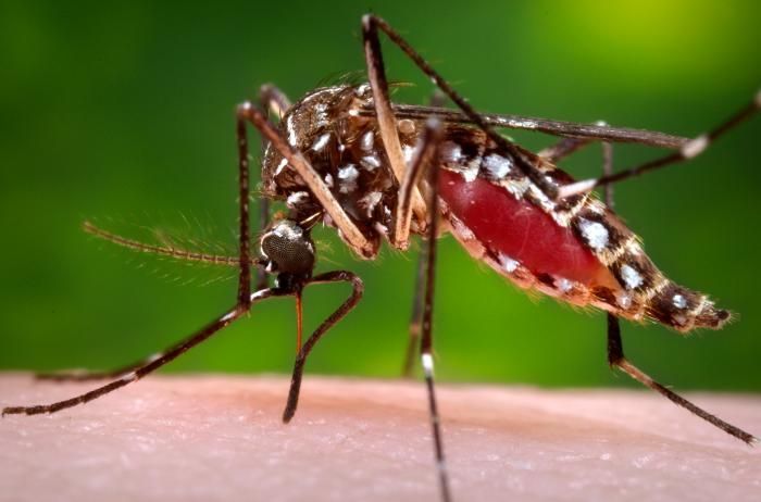
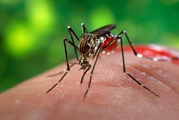
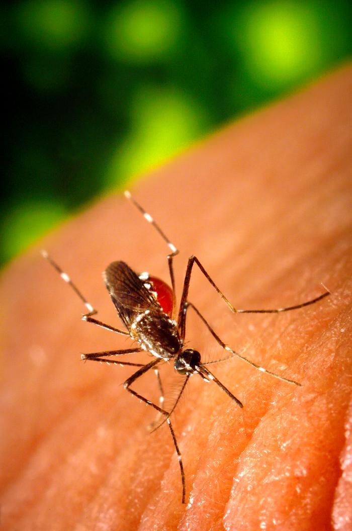
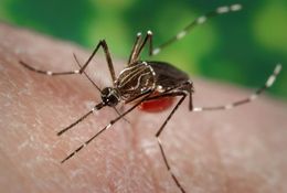
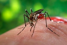
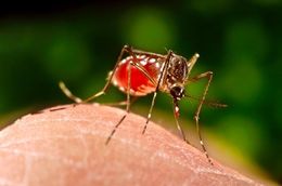
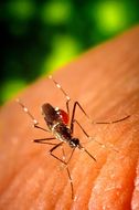
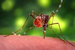
This 2006 photograph depicted a female Aedes aegypti mosquito while she was in the process of acquiring a blood meal from her human host, who in this instance, was actually the biomedical photographer, James Gathany, here at the Centers for Disease Control. The feeding apparatus consisted of a sharp, orange-colored fascicle that was covered in a soft, pliant sheath called the "labellum while not feeding. The labellum was shown here retracted as the sharp "stylets" contained within pierced the host's skin surface, thereby, allowing the insect to obtain its blood meal. The orange color of the fascicle was due to the red color of the blood as it migrated up the thin, sharp translucent tube. Note the distended abdominal exoskeleton, which being translucent, allowed the color of the ingested blood meal to be visible.Created: 2006
-
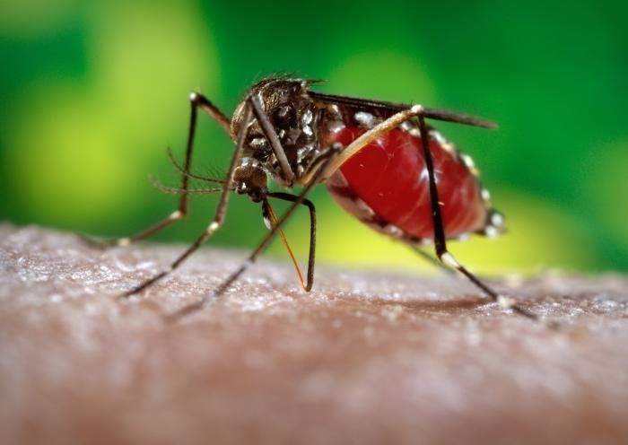
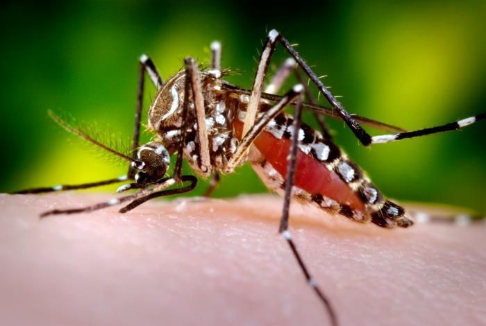
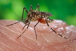
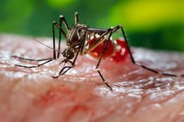
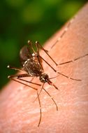
This 2006 photograph depicted a female Aedes aegypti mosquito while she was in the process of acquiring a blood meal from her human host, who in this instance, was actually the biomedical photographer, James Gathany, here at the Centers for Disease Control. The feeding apparatus consisted of a sharp, orange-colored fascicle that was covered in a soft, pliant sheath called the "labellum while not feeding. The labellum was shown here retracted as the sharp "stylets" contained within pierced the host's skin surface, thereby, allowing the insect to obtain its blood meal. The fascicle was composed of a pair of needle-sharp stylets. The larger of the two stylets, known as the "labrum", when viewed in cross-section takes on the shape of an inverted "V", and acts as a gutter, which directs the ingested host blood towards the insect's mouth. Due to the ingestion of the females blood meal, the translucent abdominal exoskeleton had taken on a reddish color.Created: 2006
-
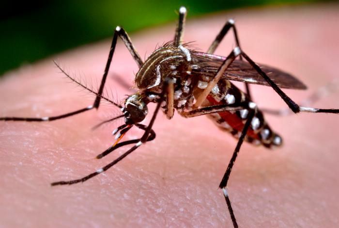
This 2006 photograph depicted a female Aedes aegypti mosquito while she was in the process of acquiring a blood meal. The feeding apparatus consisted of a sharp, orange-colored fascicle that was covered in a soft, pliant sheath called the "labellum while not feeding. The labellum was shown here retracted as the sharp "stylets" contained within pierced the host's skin surface, thereby, allowing the insect to obtain its blood meal. The orange color of the fascicle was due to the red color of the blood as it migrated up the thin, sharp translucent tube. When viewed in cross-section, the larger of the two needle-sharp stylets, known as the "labrum", takes on the shape of an inverted "V", and acts as a gutter, which directs the ingested host blood towards the insect's mouth. This females abdomen had become distended due to the blood meal she was ingesting, imparting the red coloration to her translucent abdominal exoskeleton.Created: 2006
-
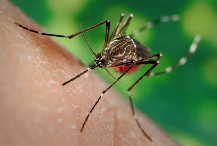
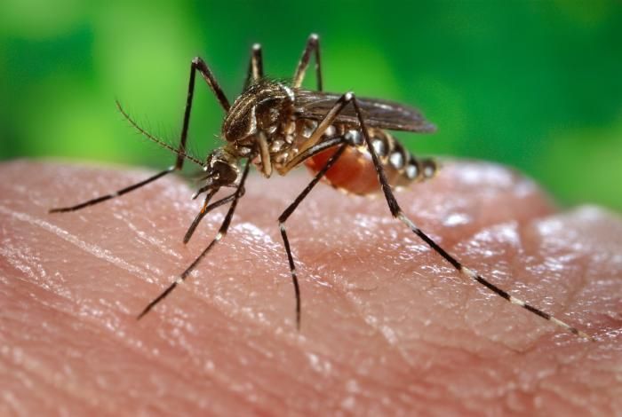
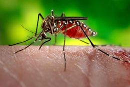
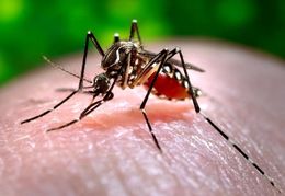
This 2006 photograph depicted a female Aedes aegypti mosquito while she was in the process of acquiring a blood meal from her human host, who in this instance, was actually the biomedical photographer, James Gathany, here at the Centers for Disease Control. The feeding apparatus consisted of a sharp, orange-colored fascicle that was covered in a soft, pliant sheath called the "labellum while not feeding. The labellum was shown here retracted as the sharp "stylets" contained within pierced the host's skin surface, thereby, allowing the insect to obtain its blood meal. The orange color of the fascicle was due to the red color of the blood as it migrated up the thin, sharp translucent tube. Though out of focus in the background, note the droplet of newly ingested blood that had been expelled, and dispersed from the distal abdominal tip merely due to over-engorgement on the hosts blood.Created: 2006
-
This 2006 photograph depicted a female Aedes aegypti mosquito while she was in the process of acquiring a blood meal from her human host, who in this instance, was actually the biomedical photographer, James Gathany, here at the Centers for Disease Control. The feeding apparatus consisted of a sharp, orange-colored fascicle that was covered in a soft, pliant sheath called the "labellum while not feeding. The labellum was shown here retracted as the sharp "stylets" contained within pierced the host's skin surface, thereby, allowing the insect to obtain its blood meal. The orange color of the fascicle was due to the red color of the blood as it migrated up the thin, sharp translucent tube. Though out of focus in the background, note the droplet of newly ingested blood that was being expelled at the distal abdominal tip merely due to over-engorgement on the hosts blood.Created: 2006
-
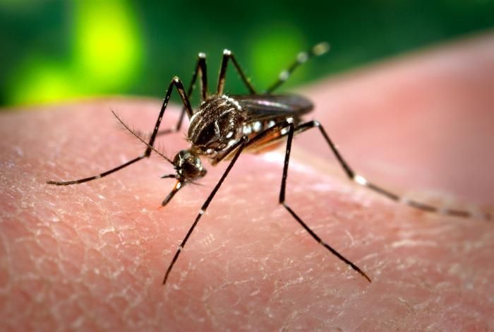
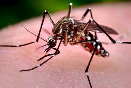
This 2006 photograph depicted a female Aedes aegypti mosquito while she was in the process of acquiring a blood meal from her human host, who in this instance, was actually the biomedical photographer, James Gathany, here at the Centers for Disease Control. The feeding apparatus consisted of a sharp, orange-colored fascicle that was covered in a soft, pliant sheath called the "labellum while not feeding. The labellum was shown here retracted as the sharp "stylets" contained within pierced the host's skin surface, thereby, allowing the insect to obtain its blood meal. The orange color of the fascicle was due to the red color of the blood as it migrated up the thin, sharp translucent tube. When viewed in cross-section, the larger of the two needle-sharp stylets, known as the "labrum", takes on the shape of an inverted "V", and acts as a gutter, which directs the ingested host blood towards the insect's mouth.Created: 2006
-
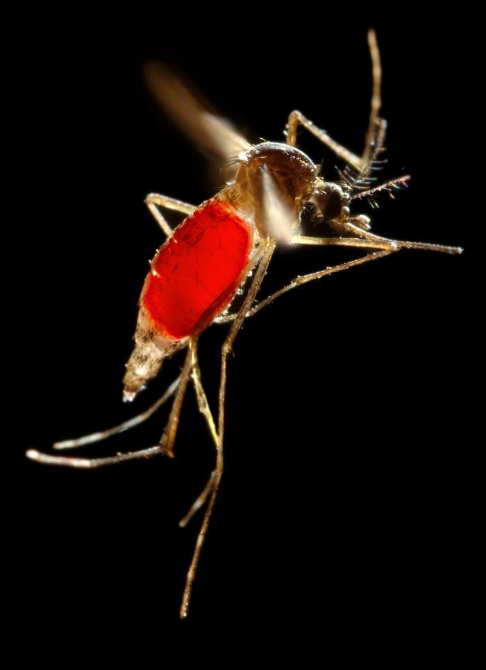
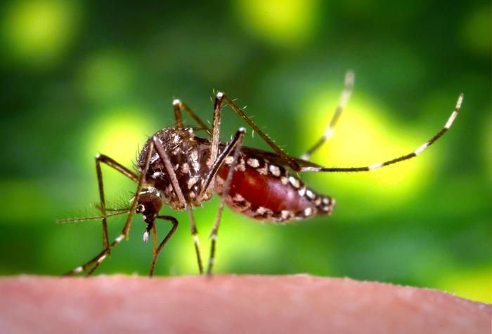
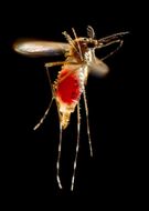
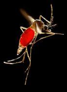
With a newly-obtained fiery red blood meal visible through her now transparent abdomen, the now heavy female Aedes aegypti mosquito takes flight as she leaves her hosts skin surface. In this case, what would normally be an unsuspecting host was actually the CDCs biomedical photographers own hand, which hed offered to the hungry mosquito so that shed alight, and be photographed while feeding. After having filled with blood, the abdomen became distended, stretching the exterior exoskeletal surface, causing it to become transparent, and allowed the collecting blood to become visible as an enlarging intra-abdominal red mass. The wings seem to be working overtime in order to keep her aloft.Created: 2006
-
With a newly-obtained fiery red blood meal visible through her transparent abdomen, the now heavy female Aedes aegypti mosquito took flight as she left her hosts skin surface. In this case, what would normally be an unsuspecting host was actually the CDCs biomedical photographers own hand, which hed offered to the hungry mosquito so that shed alight, and be photographed while feeding. As it filled with blood, the abdomen became distended, stretching the exterior exoskeletal surface, thereby, causing it to become transparent, allowing the collecting blood to become visible as an enlarging intra-abdominal red mass. The wings seem to be working overtime in order to keep her aloft.Created: 2006
-
This 2006 image depicted a female Aedes aegypti mosquito as she was completing the activity of obtaining a blood-meal from a human host through the fascicle of her feeding organ known as the proboscis. Note the droplet of host blood remaining on the tip of her proboscis after shed extracted the feeding organ from the skin surface. In this case, what would normally be an unsuspecting host was actually the CDCs biomedical photographers own hand, which hed offered to the hungry mosquito so that shed alight, and be photographed while feeding. As it filled with blood, the abdomen became distended, stretching the exterior exoskeletal surface, thereby, causing it to become transparent, allowing the collecting blood to become visible as an enlarging intra-abdominal red mass.Created: 2006
-
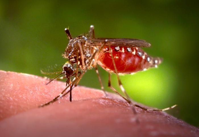
This 2006 image depicted a female Aedes aegypti mosquito as she was obtaining a blood-meal from a human host through her fascicle, which after penetrating the host's skin, had reddened in color, reflecting the bloods coloration through this tubular structure. In this case, what would normally be an unsuspecting host was actually the CDCs biomedical photographers own hand, which hed offered to the hungry mosquito so that shed alight, and be photographed while feeding. As it filled with blood, the abdomen became distended, stretching the exterior exoskeletal surface, thereby, causing it to become transparent, allowing the collecting blood to become visible as an enlarging intra-abdominal red mass, as is the case in PHIL# 9175, and 9176.Created: 2006
-
This 2006 image depicted a female Aedes aegypti mosquito as she was obtaining a blood-meal from a human host through her fascicle, which after penetrating the host's skin, has reddened in color, reflecting the bloods coloration through this tubular structure. In this case, what would normally be an unsuspecting host was actually the CDCs biomedical photographers own hand, which hed offered to the hungry mosquito so that shed alight, and be photographed while feeding. As it filled with blood, the abdomen became distended, stretching the exterior exoskeletal surface, thereby, causing it to become transparent, allowing the collecting blood to become visible as an enlarging intra-abdominal red mass.Created: 2006
-
This 2006 image depicted a female Aedes aegypti mosquito as she was obtaining a blood-meal from a human host through her fascicle, which had penetrated the host skin, was reddening in color, reflecting the bloods coloration through this tubular structure. In this case, what would normally be an unsuspecting host was actually the CDCs biomedical photographers own hand, which hed offered to the hungry mosquito so that shed alight, and be photographed while feeding. As it filled with blood, the abdomen became distended, stretching the exterior exoskeletal surface, thereby, causing it to become transparent, allowing the collecting blood to become visible as an enlarging intra-abdominal red mass.Created: 2006
-
This 2006 image depicted a female Aedes aegypti mosquito as she was obtaining a blood-meal from a human host through her fascicle, which being transparent, reflected the bloods red color. In this case, what would normally be an unsuspecting host was actually the CDCs biomedical photographers own hand, which hed offered to the hungry mosquito so that shed alight, and be photographed while feeding. As it filled with blood, the abdomen became distended, thereby, stretching the exterior exoskeletal surface, causing it to become transparent, and allowing the collecting blood to become visible as an enlarging intra-abdominal red mass, as is the case in PHIL# 9175, and 9176.Created: 2006
-
This 2006 image depicted a female Aedes aegypti mosquito as she was obtaining a blood-meal from a human host through her fascicle, which being transparent, reflected the bloods red color. In this case, what would normally be an unsuspecting host was actually the CDCs biomedical photographers own hand, which hed offered to the hungry mosquito so that shed alight, and be photographed while feeding. As it filled with blood, the abdomen became distended, stretching the exterior exoskeletal surface, thereby, causing it to become transparent, allowing the collecting blood to become visible as an enlarging intra-abdominal red mass.Created: 2006
-
This 2006 image depicted a female Aedes aegypti mosquito as she was completing the activity of obtaining a blood-meal from a human host through her fascicle, here red in color due to the blood contained therein, which shed begun to resheath in her labium. Both structures are part of her feeding organ known as the proboscis. In this case, what would normally be an unsuspecting host was actually the CDCs biomedical photographers own hand, which hed offered to the hungry mosquito so that shed alight, and be photographed while feeding. As it filled with blood, the abdomen became distended, stretching the exterior exoskeletal surface, thereby, causing it to become transparent, allowing the collecting blood to become visible as an enlarging intra-abdominal red mass, as is the case in PHIL# 9175, and 9176.Created: 2006