All Biocode files are based on field identifications to the best of the researcher’s ability at the time.
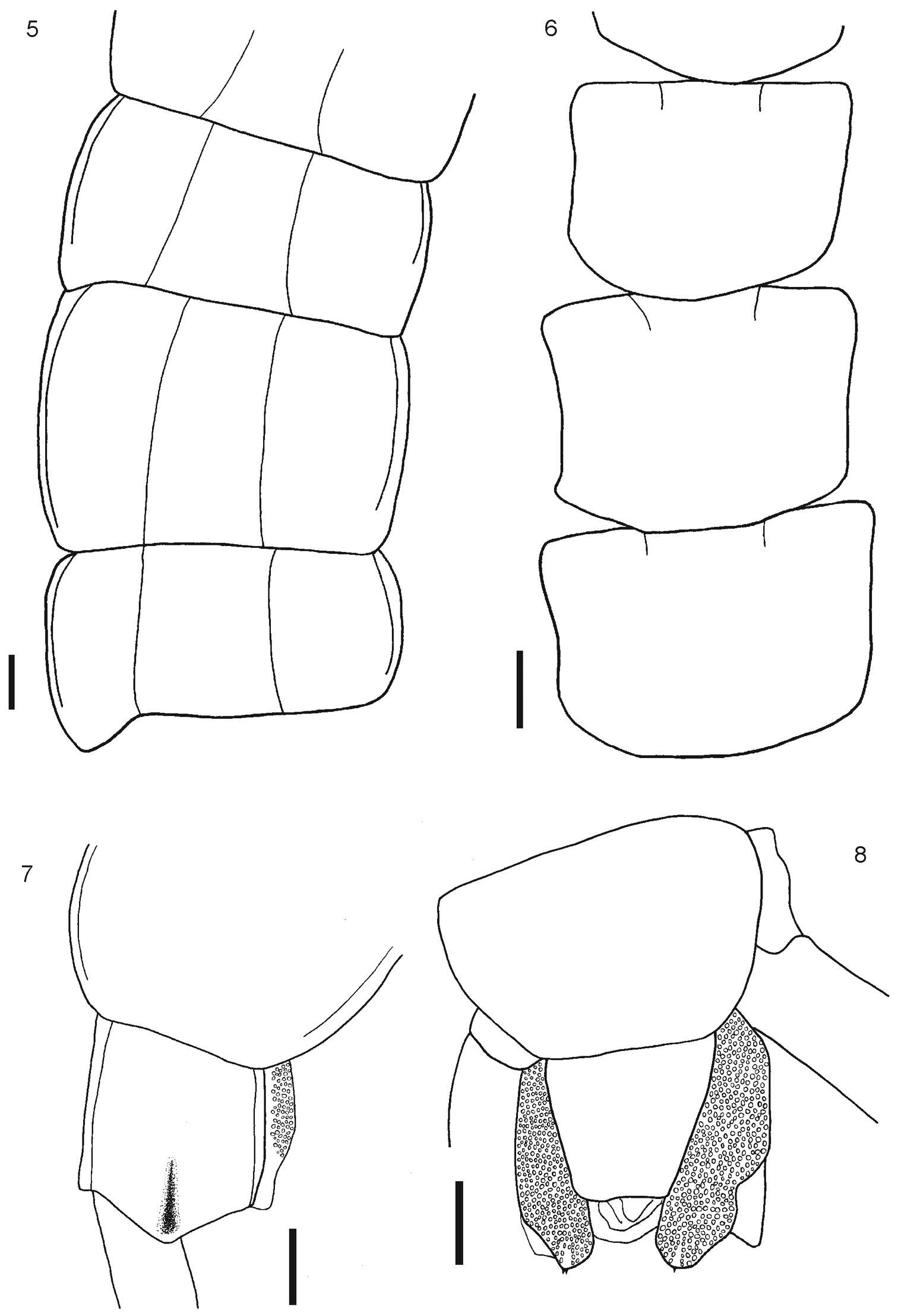
Figure 1–4.1 Rhysida celeris from Ecuador. Cephalic plate 2 Forcipular Coxosternum 3 Tooth plates 4 Forcipular trochanteroprefemur process. Scale bars 1 mm.
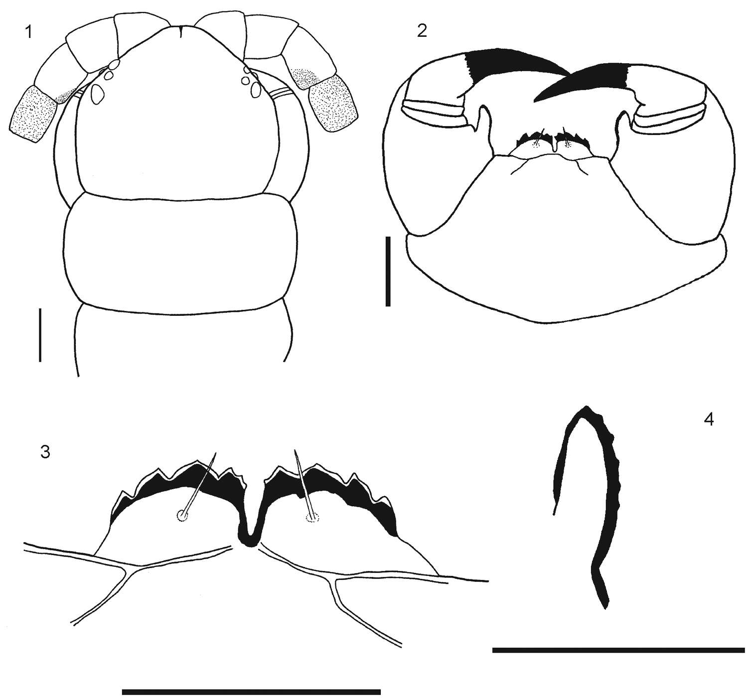
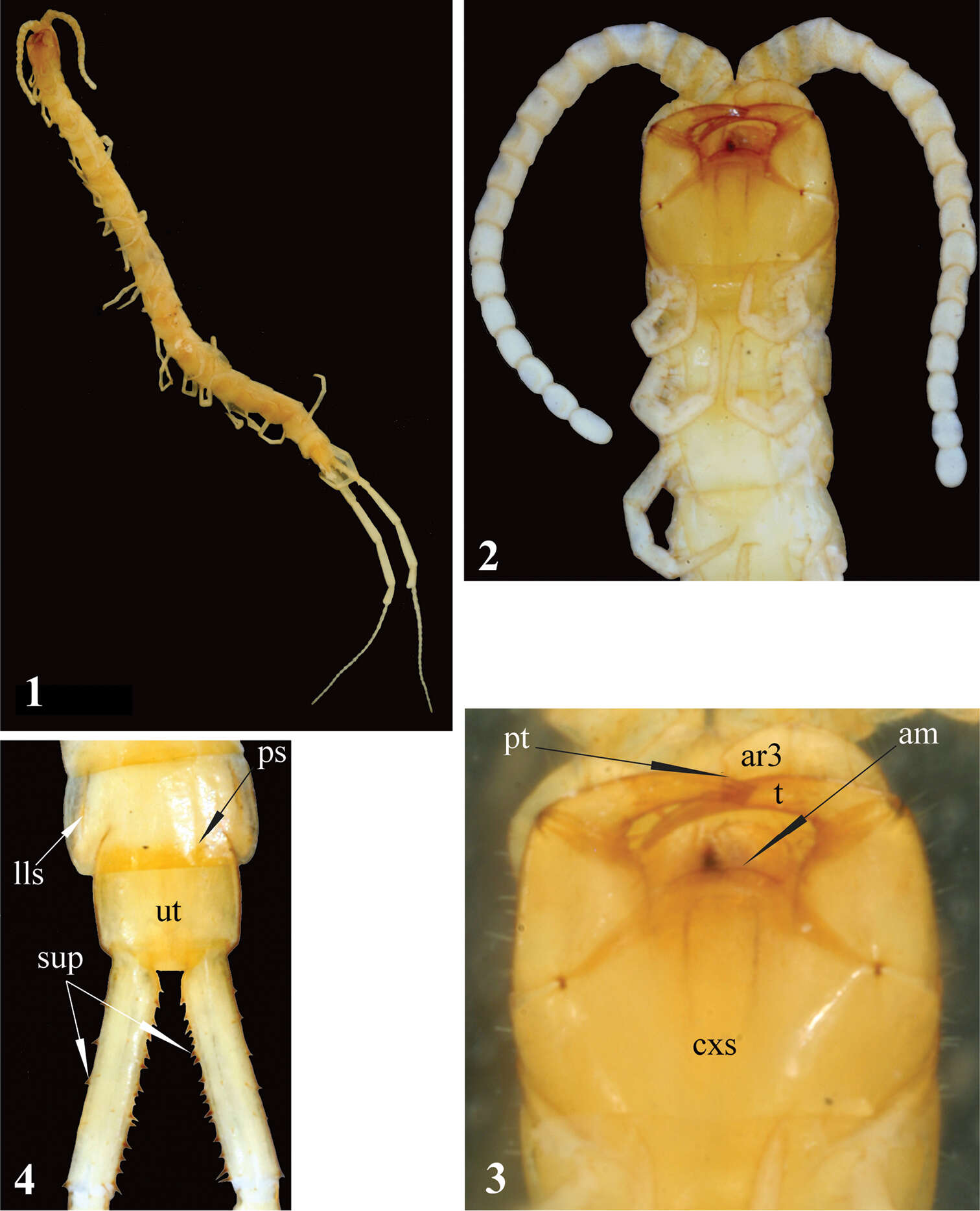
Figures 1–4.Newportia stoevi,sp. n. 1 Habitus 2 Head and anterior segments, ventral view 3 Forcipular segment, ventral view 4 Tergites 22 and 23 and prefemora of ultimate legs, dorsal view; (pt) – pretarsus of second maxilla, (ar3) – article 3 of telopodite of second maxilla, (cxs) – forcipular coxosternite, (am) – anterior margin of coxosternite, (t) – tarsungulum, (ps) – paramedian sutures, (lls) – lateral longitudinal sutures, (ut) – tergite of ultimate leg-bearing segment, (sup) – spurs of ultimate prefemur.
All Biocode files are based on field identifications to the best of the researcher’s ability at the time.
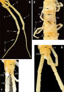
Figure 5–8. 5 Tergites 11, 12 and 13 6 Sternites 4, 5 and 6 7 Tergite 21 8 Segment 21 showing sternite 21 and coxopleuron. Scale bars 1 mm.
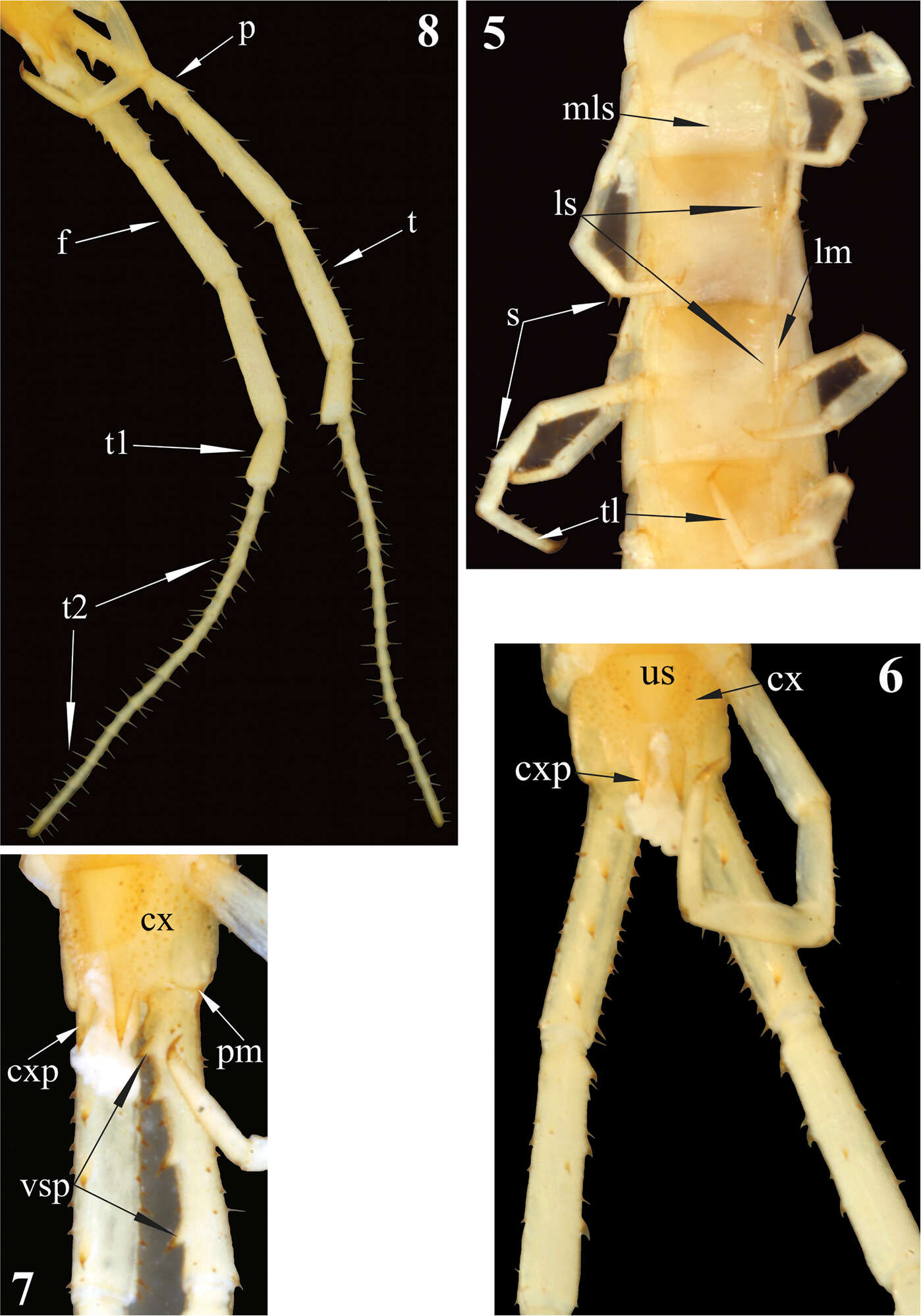
Figures 5–8.Newportia stoevi,sp. n. 5 Segments and midbody legs, ventral view 6 Posterior body end, ventral view 7 Left side of ultimate leg-bearing segment and prefemora of ultimate legs, ventro-lateral view 8 Ultimate legs, ventro-lateral view; (mls) – median longitudinal sulcus, (ls) – lateral sutures, (lm) – lateral margination, (s) – setae, (tl) – monoarticulated tarsus of locomotory leg, (us) – sternite of ultimate leg-bearing segment, (cx) – coxopleuron, (cxp) – coxopleural process, (pm) – posterior margin of pleuron of ultimate leg-bearing segment, (vsp) – ventral spinous processes of ultimate prefemur, (p) – prefemur, (f) – femur, (t) – tibia, (t1) – tarsus 1, (t2) – tarsus 2.
All Biocode files are based on field identifications to the best of the researcher’s ability at the time.
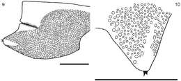
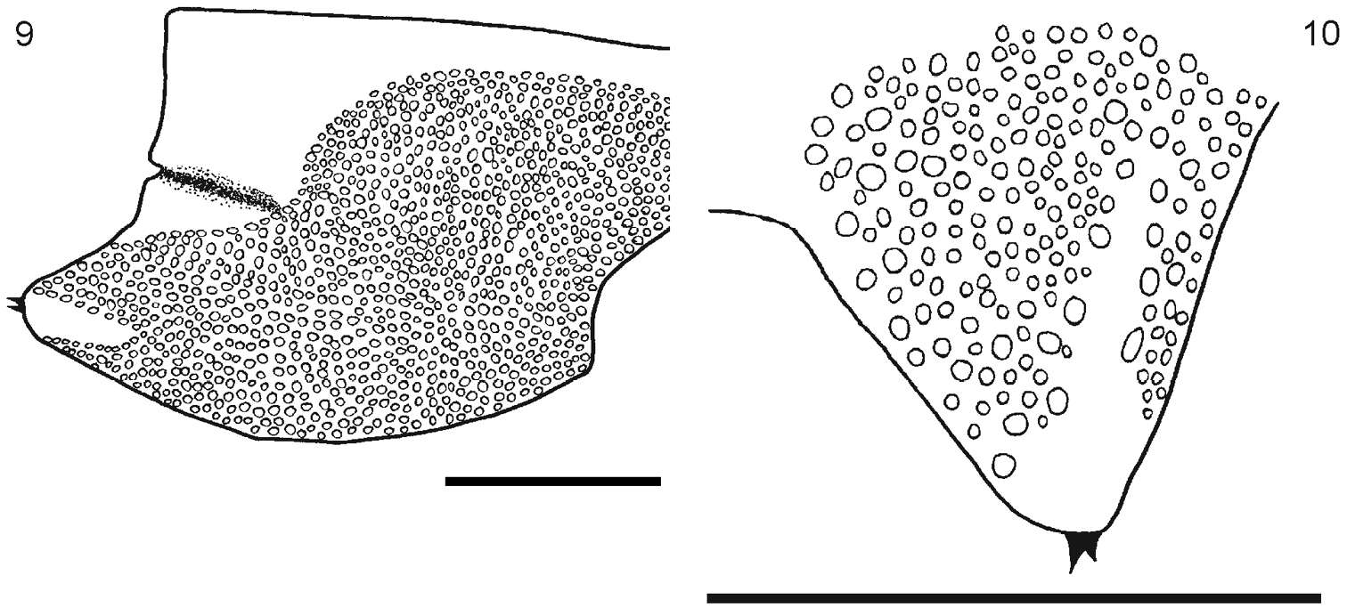
Figure 9–10. 9 Segment 21 showing the coxopleuron 10 Detail of the terminal part of the coxopleuron showing the spines. Scale bar 1 mm.
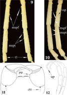
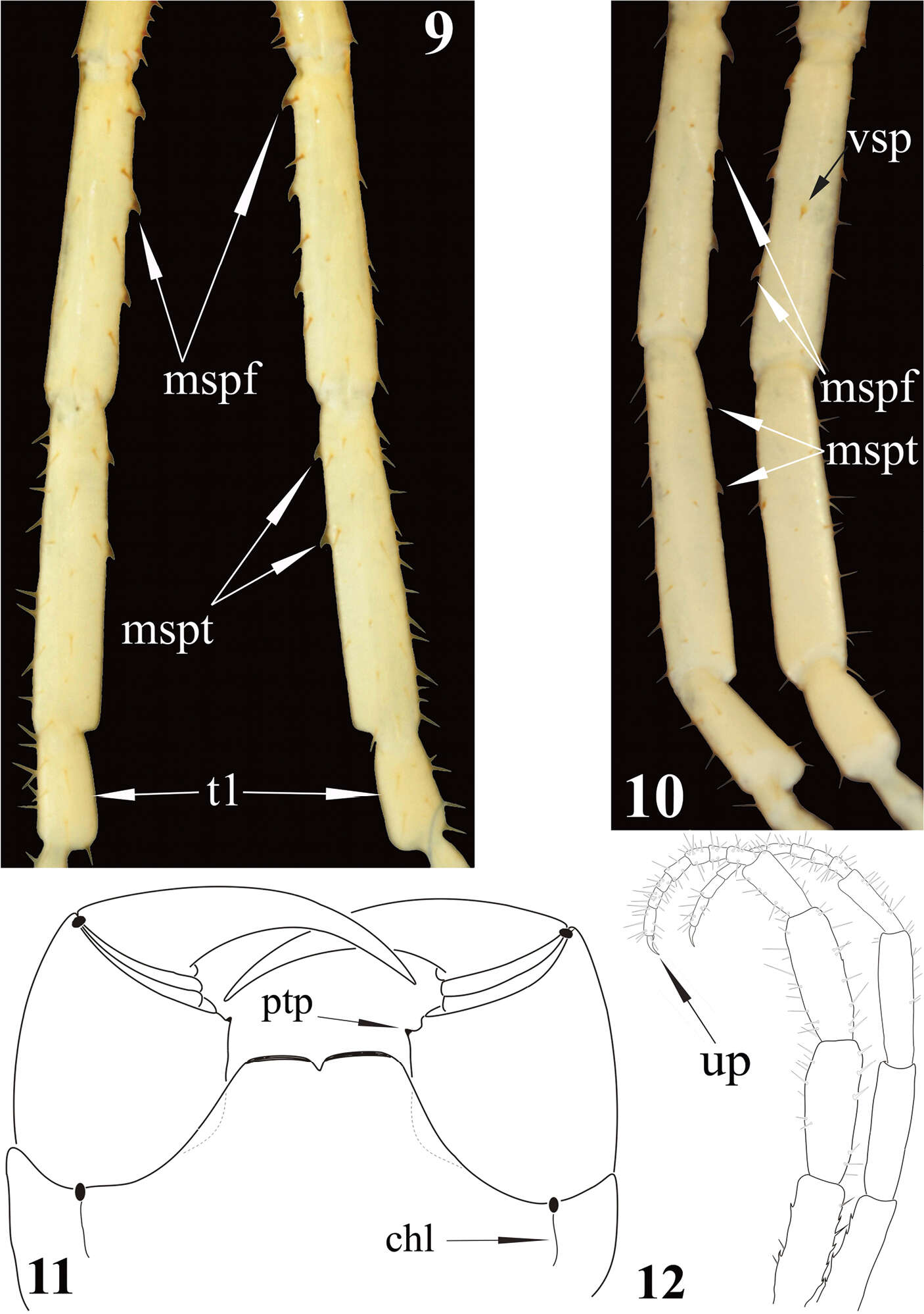
Figures 9–12.Newportia stoevi,sp. n. 9 Femora, tibiae and tarsi 1 of ultimate legs, dorsal view 10 Femora, tibiae and tarsi 1 of ultimate legs, ventral view; Newportia divergens Chamberlin, 1922 11 Forcipular segment, ventral view (after Schileyko and Minelli 1998); Newportia unguifer Chamberlin, 1921 12 Ultimate legs, dorso-lateral view (after Schileyko and Minelli 1998); (mspf) – medial spinous processes of ultimate femur, (mspt) – medial spinous processes of ultimate tibia, (vsp) – ventral spinous process of ultimate femur, (t1) – tarsus 1, (up) – ultimate pretarsus, (chl) – chitin-lines, (ptp) – process of trochanteroprefemur.
All Biocode files are based on field identifications to the best of the researcher’s ability at the time.
All Biocode files are based on field identifications to the best of the researcher’s ability at the time.
All Biocode files are based on field identifications to the best of the researcher’s ability at the time.
All Biocode files are based on field identifications to the best of the researcher’s ability at the time.
All Biocode files are based on field identifications to the best of the researcher’s ability at the time.
All Biocode files are based on field identifications to the best of the researcher’s ability at the time.
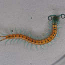
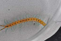
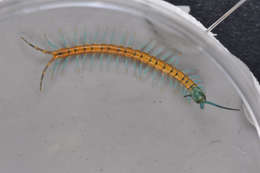
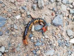
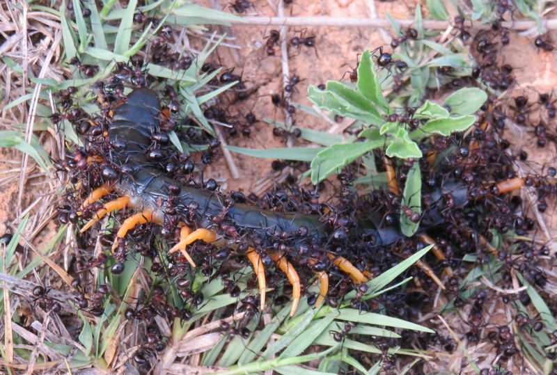
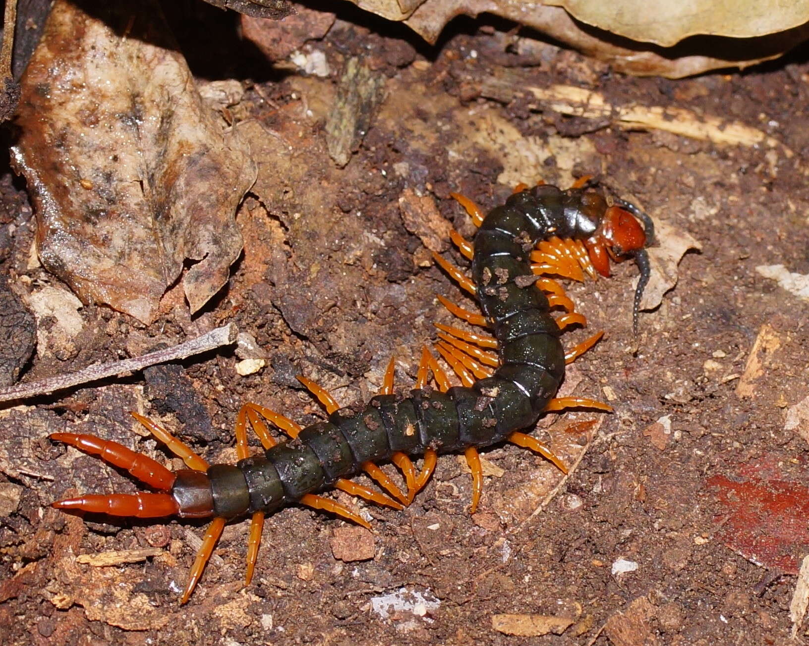
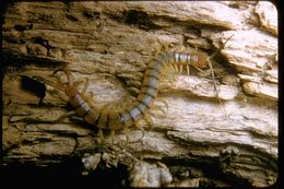
Being attacked by ants. Tentative i.d.
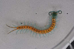
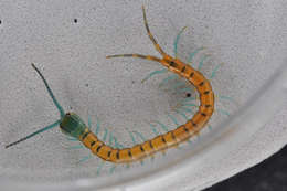
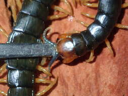
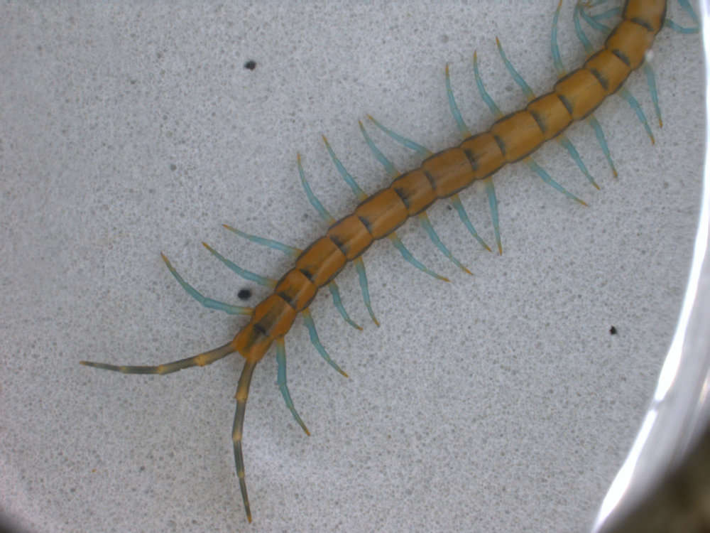
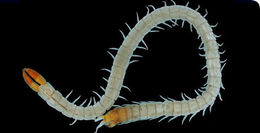
Tentative i.d.
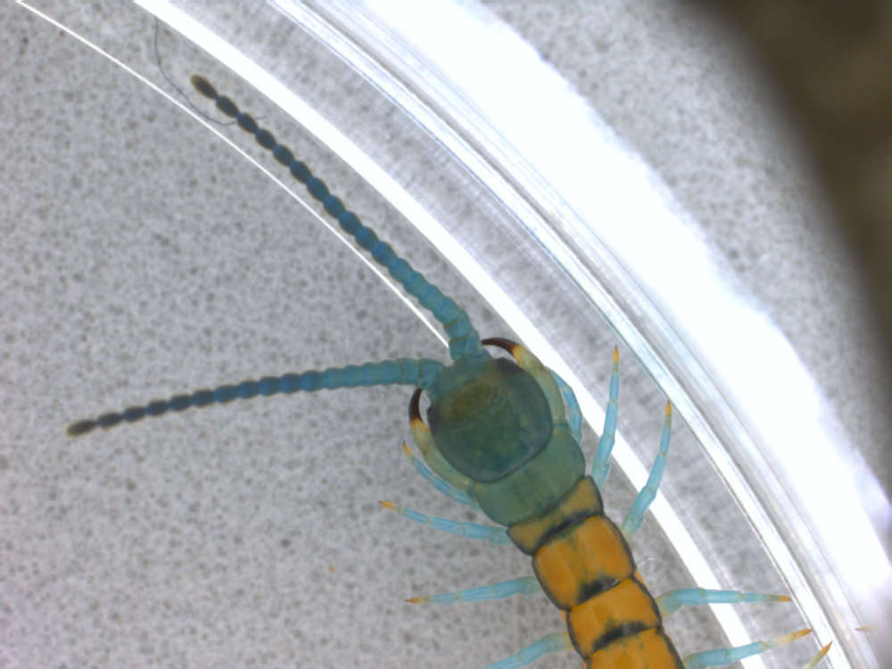
2005 California Academy of Sciences
CalPhotos
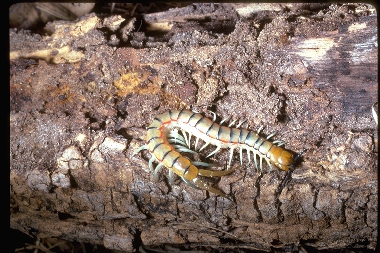
2007 California Academy of Sciences
CalPhotos
Habitus of Scolopendropsis duplicata . An individual with 43 trunk segments (a paratype deposited in the Instituto Butantan, Sao Paolo). Scale bar 5mm. Photo by Amazonas Chagas, Jr.
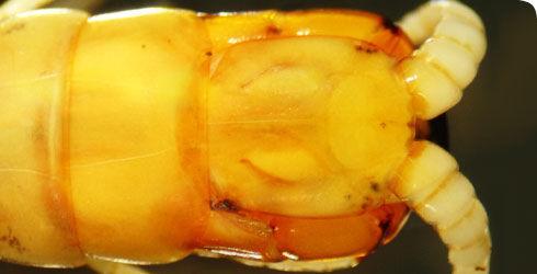
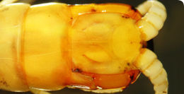
The head and anterior trunk segments of Scolopendropsis duplicata in dorsal view, showing the relatively small size of the cephalic plate, distally tapering antennae, and the presence of a longitudinal median suture along the posterior half of the cephalic plate. Photo by Amazonas Chagas, Jr.