-
-
-
-
-
-
-
-
-
-
-
-
-
-
-
-
-
-
Ribadelago, Castille and Leon, Spain
-
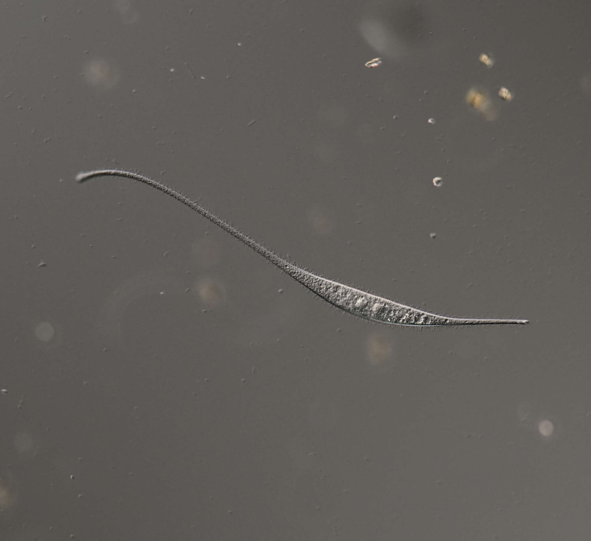
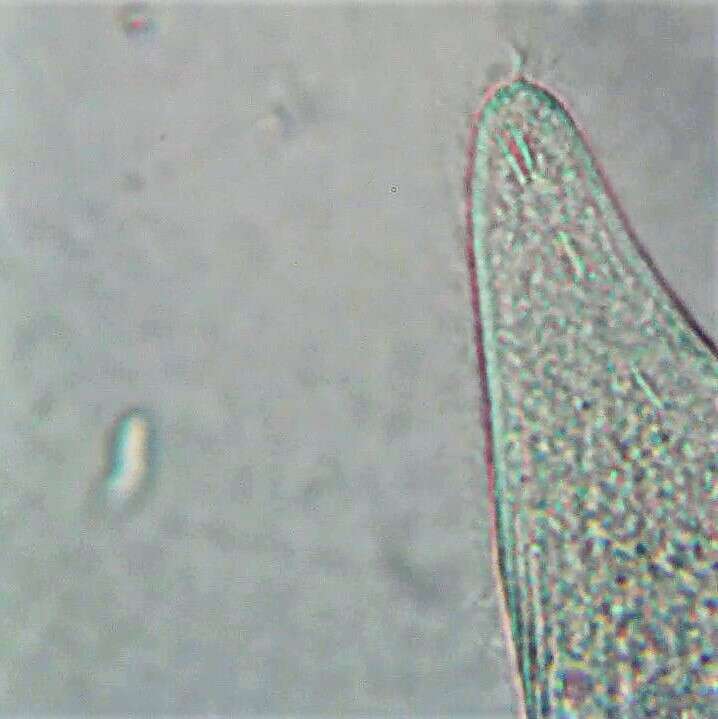
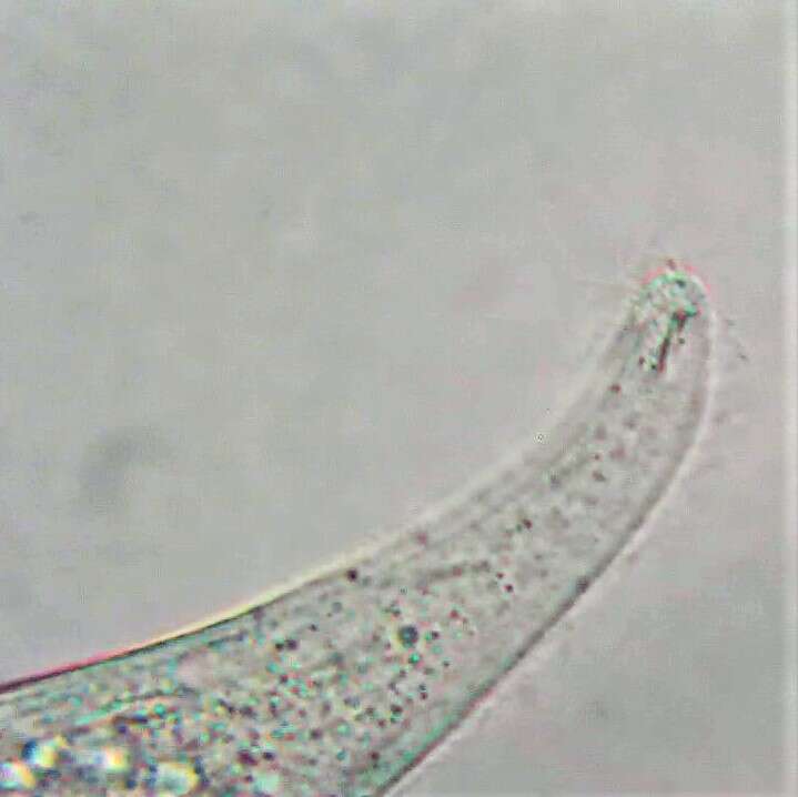
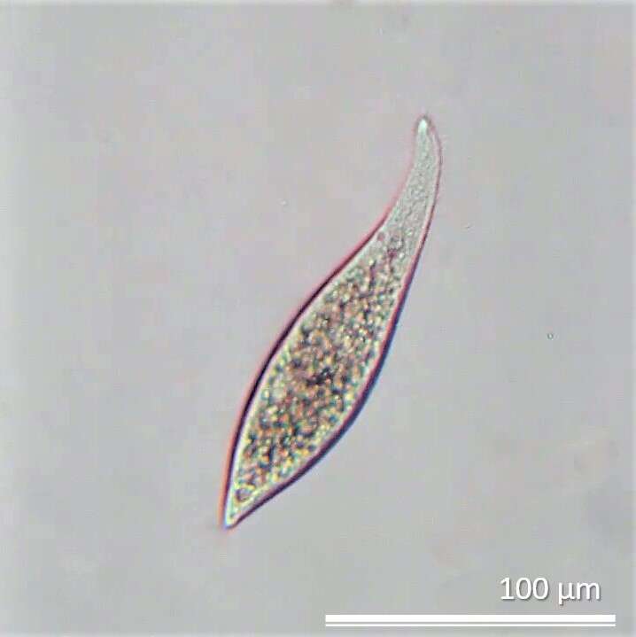
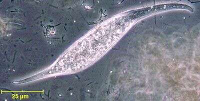
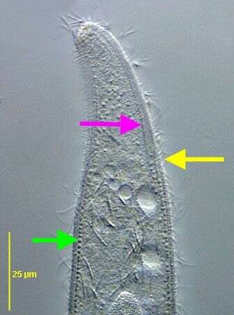
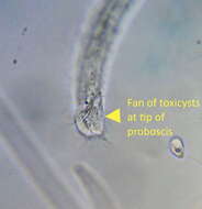
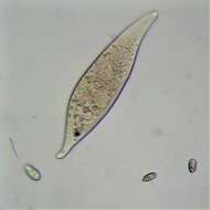
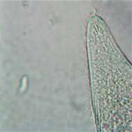
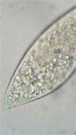
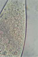
Portrait of Amphileptus, a widely distributed haptorid ciliate with posterior drawn out to a tapered point and elongate anterior neck region. Prominent refractile trichites are clustered at the anterior end of the neck region. Right side longitudinal kineties converge on one another anteriorly and posteriorly in a diamond shaped pattern unlike the parallel kineties of the similar genus Litonotus. The mouth is slit-like and difficult to see, located on the convex aspect of the neck region. Two spherical macronuclei. Multiple contractile vacuoles along dorsal and ventral margins. Some species attach to and prey on stalked peritrich ciliates. From freshwater pond near Boise, Idaho. Phase contrast.
-
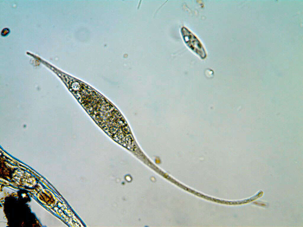
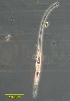
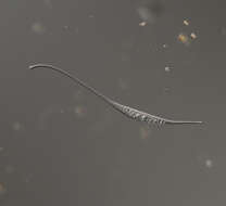
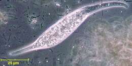
Portrait of Amphileptus filum (Gruber,1884) Gruber, 1888, a markedly elongated haptorid ciliate. Distinguished from the very similar Litonotus Cygnus by the cluster of brightly refractile trichites at the anterior tip of the neck region and the anteriorly and posteriorly converging kineties. From freshwater pond near Boise, Idaho. Phase contrast.
-
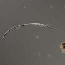
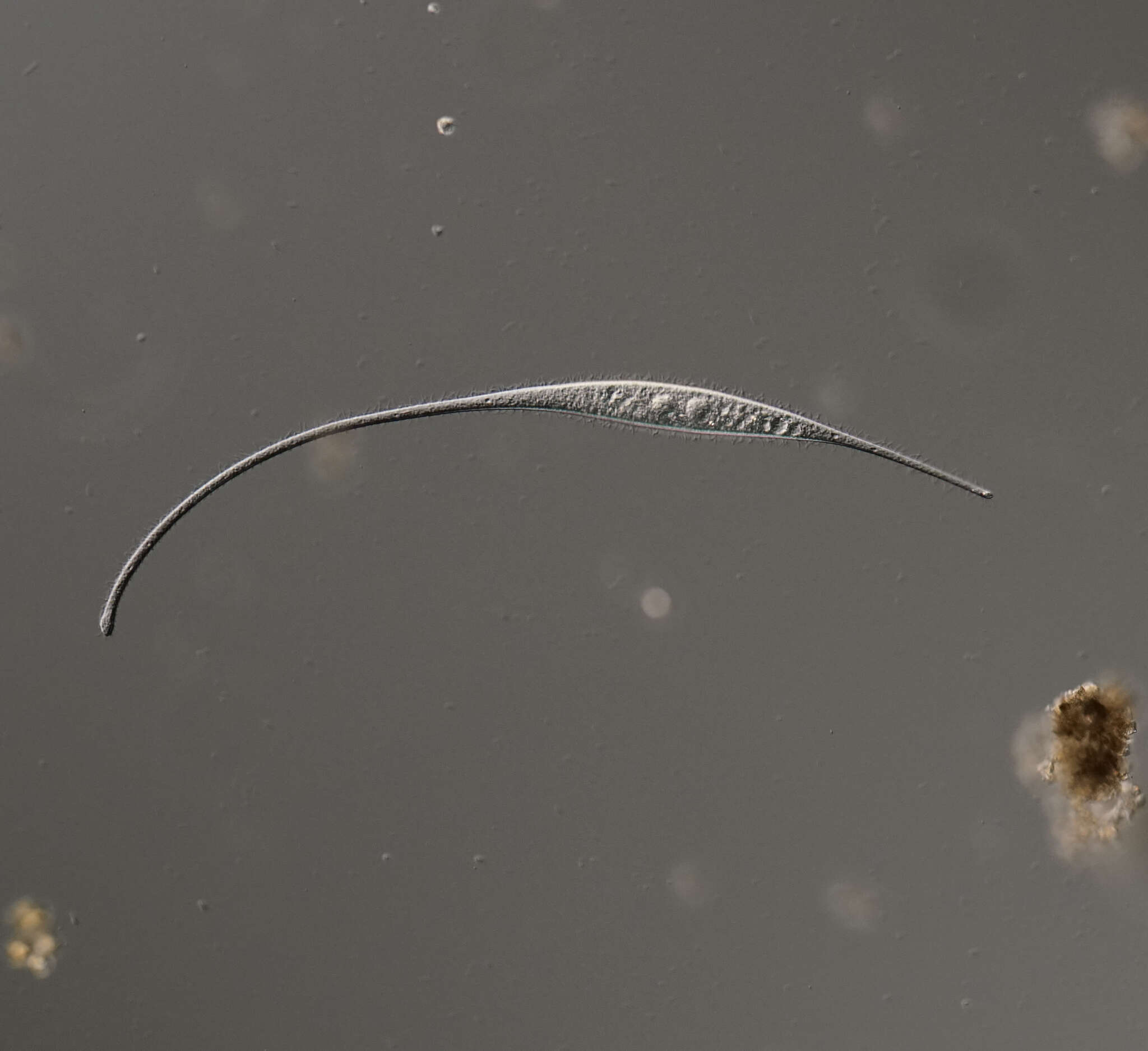
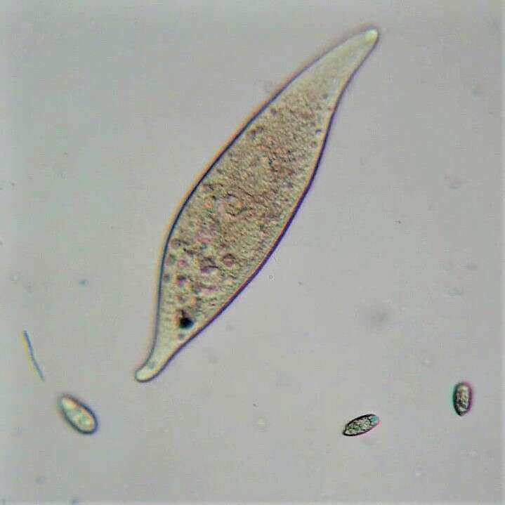
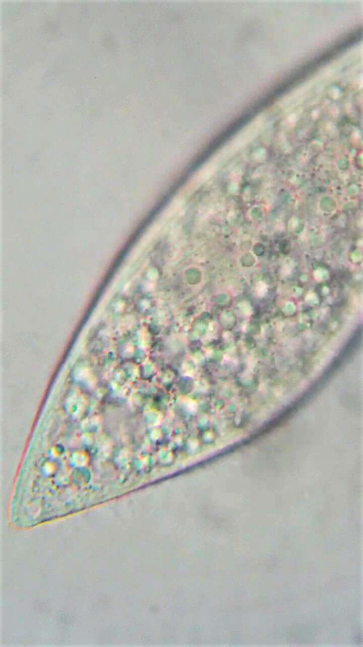
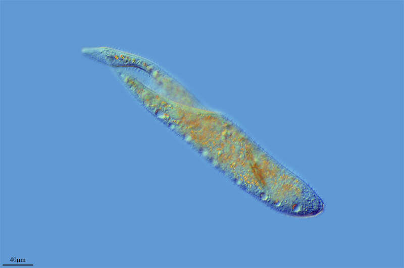
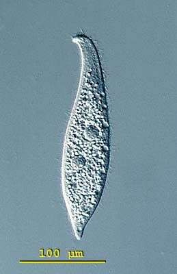
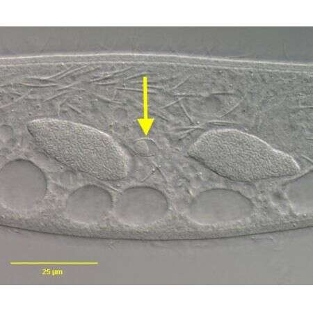
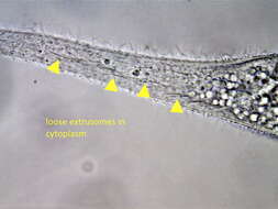
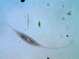
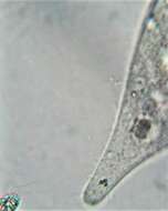
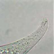
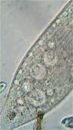
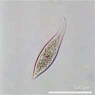
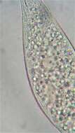
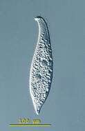
Amphileptus (am-fee-lep-tus) pleurosigma. The body of the members of the genus Amphileptus is laterally compressed and elongate. The oral aperture is a slit on the convex edge of the neck region, and extends less than halfway down the body. Ciliation is present on both lateral surfaces although there is a tendency to some reduction on the left surface. Ciliation on the right surface is extensive and forms longitudinal rows which converge on each other in the anterior region. Trichocysts are common - particularly in neck. Macronucleus in 2 to 4 spherical parts with single micronucleus placed between macronuclei. Many contractile vacuoles occur along both dorsal and ventral edges. Lives in fresh water ponds and lakes. Free swimming specimen of Amphileptus pleurosigma. The two macronuclei and the contractile vacuoles located at the edges are visible. The S-shaped body and the neck-like anterior end are characteristic. Measuring 228 microns. Differential interference contrast.
-
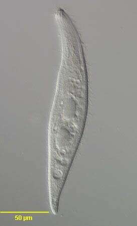
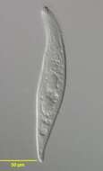
in vivo portrait of the pleurostomatid ciliate, Amphileptus pleurosigma (Stokes,1884) Foissner, 1984. DIC.
-
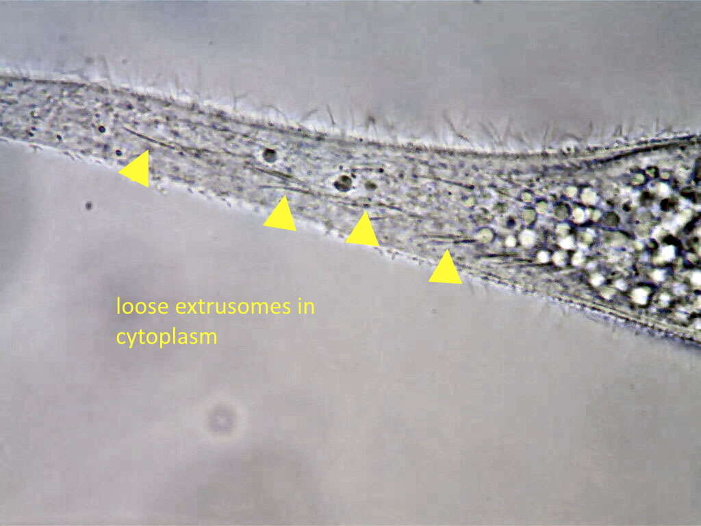
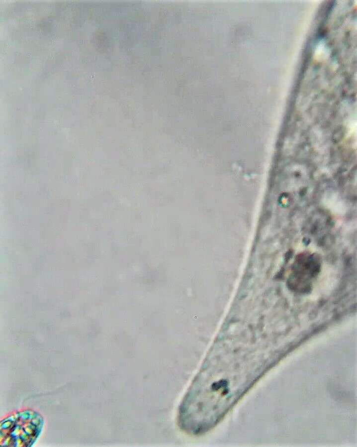
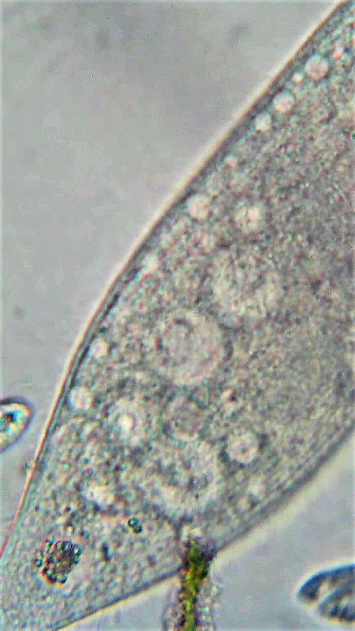
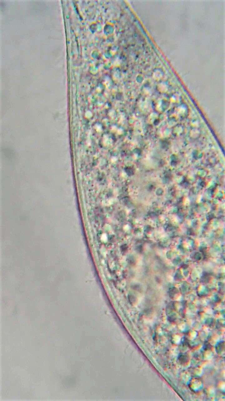
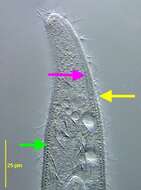
The green arrow indicates a uniform subpellicular layer of globular structures,probably mitochondria.The yellow arrow indicates the posterior end of the slit-like oral aperture.The pink arrow indicates the right perioral kinety.
-
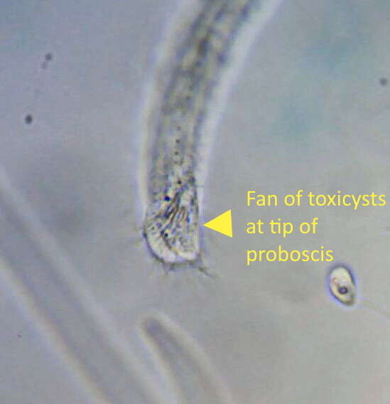
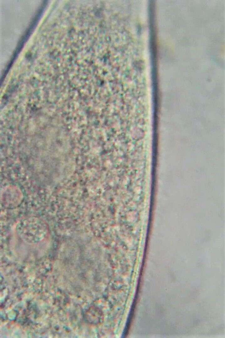
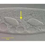
The yellow arrow indicates the micronucleus in a membranous envelope between the two granular macroniclei. DIC.