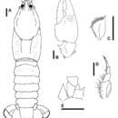
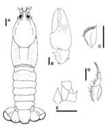
Figure 2.Euastacus morgani sp. n. Dorsal view of body A and cheliped B, holotype specimen, AM P.84263. Dorsal view of antennal squame C, lateral view of third maxilliped D and ventral view of left hand side of cephalon showing interantennal scale E, specimen ACP 1103. Scale bars = 5 mm.