-

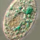
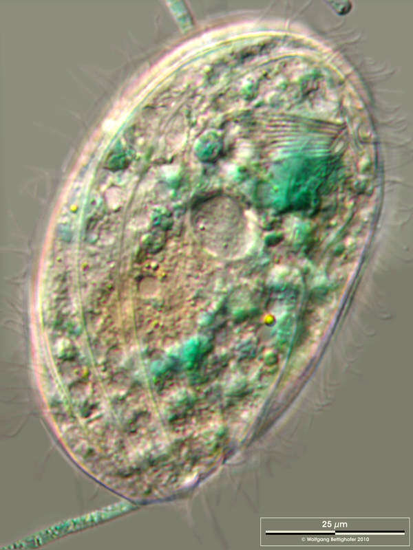
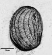
Portrait (ventrolateral view) of the microthoracid ciliate Pseudomicrothorax dubius (Maupas, 1883) Penard, 1922.The cell outline is oval with right edge more curved than left edge. Laterally compressed. The right and left sides of the inflexible pellicle have curved longitudinal ciliated grooves separated by broader flat ridges. Somatic ciliature restricted to 13-14 longitudinal kineties more on the right than the left surface. The oral aperture is in anterior 1/3 of body in a shallow depression. The cytopharynx is supported by fine transverse trichites. There are 3 short membranelles on the left of the cytostome and a short undulating membrane on its right. There is a small group of unciliated basal bodies posterior to the undulating membrane. This is a primordial stomatogenic field. There are many fusiform peripheral extrusomes. Macronucleus elongate ovoid, centrally placed with single micronucleus. The contractile vacuole is centrally located. P. dubius feeds on cyanobacteria giving it a green to green-blue color. Collected from a freshwater stream near Boise, Idaho. DIC.
-

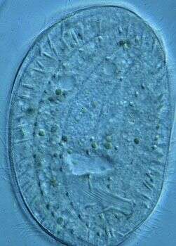
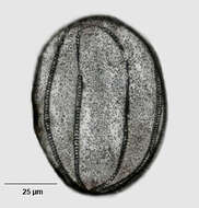
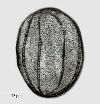
Infraciliature (ventrolateral view) of the microthoracid ciliate Pseudomicrothorax dubius (Maupas, 1883) Penard, 1922.The cell outline is oval with right edge more curved than left edge. Laterally compressed. The right and left sides of the inflexible pellicle have curved longitudinal ciliated grooves separated by broader flat ridges. Somatic ciliature restricted to 13-14 longitudinal kineties more on the right than the left surface. The oral aperture is in anterior 1/3 of body in a shallow depression. The cytopharynx is supported by fine transverse trichites. There are 3 short membranelles on the left of the cytostome and a short undulating membrane on its right. There is a small group of unciliated basal bodies posterior to the undulating membrane. This is a primordial stomatogenic field. There are many fusiform peripheral extrusomes. Macronucleus elongate ovoid, centrally placed with single micronucleus. The contractile vacuole is centrally located. P. dubius feeds on cyanobacteria giving it a green to green-blue color.Stained by the dry silver nitrate technic (see Foissner, W.Europ. J. Protistol.27,313-330;1991).Collected from a freshwater stream near Boise, Idaho.Brightfield.
-
Dorsal infraciliature of the microthoracid ciliate Pseudomicrothorax dubius (Maupas, 1883) Penard, 1922.The cell outline is oval with right edge more curved than left edge. Laterally compressed. The right and left sides of the inflexible pellicle have curved longitudinal ciliated grooves separated by broader flat ridges. Somatic ciliature restricted to 13-14 longitudinal kineties more on the right than the left surface. The oral aperture is in anterior 1/3 of body in a shallow depression. The cytopharynx is supported by fine transverse trichites. There are 3 short membranelles on the left of the cytostome and a short undulating membrane on its right. There is a small group of unciliated basal bodies posterior to the undulating membrane. This is a primordial stomatogenic field. There are many fusiform peripheral extrusomes. Macronucleus elongate ovoid, centrally placed with single micronucleus. The contractile vacuole is centrally located. P. dubius feeds on cyanobacteria giving it a green to green-blue color.Stained by the dry silver nitrate technic(see Foissner, W.Europ. J. Protistol.27,313-330;1991) Collected from a freshwater stream near Boise, Idaho. Brightfield.
-
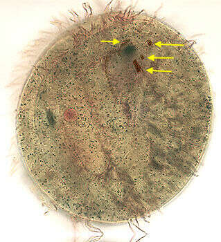
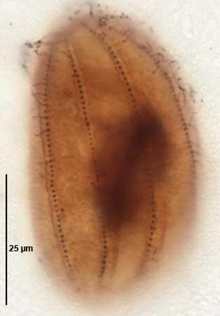
Ventral infraciliature of the nassulid ciliate Pseudomicrothorax (Mermod,1914) Penard,1922.The three arrows to the viewer's right indicate the 3 nassulid adoral membranelles. The small undulating membrane is indicated by the arrow to the viewer's left. The cytopharynx is the dark spot between the arrows.Stained by the silver carbonate technique (see Foissner, W. Europ. J. Protistol., 27:313-330;1991).Brightfield.
-
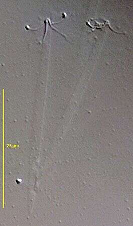
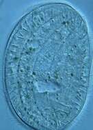
Anterior is to the bottom of the image. The mouth, with supporting rods, can be seen to the lower right of the image. Stuff extrusomes are located near the cell surface.
-
-
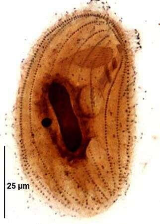
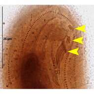
Ventral infraciliature of the nassulid ciliate Pseudomicrothorax dubius (Mermod,1914) Penard,1922.The arrowheads indicate the 3 nassulid adoral membranelles.Protargol A (see Foissner, W. Europ. J. Protistol., 27:313-330;1991).Brightfield.
-
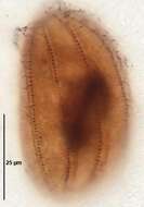
Dorsal kineties of Pseudomicrothorax dubius (Mermod,1914) Penard,1922.Protargol A (see Foissner, W. Europ. J. Protistol., 27:313-330;1991).Brightfield.
-
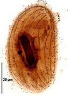
Ventral infraciliature of Pseudomicrothorax dubius (Mermod,1914) Penard,1922.Protargol A (see Foissner, W. Europ. J. Protistol., 27:313-330;1991).Brightfield.
-
Scale bar indicates 25 µm.Specimen from the protists culture of FU Berlin.Microscope Zeiss Universal, camera Olympus C7070WZ.© Wolfgang Bettighofer,images under Creative Commons License V 3.0 (CC BY-NC-SA).For permission to use of (high resolution) images please contact
postmaster@protisten.de.For further information about the image, please click here:
Link to protisten.de page