-
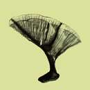
Whole leaf scale, transmission electron micrograph.
-
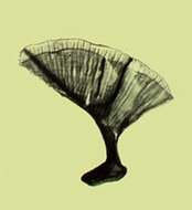
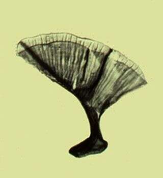
Spine scale, whole mount, transmission electron micrograph.
-
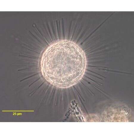
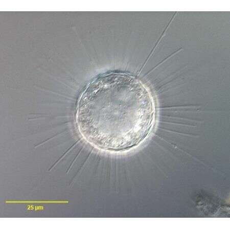
Acanthocystis penardi (WAILES,1925).
-
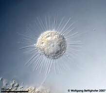
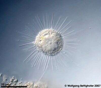
Acanthocystis penardi (WAILES,1925).DIC.
-
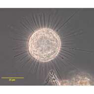
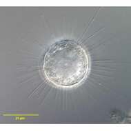
Acanthocystis penardi shows numerous siliceous spines and scales inserting and lying respectively upon the surface of the cell forming the periplast. The central body from which all the axopodia originate is clearly visible. Scale bar indicates 25 µm. Sample from sphagnum pond Dosenmoor near Neumuenster (Schleswig-Holstein, Germany). Images were taken using Zeiss Universal with Olympus C7070 CCD camera.
-
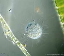
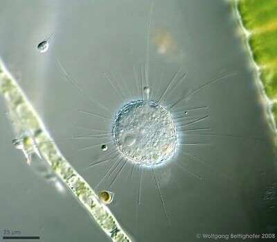
Acanthocystis penardi shows numerous siliceous spines and scales inserting and lying respectively upon the surface of the cell forming the periplast. The nucleus and several narrow axopodia are clearly visible, also a free swimming Salpingoeca species and a Chrysophyta called Chrysopyxis inaequalis fixed on a Tribonema filament with a mucilaginous string. Scale bar indicates 25 µm. Sample from sphagnum pond Dosenmoor near Neumuenster (Schleswig-Holstein, Germany). Images were taken using Zeiss Universal with Olympus C7070 CCD camera.Image under Creative Commons License V 3.0 (CC BY-NC-SA).