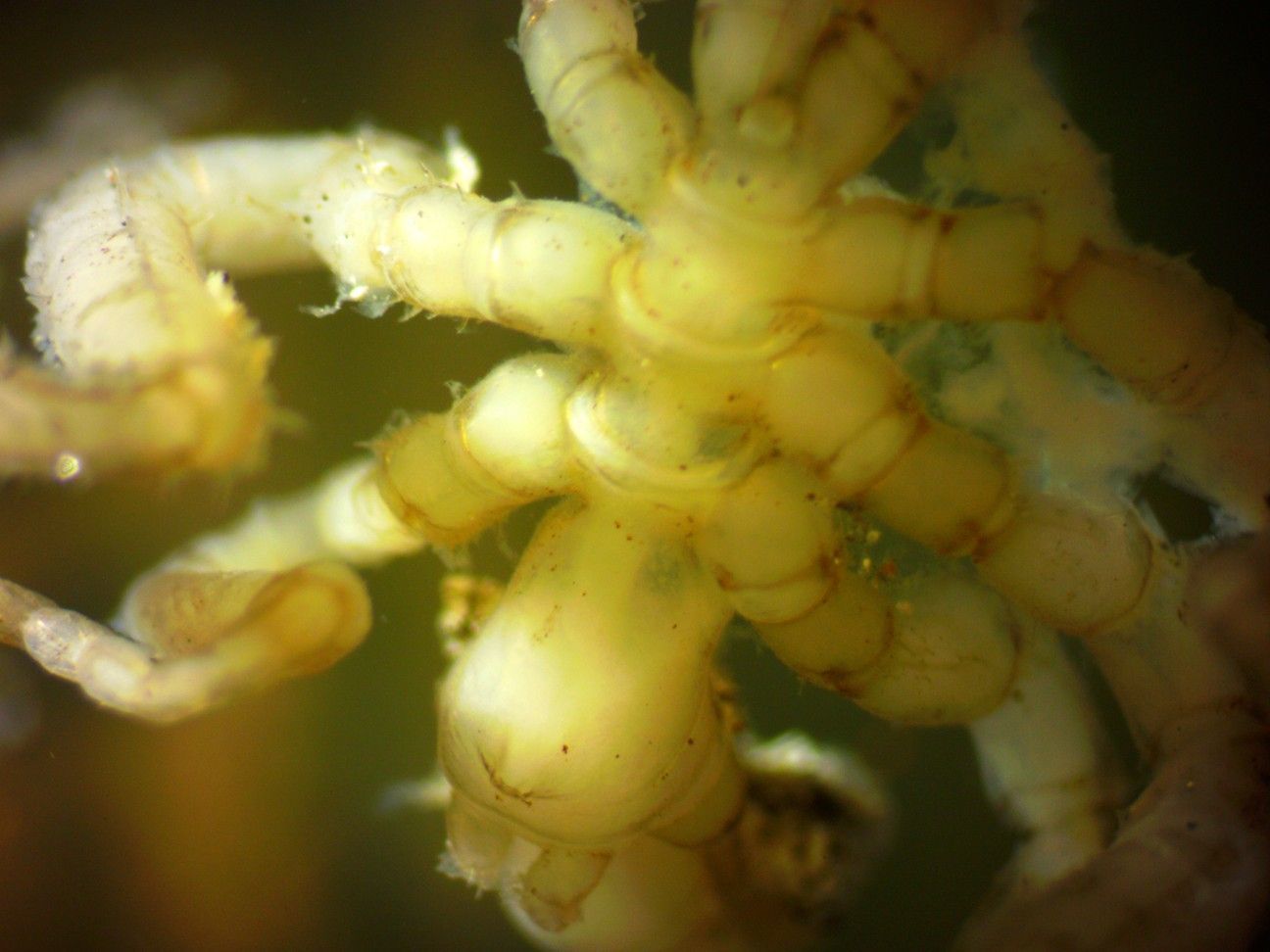
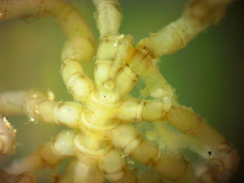
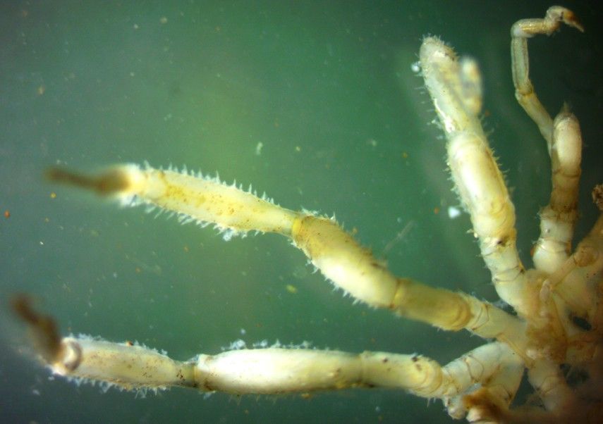
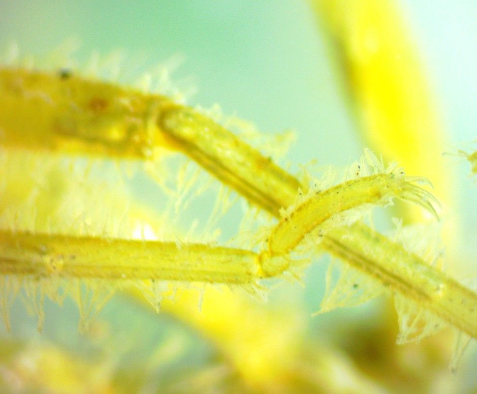
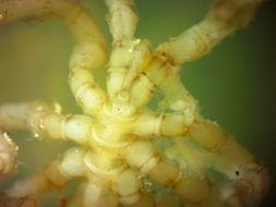
This dorsal view of the trunk and base of the head (cephalon) of this female shows several features. The animal is facing the bottom of the photo. Notice how the posterior ends of the trunk segments flare out. Other members of this genus also have dorsal tubercles arising from this flare but this species does not. Each trunk segment has lateral extensions to which the legs are attached. The three short coxae (basal segments) of most legs can be seen, then a longer femur which often extends upward while walking. The first and second tibia usually extend downward from the end of the femur so the animal walks with legs curved out to the sides similar to spiders. The ocular tubercle can be seen dorsally on the neck (base of the cephalon) near the bottom of the photo but note that it has no eyes. The stubby chelifores with undeveloped chelae can be seen just below the ocular tubercle. The base of the left palp can be seen exiting the cephalon to the right of the chelifores.
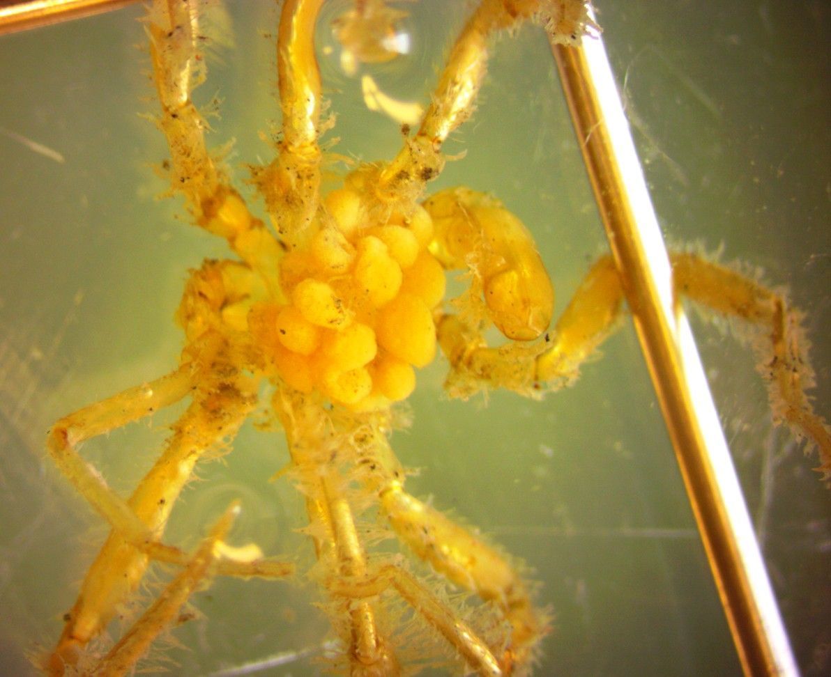
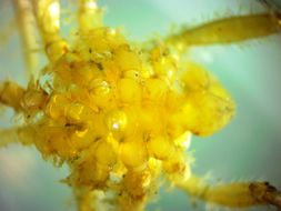
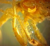
This dorsal view of the rear portion of the same animal (female) shows the abdomen, which is larger than seen on most pycnogonids, arising dorsally on the last thoracic segment and curving down between the hind legs. This dorsal view of a male shows similar features. This male is carrying a large mass of egg clusters ventrally to the trunk. Anterior is to the right.
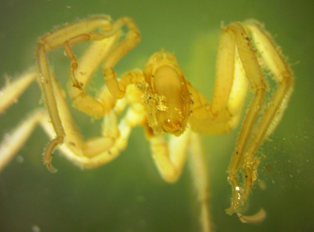
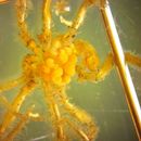
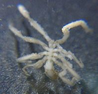
A female Ammothea verenae pycnogonid, about 1 cm long from tip of proboscis to tip of abdomen. Collected by Kirt Onthank from Endeavor hydrothermal vents on Juan de Fuca Ridge off Washington. The large proboscis is at the bottom of the photo and the small abdomen is at the top. Alcohol-preserved specimen. (Photo by: Dave Cowles, Feb 2014)