-
-

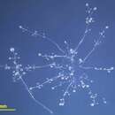
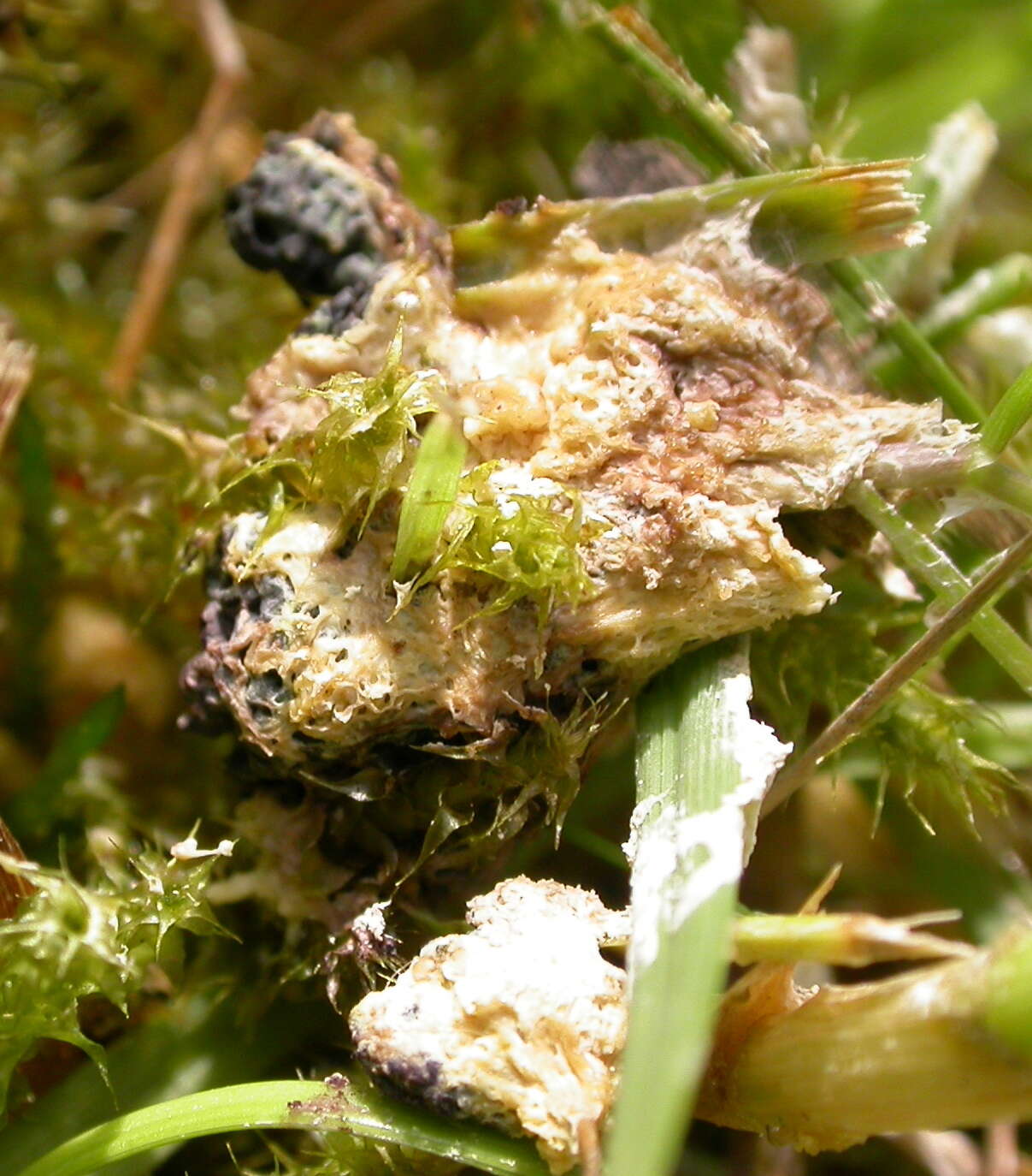
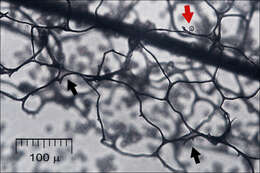
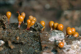
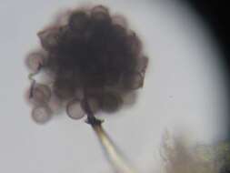
Stemonitis flavogenita E.JahnEN: ?, DE: ?Slo.: ?Membranous expansions in the capillitium (black arrows) and a single spore still attached to the capillitium (red arrow).Dat.: Aug. 11. 2014Lat.: 46.36119 Long.: 13.70107Code: Bot_823/2014_DSC2764Habitat: old partly overgrown pasture, near mixed wood edge, moderately southeast inclined foot of a mountain; open, dry, sunny place; shallow, skeletal, calcareous ground, exposed to direct rain, average precipitations ~ 3.000 mm/year, average temperature 7-9 deg C, elevation 630 m (2.070 feet), alpine phytogeographical region. Substratum: north side of a stump of Picea abies (on decorticated part) cut down three years ago.Place: Lower Trenta valley, between villages Soa and Trenta, upper part of 'Na melu' place, East Julian Alps, Posoje, Slovenia ECComment: Highly distinctive traits of Stemonitis flavogenita are typically zig-zag bent columella with a kind of plate at the end just before the end of sporangia (Ref.:1). It is not gradually tapering toward the end of sporocarp as with other species of genus Stemonitis. It is also distinguished by the presence of membranous expansions in the capillitium. Also agreement of macroscopic properties fit well to literature, so I hope the determination is correct. This observation may be interesting since this species is listed neither in Boletus Informaticus data base, Mycotheca and lichen herbarium (LJU-Li) of Slovenian Forestry Institute nor in official Slovenian fungi checklist.Sporocarp color rusty, oac719; spores on mass chocolate brown, oac635. Stalks 2.5 to 3 mm long, total length of sporangia 9 mm, all of them were fairly the same length, very closely tufted, their tips blunt. The whole clump had 14 mm in diameter.Spores finely warted, globose. Dimensions: 8,4 [8,8 ; 9] 9,5 x 8,2 [8,6 ; 8,7] 9,1 microns; Q = 1 [1,0] 1,1; N = 32; C = 95%; Me = 8,9 x 8,7 microns; Qe = 1. Olympus CH20, NEA 100x/1.25, magnification 1.000 x, oil (spores), NEA 10x/0.25, magnification 100x (all other pictures); in water. AmScope MA500 digital camera. Ref.:(1) B. Ing, The Myxomycetes of Britain and Ireland,The Richmond Publ. Co.Ltd, (1999), p 199. (2) S.L.Stephenson and H.Stempen, Myxomycetes, Timber Press Inc.(2000), p 153.(3)
www.discoverlife.org/mp/20q?search=Stemonitis+flavogenita (4)
www.myxomycetes.wolf-5.cyberdusk.pl/index.php?wyb=gal&... (6)
www.myxomycetes.net/index.php (7)
myxo.be/pdf/Stemonitis%20flavogenita%20Stemonitopsis%20pe... (8)
www.bookiejar.com/Content/Books/7ccbe2a1-12a9-41fa-a3ff-0... a(9)
www.shitennoji.ac.jp/ibu/images/toshokan/kiyo45-15.pdf
-
Briantspuddle, England, United Kingdom
-
Washington, District of Columbia, United States
-
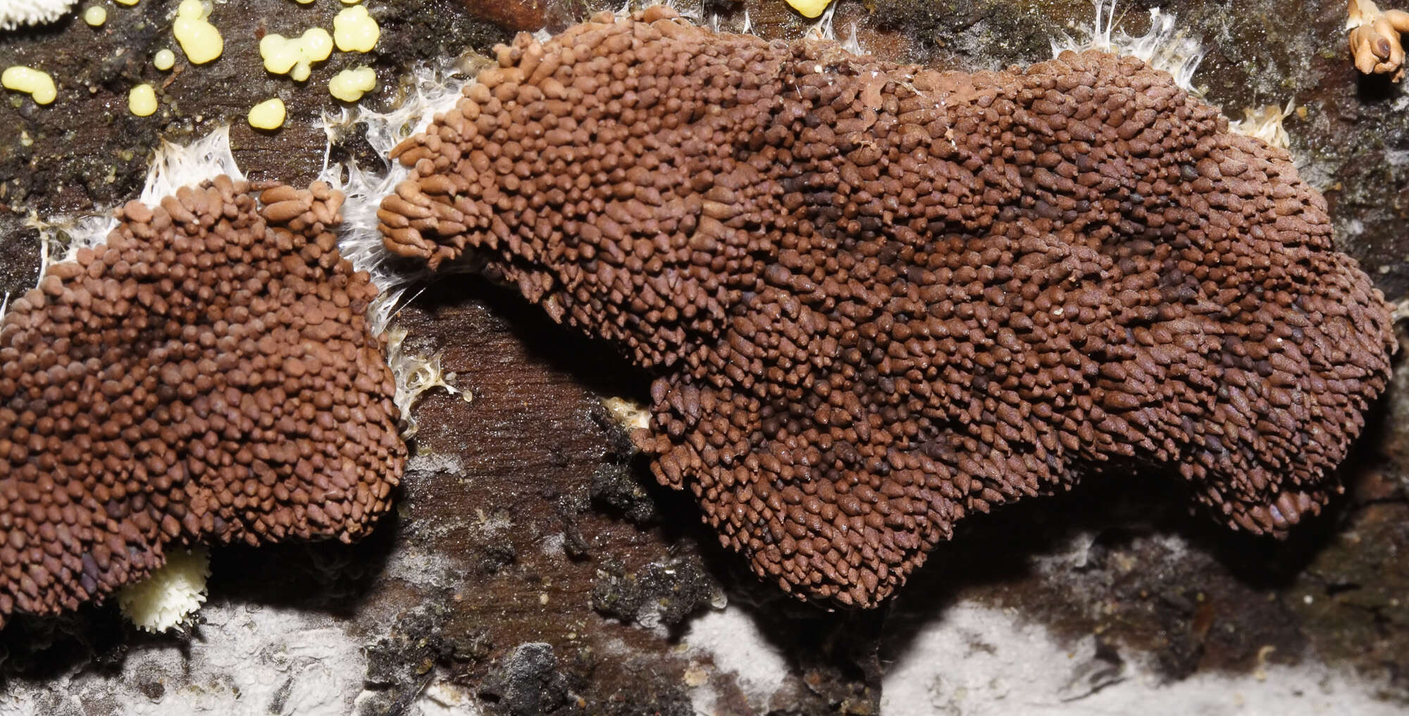
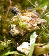
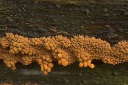
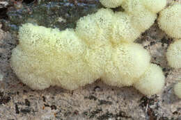
This is the vegetative stage of a bacteria-eating slime mold, preparing to form spores. Sunshine Coast, British Columbia. Trichiaceae Family
-
Steiglitz, Victoria, Australia
-
Hemitrichia serpulaPretzel slime moldSlo.: ?Date: Sept. 11. 2009Lat.: 46,33481 Long.: 13,53083Code: Bot_377/2009-3314Habitat: mixed woodland, nearly flat ground, cretaceous clastic rock (flysh), rain protected by trees canopies, in full shade, average precipitations ~3.000 mm/year, average temperature 8-10 deg C, altitude 445 m (1.450 feet), alpine phytogeographical region. Substratum: fallen old deciduous tree, probably Acer sp., partly debarked, covered with mosses.Place: West of Bovec, near the trail to Pluna village, East Julian Alps, Posoje, Slovenia EC
-

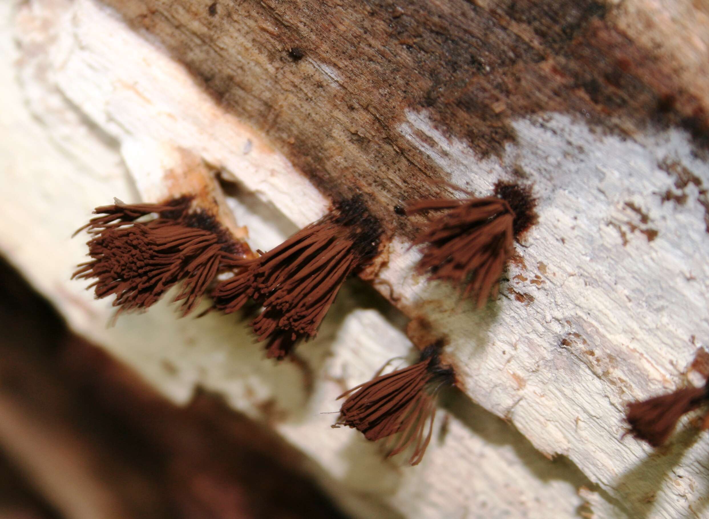
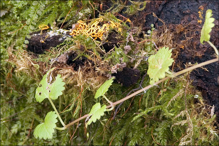
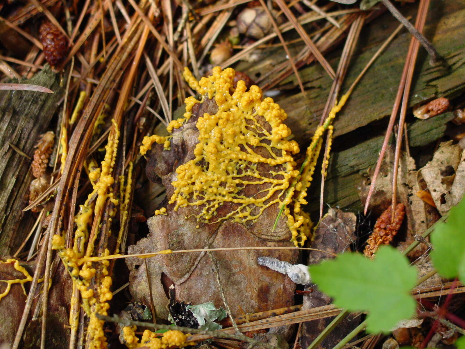
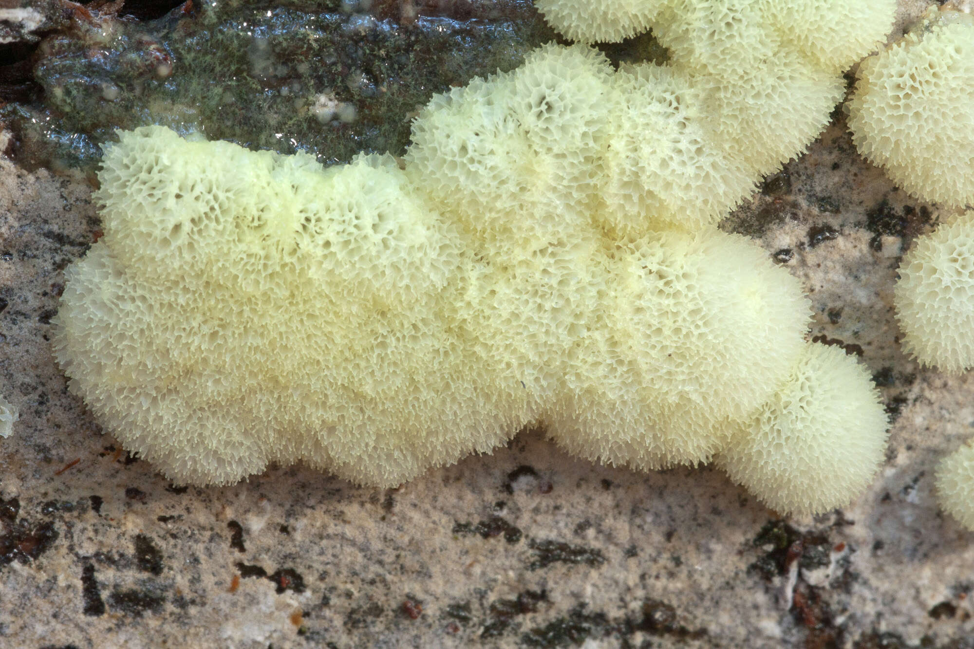
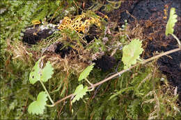
Hemitrichia cf clavata (Pers.) Rostaf., syn.: Trichia clavata Pers., Hyporhamma clavatum (Pers.) LadoEN: Yellow-Fuzz Cone Slime, DE: Gelber ScheinhaarstublingSlo.: kitajska zlatovkaDat.: Oct. 5. 2009Lat.: 46.33439 Long.: 13.48114Code: Bot_388/2009_DSC5885Habitat: Mixed, predominantly Fagus sylvatica forest, in shade, partly protected from direct rain by tree canopies, average precipitations ~3.000 mm/year, average temperature 3 -5 deg C, elevation 1.330 m (4.400 feet), alpine phytogeographical region. Substratum: fallen, heavily rotted trunk of a deciduous tree, probably Fagus sylvatica. Place: Bovec basin, Gozdec forest, above the road from Mt.Kanin cable car station B to the foot of Mt. Kopa, East Julian Alps, Posoje, Slovenia EC. Comment: There was no microscopy done for this observation, hence the determination is unreliable. Myxomycetes pass through very different shapes and colors during development of their sporocarps. In most cases it is impossible to determine them to species level without microscopic observation of their spores and structure, at least for me. These pictures show immature sporocarps. Sporocysts have not yet opened, capillitium and spore mass are not yet visible. Also stalks are mostly not yet developed. This makes determination even more difficult. Nevertheless, the size and the shape of the oldest sporocarps and stalks with reddish tint points toward Hemitrichia clavata. But, it is possible that the pictures show other Hemitrichia or Trichia species like similar Trichia decipiens or Hemitrichia calyculata. Picture #10 shows eventually another species. On picture #8 one can see also two species of (probably) Ascomycetes - a larger jelly fungus and smaller black blobs (both lower left). What a picturesque life one can find just on a small piece of rotten wood! A white 'mycelium' shown on the same picture (and others - upper left) probably belongs to another kind of fungus. Although Hemitrichia clavata has white plasmodium these fibers don't seem to me to be its plasmodium. Ref.:(1) B. Ing, The Myxomycetes of Britain and Ireland,The Richmond Publ. Co.Ltd, (1999), p 127.(2) S.L.Stephenson and H.Stempen, Myxomycetes, Timber Press Inc.(2000), p 124.(x) M. Poulain, M. Meyer, J. Borronet, Les Myxomycetes, FMBDS (2011), Vol.1., p 371; Vol.2. p 130.(3) S. Behri, Raznolikost Pravih Sluzavk (Myxomycetes) v okolici Mengea, (in Slovene) (True Slime Molds (Myxomycetes) Diversity in the Vicinity of Menge), Graduation thesis, University Studies, University in Ljubljana, Biotechnical Faculty, Biology department (2015), p 86.(4) H. Neubert, W. Nowotny, K. Baumann - H. Marx, Die Myxomyceten Deutschlands und des angrenzenden Alpenraumes unter besonderen Bercksichtigung sterreichs, Vol.2.,Karlheinz Baumann Verlag, (1993, 1995, 2000), p 232.
-
Three Lakes, Wisconsin, United States
-
Clastoderma
-
Castel Fusano, Lazio, Italy
-
Castel Fusano, Lazio, Italy
-

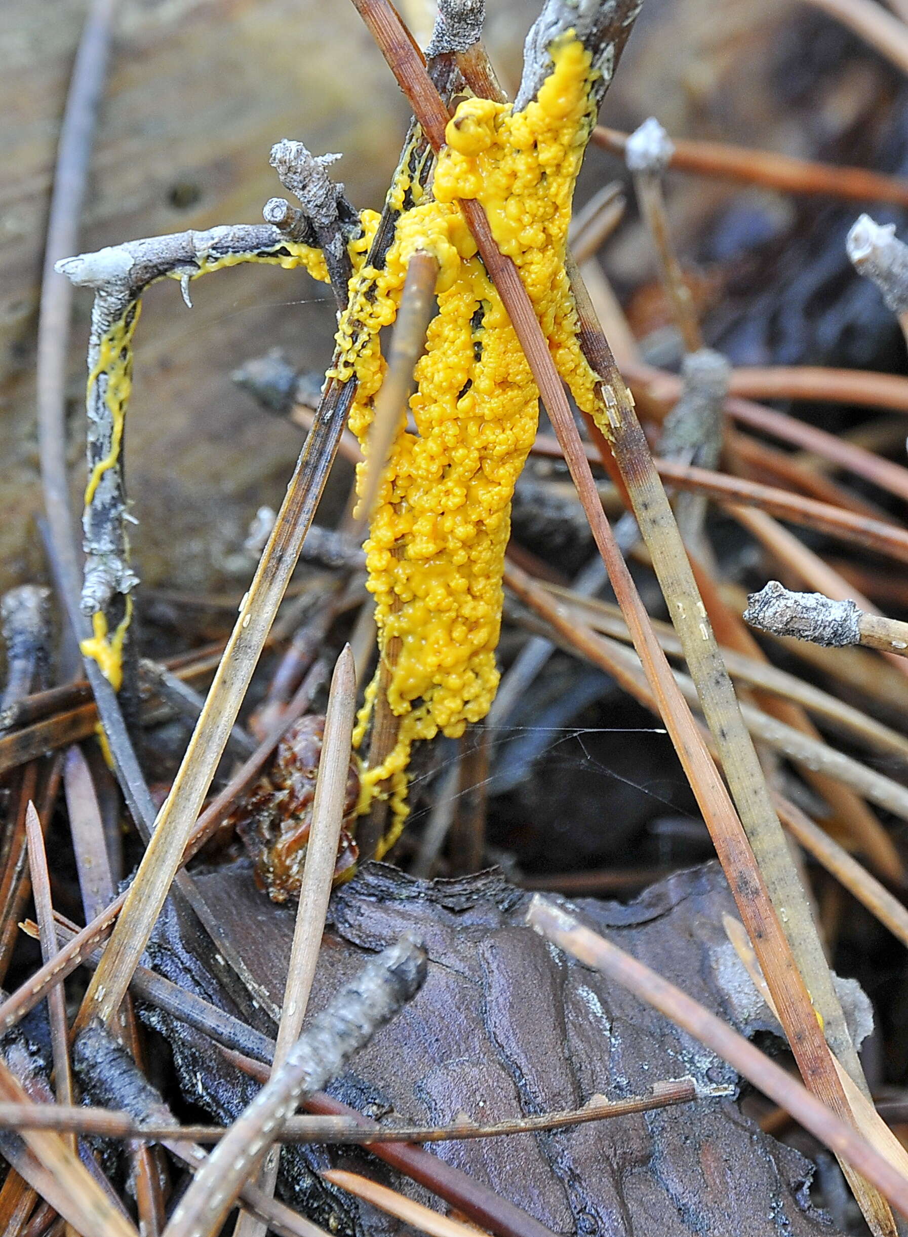
Fuligo septica (L.)Wigg., syn. Mucorsepticus L., Reticularia septica (L.) With., Aethalium septicum (L.) Fr., Fuligo varians Sommerf.Scrambled-egg slime, Dog vomit slime mold, Flowers of Tan, DE: Gelbe Lohblte, HexenbutterSlo.: reslov cvetThe aethalium picture taken on July 21. Dat.: July 21. 2014Code: Bot_815/2014_DSC2009 Lat.: 46.36114 Long.: 13.70122Habitat: old partly tree overgrown pasture, near mixed wood edge; moderately southeast inclined foot of an old overgrown scree slope; open, dry, sunny place; shallow, skeletal, calcareous ground, exposed to direct rain, average precipitations ~ 3.000 mm/year, average temperature 7-9 deg C, elevation 630 m (2.070 feet), alpine phytogeographical region. Substratum: a stump of Picea abies cut down three years ago.Place: Lower Trenta valley, between villages Soa and Trenta, upper part of 'Na Melu' place, East Julian Alps, Posoje, Slovenia EC.Comment: Myxomycetes are poorly known yet very interesting creatures. For decades they have been shuffled back and forth between the animal and plants kingdoms until recognized as separate creatures. They are not animals because they proliferate by spores. They are also not plants since they crumble around (an animal like ability) and fix themselves firmly to substrate only at the end of their life cycle. They don't produce their own food like plants but feed by 'hunting' (actually engulfing) bacteria and tiny bits of other organic matter, which is another animal like feature. The first stage in their development cycle, which is observable in the field, is called plasmodium. Earlier stages (from myxoflagellates, myxoamoebae, to zygote) are microscopic and can be observed only in labs. Plasmodium is a single giant living cell, a clump of protoplasm filled with thousands of cell nuclei, crawling around, eating bacteria and growing. Some of such plasmodia are the largest cells of living creatures known. In some species they can measure several meters across or weight up to 20 kg. That it much larger than ostrich's eggs, which are popularly considered as 'largest living cells'! Plasmodia could be found in the field, some are even brightly colored and easy to spot; however, it is almost impossible to determine to which species they belong. Plasmodium of Fuligo septica is commonly described like disgusting mucus, spilled scrambled eggs, dogs vomiting and other 'benevolent' portrayals.When the time is right or delicate environmental conditions required for growth worsen plasmodia eventually evolves (usually almost completely) into sporocarps of different forms. These are bodies producing spores and then vanishing. In genus Fuligo sporocarp is a cushion like aethalium sitting on a thin whitish, 'fibrous' layer called hypothallus (Fig.14.). These'cushions' are what one usually finds in the field. But, other Myxomycetes develop also many other forms of sporocarps full of beauty, delicacy and imagination. Aethalia of Fuligo septica are usually covered with a kind of crust called cortex, which is brittle and soon crumbles away. In humid conditions it may not fully develop (Ref.1). Inside a mature aethalium there is a mesh of thin tubes or fibers called capillitium and zillions of dark brown spores. Fuligo septica has characteristic nodes on capillitial tubes, which are clearly seen on Fig. 4M. In due course the aethalium decomposes almost entirely into spore mass (Fig.17., 18.), which are sooner or later blown or washed away (Fig.20. taken about three weeks after the first photo). Size and shape of spores and structure of their surface are important traits for species determination. Cushion-shaped aethalium measured approximately 14 x 5 cm and was about 3 cm thick. Spores are minutely warty and globose to subglobose. Dimensions: 8 [8,4 ; 8,7] 9,1 x 7,4 [8 ; 8,2] 8,7 microns; Q = [1 ; 1,07] 1,1; N = 25; C = 95%; Me = 8,5 x 8,1 microns; Qe = 1,05. Olympus CH20, NEA 100x/1.25, magnification 1.000 x, oil (spores), NEA 40x/0.65, magnification 400x (capillitium), NEA 10x/0.25, magnification 100x (hypothallus); in water, living material. AmScope MA500 digital camera. Spore sample taken on July 23. 2014.Herbarium: Mycotheca and lichen herbarium (LJU-Li) of Slovenian Forestry Institute, Vena pot 2, Ljubljana, Index Herbariorum LJFRef.:(1) B. Ing, The Myxomycetes of Britain and Ireland, The Richmond Publ. Co.Ltd, (1999), p 246(2) S.L.Stephenson and H.Stempen, Myxomycetes, Timber Press Inc.(2000), p 123(3)
www.hiddenforest.co.nz/slime/family/physaraceae/physa02.htm 6-9(4) M. Poulain, M. Meyer, J. Bozzonet, Les Myxomycetes, FMBDS (2011), Vol.1., p128; Vol.2., p168.
-
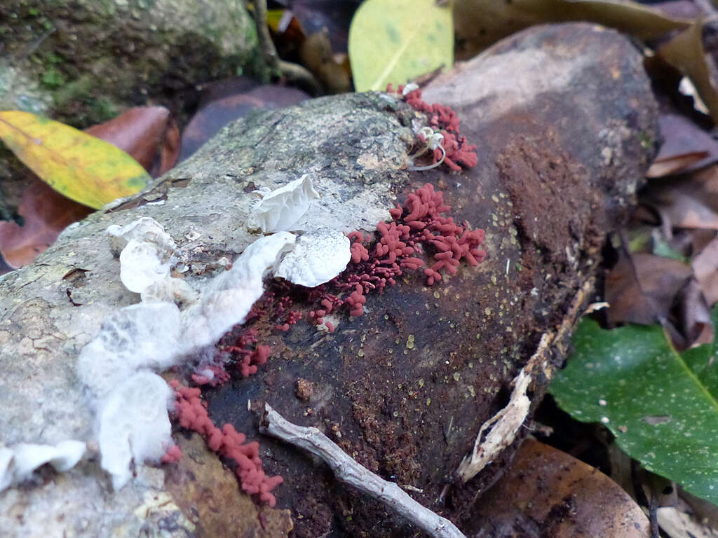
Unfortunately, I couldn't get very close to this log. It was on the other side of the track's fence. The log had a lot of interesting things on it, including this lovely red-brown slime mould. There were also some very tiny yellowish cup fungi on the side of the log that I really would have liked to have a closer look at. There was also an interesting Campanella species. Sigh.
IDENTIFYING AUSTRALIAN RAINFOREST PLANTS,TREES & FUNGI - Flick Group -->
DATABASE INDEX -
TAGS
-

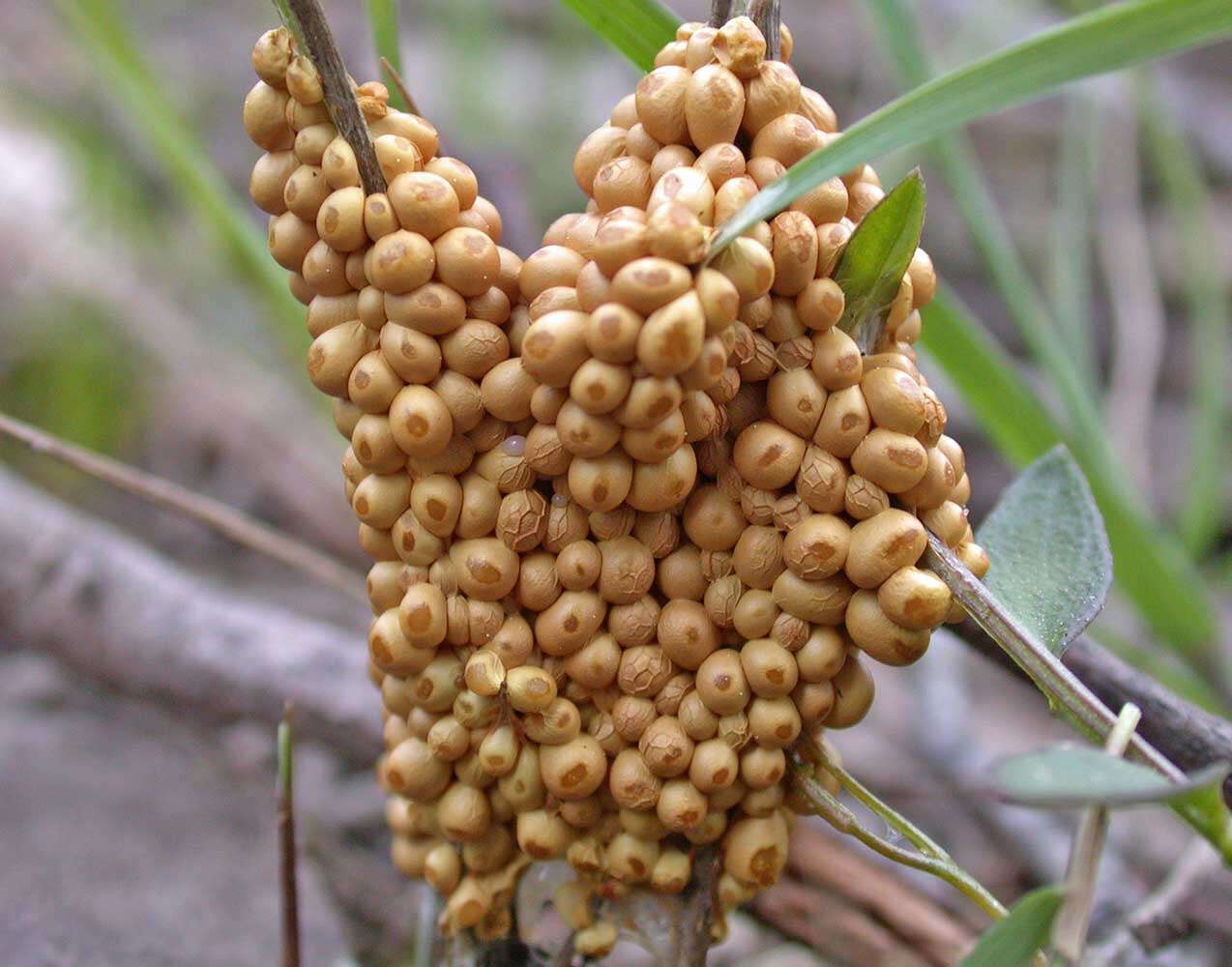
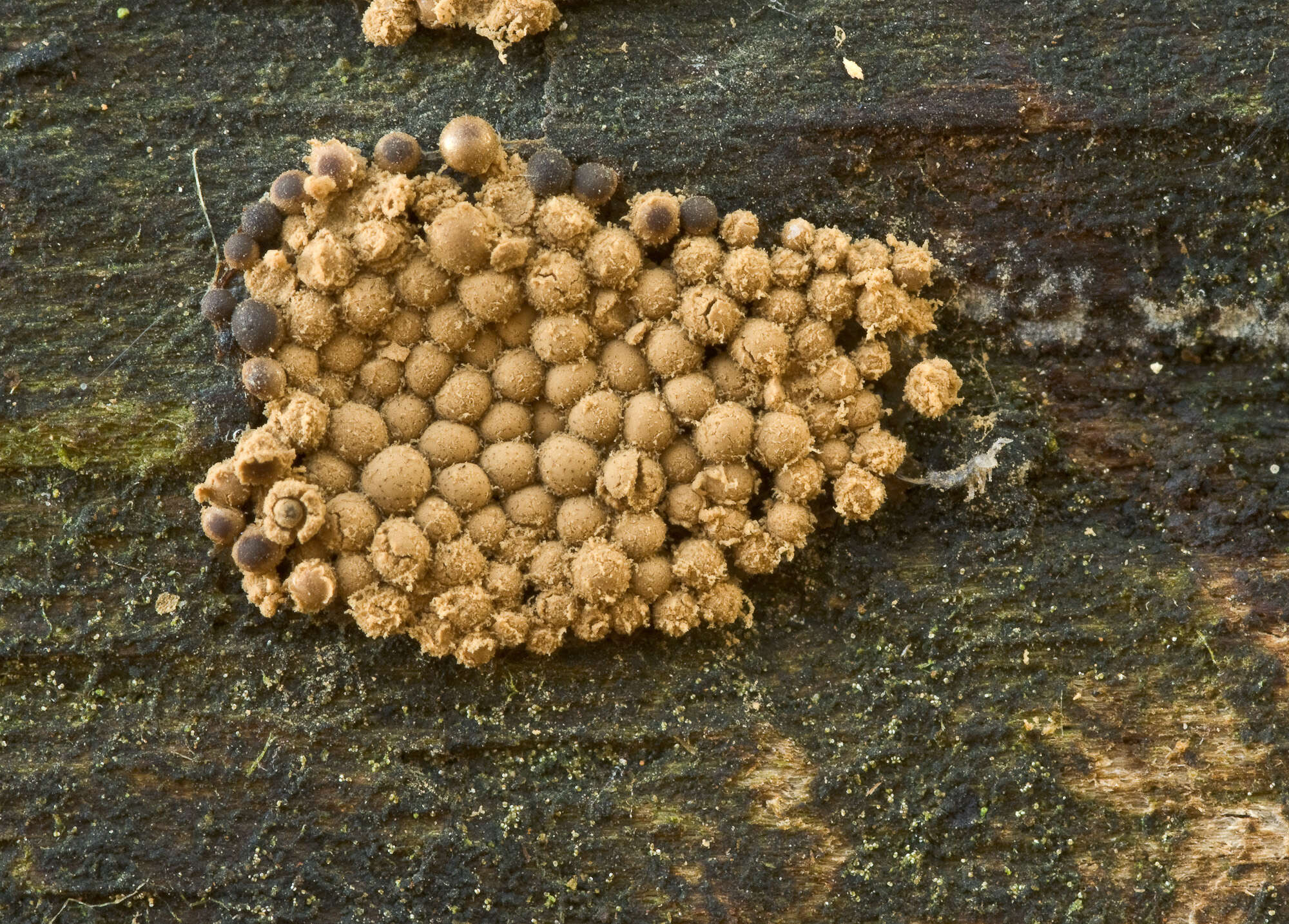
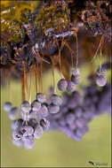
Cribraria vulgaris Schrad, syn.: Cribraria vulgaris var. genuina Rostaf., Cribraria vulgaris var. vulgaris Schrad. Date: June 15. 2009Lat.: 46.33506 Long.: 13.53008Code: Bot_356/2009-DSC0347Habitat: mixed wood in a mountain ravine, moderately inclined mountain slope, southwest aspect; cretaceous clastic rock (flysh); protected from direct rain by trees canopies and tall herbs, in shade, very humid place; precipitations ~3.000 mm/year, average temperature 8-10 deg C, altitude 450 m (1.500 feet), alpine phytogeographical region.Substratum: fallen, debarked and completely rotten deciduous tree trunk, hidden in tall herbs. Place: Bovec basin, west of Bovec, near the trail to Pluna village, East Julian Alps, Posoje, Slovenia ECComment: My initial tentative determination in 2009 Cribraria intricata was wrong. Based on new literature I've obtained the best fit seems to be Cibraria vulgaris. Fitting dimensions of sporocarps, distinct, well defined cup-shaped calyculus with irregular teeth, density of peridium mesh, color of sporangia and stalk tapering upwards, substratum and large colony speak in favor of this determination. Species has been already found in Slovenia (Ref.4) contrary to Cribraria intricata. However, determination with certainty would still require microscopic verification of traits. Unfortunately this hasn't been done.Ref: (1) B. Ing, The Myxomycetes of Britain and Ireland,The Richmond Publ. Co.Ltd, (1999), p 103(2) S.L.Stephenson and H.Stempen, Myxomycetes, Timber Press Inc.(2000), p 80.(3) M. Poulain, M. Meyer, J. Borronet, Les Myxomycetes, FMBDS (2011), Vol.1., p 307; Vol.2. p 19.(4) S. Behri, Raznolikost Pravih Sluzavk (Myxomycetes) v okolici Mengea, (in Slovene) (True Slime Molds (Myxomicetes) Diversity in Vicinity of Menge) (in Slovene), Graduation Thesis, University Studies, University in Ljubljana, Biotechnical Faculty, Biology department (2015), p70.(5)
www.br.fgov.be/cgi-bin/RESEARCH/COLLECTIONS/HERBARIUMS/FU...(6)
www.micobotanicajaen.com/Revista/Articulos/FMorenoG/Myxom...
-
Castel Fusano, Lazio, Italy
-
Castel Fusano, Lazio, Italy
-
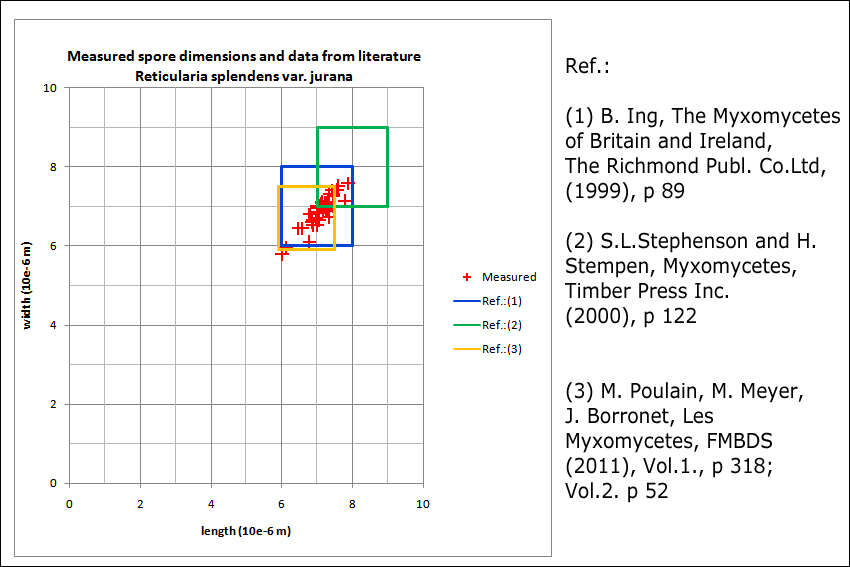
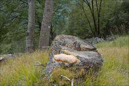
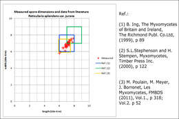
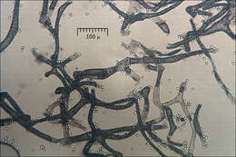
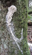
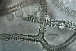
Reticularia splendens var. jurana (Meyl.) Kowalski, syn.: Enteridium juranum (Meyl.) Mornand, Enteridium splendens var. juranum (Meyl.) Hrk., Reticularia jurana Meyl., Reticularia lycoperdon var. jurana (Meyl.) G. ListerE: no name, DE: no nameSlo.: no nameLat.: 46.35884 Long.: 13.69819Dat.: Oct. 21. 2016Code: Bot_1021/2016_DSC5937Habitat: pasture, at the edge of the mixed forest, Fagus sylvatica, Ostrya carpinifolia and Picea abies dominant trees; moderately inclined slope at the foot of mountains, south east aspect; colluvial, skeletal, calcareous ground; mostly sunny place; relatively warm and dry place; exposed to direct rain, average precipitation ~ 3.000 mm/year, average temperature 7-9 deg C, elevation 615 m (2.020 feet), alpine phytogeographical region. Substratum: dead, thick, partly decorticated branch of Ostrya carpinifolia laying on ground; fund on top side of it; wood still firm, hard to cut; in its initial state of disintegration.Place: Lower Trenta valley, between villages Soa and Trenta, right bank of river Soa; pasture west of Strgulc abandoned farm house, Soa 47, East Julian Alps, Posoje, Slovenia EC. Comment: Three aethaila found in a circle of about 0.4 m diameter. This myxomicete can be recognized by its vividly pink (not yet mature) aethalia, white hypothallus, characteristic spores, which are distinctly reticulate only on about 2/3 of their surface and persistent pseudocapillitium consisting of membranous perforated plates and threads. Reticularia splendens var. jurana can be distinguished from Reticularia splendens var. splendens by smaller size of aethalia and flaccid cortex.To my knowledge this is the second or third find of this species in Slovenia and the first one in the Julian Alps. Spores coarsely reticulated on about 2/3 of their surface and minutely warted (barely seen with my equipment) on the rest of it; globose to subglobose. Dimensions (without reticule and warts): 6,3 [7 ; 7,2] 7,8 x 6,1 [6,8 ; 7] 7,6 microns; Q = 1 [1,0] 1,1; N = 40; C = 95%; Me = 7,1 x 6,9 microns; Qe = 1. Reticule > 1 micron high. Olympus CH20, NEA 100x/1.25, magnification 1.000 x, oil (spores), NEA 40x/0.65, magnification 400x and NEA 10x/0.25, magnification 100x (pseudocapillitium); fresh material; in water. AmScope MA500 digital camera. Spores were taken from the centre of the second aethalium (Fig. 5, larger one) by soft brush.Ref.:(1) B. Ing, The Myxomycetes of Britain and Ireland,The Richmond Publ. Co.Ltd, (1999), p 89 (2) S.L.Stephenson and H.Stempen, Myxomycetes, Timber Press Inc.(2000), p 122(3) M. Poulain, M. Meyer, J. Borronet, Les Myxomycetes, FMBDS (2011), Vol.1., p 318; Vol.2. p 52.(4)
www.repository.naturalis.nl/document/572530 (5)
www.zdravgozd.si/bi_karta_sre.aspx?idorg=d0561d66-42d2-4f...
-
Castel Fusano, Lazio, Italy
-
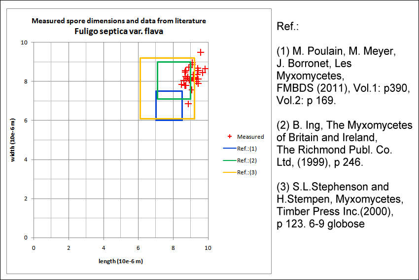
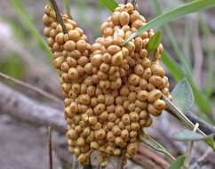
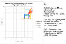
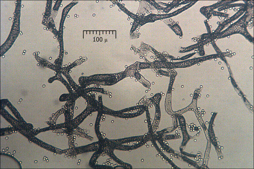
Fuligo septica var. flava (Pers.) Morgan, syn.: Mucor septicus L., Aethalium flavum (Pers) LinkFlowers of Tan, DE: Gelbe LohblteSlo.: reslov cvet, rumeni razliekDat.: Sept. 05. 2014Lat.: 46.35965 Long.: 13.70116Code: Bot_832/2014_DSC3621Habitat: mixed wood, Fagus sylvatica and Picea abies dominant, moderately southeast inclined mountain slope, shallow, skeletal calcareous ground, old overgrown slope and moraine scree with larger rocks and boulders, in shade, relatively warm place, partly protected from direct rain by tree canopies, average precipitation ~ 3.000 mm/year, average temperature 7 - 9 deg C, elevation 600 m (1.970 feet), alpine phytogeographical region. Substratum: debarked trunk of Picea abies lying on ground in its late disintegration stage.Place: Lower Trenta valley, between villages Soa and Trenta, next to the trail from Trenta 2b cottage to abandoned farmhouse 'Strgulc', East Julian Alps, Posoje, Slovenia EC.Comment: Fuligo septica is probably the most common and widely known Myxomicete. The latest monograph on Myxomycetes I have (Ref.:1) describes six varieties of this species, which differ mostly in cortex structure (single versus double layered) and color of different parts of sporocarp and plasmodium. Fuligo septica var. flava should have vivid yellow aethalia, yellow inner lime and yellow plasmodium. Two days before I took these pictures I had seen the plasmodium, which was in a form of vividly yellow colored patch of densely packed small half-spheres. The rest of traits of Fuligo septica var. flava also fit well to my observation.Spores minutely warty, globose to subglobose. Dimensions: 8 [8,4 ; 8,7] 9,1 x 7,4 [8 ; 8,2] 8,7 microns; Q = [1 ; 1,07] 1,1; N = 25; C = 95%; Me = 8,5 x 8,1 microns; Qe = 1,1. Olympus CH20, NEA 100x/1.25, magnification 1.000 x, oil (spores), NEA 40x/0.65, magnification 400x (capillitium, calcareous granules); in water; live material. AmScope MA500 digital camera.Herbarium: Mycotheca and lichen herbarium (LJU-Li) of Slovenian Forestry Institute, Vena pot 2, Ljubljana, Index Herbariorum LJFRef.:(1) M. Poulain, M. Meyer, J. Borronet, Les Myxomycetes, FMBDS (2011), Vol.1: p390, Vol.2: (2) B. Ing, The Myxomycetes of Britain and Ireland, The Richmond Publ. Co.Ltd, (1999), p 246. (3) S.L.Stephenson and H.Stempen, Myxomycetes, Timber Press Inc.(2000), p 123.
-
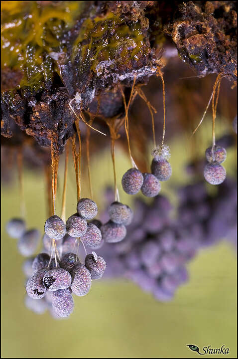
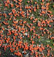
Lycogala epidendrum (L.) Fr.Wolf's Milk, Groening's Slime, DE: BlutmilchpilzSlo.: razbarvana grahovkaDat.: Nov. 9. 2017Lat.: 46.36014 Long.: 13.70435Code: Bot_1096/2017_DSC9621Picture file names: from Lycogala-epidendrum_raw_20 to Lycogala-epidendrum_raw_24.Habitat: mountain pasture; slightly inclined terrain, southeast aspect; colluvial/glacial, calcareous ground; full sun, dry place; elevation 575 m (1.900 feet); average precipitations ~ 3.000 mm/year, average temperature 7-9 deg C, alpine phytogeographical region. Substratum: a pile of partly rotten stump of Picea abies, mostly still in bark.Place: Lower Trenta valley, right bank of river Soa; between villages Soa and Trenta; near Trenta 2 farm house, East Julian Alps, Posoje, Slovenia EC.Comment: Average diameter of seven aethalia found was somewhat small (AVG = 4.5 mm, SD = 0.6 mm) compared to data from literature (the smallest had only 2.2 mm in diameter); however all other macroscopic traits fit well to Lycogala epidendrum species descriptions. Microscopically spore dimensions, their shape and reticulated surface, all fit to this species. Also pseudocapillitium diameter, its surface with conspicuous transverse faults and its club shaped free ends fit well. Spore mass grayish with pink tint. Spores reticulated, globose to subglobose. Dimensions: (6,7) 7 - 7,5 (7,9) x (6,5) 6,8 - 7,3 (7,5) microns; Q = 1 - 1,06 (1,1); N = 35; Me = 7,3 x 7,1 microns; Qe = 1. Olympus CH20, NEA 100x/1.25, magnification 1.000 x, oil (spores), NEA 40x/0.65, magnification 400x (pseudocapillitium), NEA 10x/0.25, magnification 100x (pseudocapillitium); in water; fresh material. AmScope MA500 digital camera.Ref.:(1) B. Ing, The Myxomycetes of Britain and Ireland,The Richmond Publ. Co.Ltd, (1999), p 91. (2) S.L.Stephenson and H.Stempen, Myxomycetes, Timber Press Inc.(2000), p 135. (3) M. Poulain, M. Meyer, J. Borronet, Les Myxomycetes, FMBDS (2011), Vol.1., p 321; Vol.2. p 75. (4) S. Behri, Raznolikost Pravih Sluzavk (Myxomycetes) v okolici Mengea, (in Slovene) (True Slime Molds (Myxomicetes) Diversity in Vicinity of Menge) (in Slovene), Graduation Thesis, University Studies, University in Ljubljana, Biotechnical Faculty, Biology department (2015), p 74. Baumann - H. Marx, Die Myxomyceten Deutschlands und des angrenzenden Alpenraumes unter besonderen Bercksichtigung sterreichs, Vol.1., Karlheinz Baumann Verlag, (1993, 1995, 2000), p 135.
-
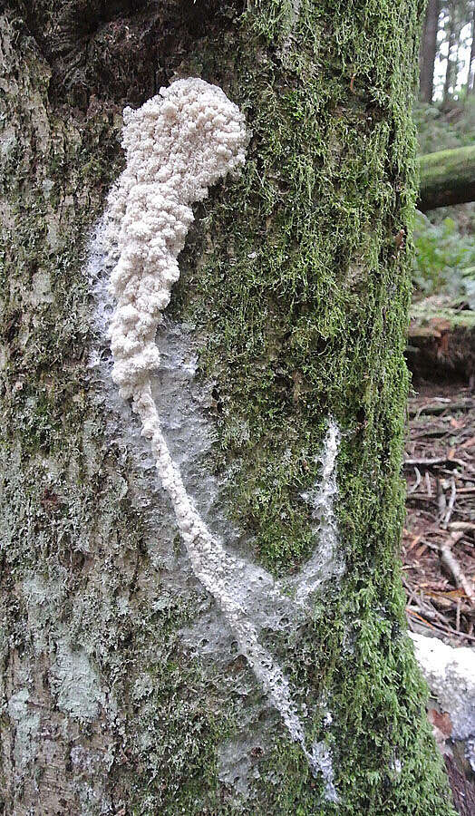
In Spring on Mt. Elphinstone, British Columbia. Stemonitidaceae Famly
-

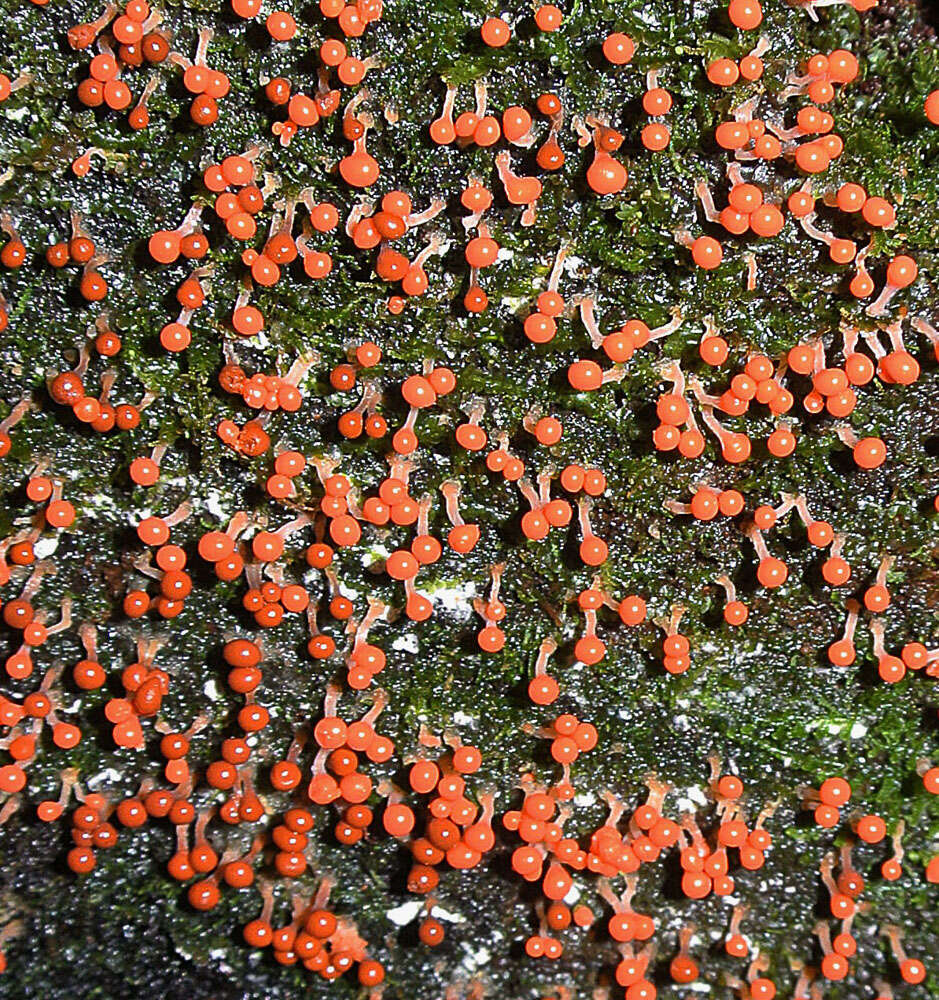
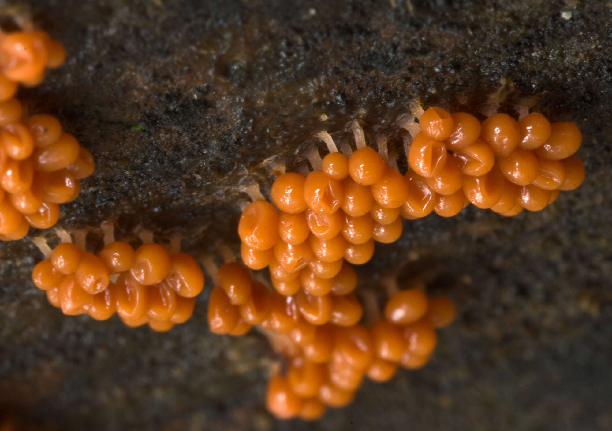
Arcyria obvelata, syn.: Arcyria nutans (Bull) GrevCapillitium threds at magnification 1000x.Dat.: Sept. 26. 2013Lat.: 46.36151Long.: 13.70434Code: Bot_753/2013_DSC8059Habitat: Overgrown former grassland; dominant trees Ailanthus altissima, Fraxinus ornus, Corylus avellana, Fagus sylvatica, Juglans regia, Tilia sp., Prunus domestica; next to an abandoned farmhouse; flat terrain, calcareous ground; full shade, quite humid and relatively warm place, partly protected from direct rain by tree canopies; average precipitation ~ 3.000 mm/year, average temperature 7-9 deg C, elevation 590 m (1.950 feet), alpine phytogeographical region. Substratum: vertical surface of dead, still standing trunk of Juglans regia partly still in bark fully covered by a Polyporaceae, probably Inonotus sp.; about 1 m (three feet) above ground, northeast oriented surface of the trunk.Place: Lower Trenta valley, between villages Soa and Trenta, near abandoned homestead 'Koc', Trenta 3, East Julian Alps, Posoje, Slovenia EC Comment: Long, hanging and very shortly stipitate sporocarps distinguish this species from others in Myxomycetes genus Arcyria. Sporocarps 8 - 13 mm long, about 1 mm diameter, flexible. Stalk very short, hard to observe, almost sessile sporocarps; sporocarps ocher-yellow, oac848; SP abundant, ocher-yellow, oac 856.Spores subglobose and almost smooth, scattered warts hardly visible with my equipment. Dimensions: 8.7 (SD = 0.3) x 8.3 (SD = 0,2) , Q = 1.05 (SD = 0.02), n = 30. Olympus CH20, NEA 100x/1.25, magnification 1.000 x, oil, (picture of spores and capillitium threads). Bausch & Lomb 4/0.10, magnification 40x, in water (picture of capillitium). AmScope MA500 digital camera.Ref.:(1) B. Ing, The Myxomycetes of Britain and Ireland, The Richmond Publ. Co.Ltd, (1999), p 113. (2)
www.bcrc.firdi.org.tw/fungi/fungal_detail.jsp?id=FU200802...(3) http//hiddenforest.co.nz/slime/family/arcyriaceae/arcyr03.htm
-
Zaragoza: Aragn (Espaa)Reino: ProtozoaFilo: MycetozoaClase: MyxomycetesOrden: PhysaralesFamilia: Physaraceae