Reticularia-splendens-var-jurana_spore_stat_2M
Description:
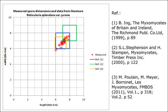
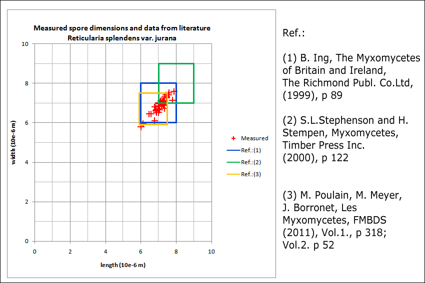
Reticularia splendens var. jurana (Meyl.) Kowalski, syn.: Enteridium juranum (Meyl.) Mornand, Enteridium splendens var. juranum (Meyl.) Hrk., Reticularia jurana Meyl., Reticularia lycoperdon var. jurana (Meyl.) G. ListerE: no name, DE: no nameSlo.: no nameLat.: 46.35884 Long.: 13.69819Dat.: Oct. 21. 2016Code: Bot_1021/2016_DSC5937Habitat: pasture, at the edge of the mixed forest, Fagus sylvatica, Ostrya carpinifolia and Picea abies dominant trees; moderately inclined slope at the foot of mountains, south east aspect; colluvial, skeletal, calcareous ground; mostly sunny place; relatively warm and dry place; exposed to direct rain, average precipitation ~ 3.000 mm/year, average temperature 7-9 deg C, elevation 615 m (2.020 feet), alpine phytogeographical region. Substratum: dead, thick, partly decorticated branch of Ostrya carpinifolia laying on ground; fund on top side of it; wood still firm, hard to cut; in its initial state of disintegration.Place: Lower Trenta valley, between villages Soa and Trenta, right bank of river Soa; pasture west of Strgulc abandoned farm house, Soa 47, East Julian Alps, Posoje, Slovenia EC. Comment: Three aethaila found in a circle of about 0.4 m diameter. This myxomicete can be recognized by its vividly pink (not yet mature) aethalia, white hypothallus, characteristic spores, which are distinctly reticulate only on about 2/3 of their surface and persistent pseudocapillitium consisting of membranous perforated plates and threads. Reticularia splendens var. jurana can be distinguished from Reticularia splendens var. splendens by smaller size of aethalia and flaccid cortex.To my knowledge this is the second or third find of this species in Slovenia and the first one in the Julian Alps. Spores coarsely reticulated on about 2/3 of their surface and minutely warted (barely seen with my equipment) on the rest of it; globose to subglobose. Dimensions (without reticule and warts): 6,3 [7 ; 7,2] 7,8 x 6,1 [6,8 ; 7] 7,6 microns; Q = 1 [1,0] 1,1; N = 40; C = 95%; Me = 7,1 x 6,9 microns; Qe = 1. Reticule > 1 micron high. Olympus CH20, NEA 100x/1.25, magnification 1.000 x, oil (spores), NEA 40x/0.65, magnification 400x and NEA 10x/0.25, magnification 100x (pseudocapillitium); fresh material; in water. AmScope MA500 digital camera. Spores were taken from the centre of the second aethalium (Fig. 5, larger one) by soft brush.Ref.:(1) B. Ing, The Myxomycetes of Britain and Ireland,The Richmond Publ. Co.Ltd, (1999), p 89 (2) S.L.Stephenson and H.Stempen, Myxomycetes, Timber Press Inc.(2000), p 122(3) M. Poulain, M. Meyer, J. Borronet, Les Myxomycetes, FMBDS (2011), Vol.1., p 318; Vol.2. p 52.(4) www.repository.naturalis.nl/document/572530 (5) www.zdravgozd.si/bi_karta_sre.aspx?idorg=d0561d66-42d2-4f...
Included On The Following Pages:
- Life (creatures)
- Cellular (cellular organisms)
- Eukaryota (eukaryotes)
- Amoebozoa (amoeboid protists)
- Evosea
- Eumycetozoa (slime molds)
- Myxogastria (plasmodial slime molds)
- Lucisporidia
- Liceales
- Reticulariaceae
- Reticularia
- Reticularia jurana
This image is not featured in any collections.
Source Information
- license
- cc-by-nc-sa
- copyright
- Amadej Trnkoczy
- photographer
- Amadej Trnkoczy
- original
- original media file
- visit source
- partner site
- Flickr Group
- ID