-
-
-
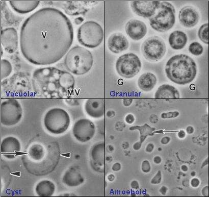
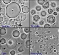
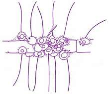
Description: Image shows four common forms of Blastocystis hominis - vacuolar, granular, amoeboid, and cyst forms. Image created by Valentia Lim Zhining on May 14th, 2006. Date: 14 May 2006. Source: Own work. Author: Valentia Lim
Valzn. Permission (
Reusing this file): : Permission is granted to copy, distribute and/or modify this document under the terms of the
GNU Free Documentation License, Version 1.2 or any later version published by the
Free Software Foundation; with no Invariant Sections, no Front-Cover Texts, and no Back-Cover Texts. A copy of the license is included in the section entitled
GNU Free Documentation License.http://www.gnu.org/copyleft/fdl.htmlGFDLGNU Free Documentation Licensetruetrue. : This file is licensed under the
Creative Commons Attribution-Share Alike 2.5 Generic license.:. https://creativecommons.org/licenses/by-sa/2.5 CC BY-SA 2.5 Creative Commons Attribution-Share Alike 2.5 truetrue. I, the copyright holder of this work, hereby publish it under the following licenses: : Permission is granted to copy, distribute and/or modify this document under the terms of the
GNU Free Documentation License, Version 1.2 or any later version published by the
Free Software Foundation; with no Invariant Sections, no Front-Cover Texts, and no Back-Cover Texts. A copy of the license is included in the section entitled
GNU Free Documentation License.http://www.gnu.org/copyleft/fdl.htmlGFDLGNU Free Documentation Licensetruetrue. : This file is licensed under the
Creative Commons Attribution-Share Alike 3.0 Unported license.:.. This licensing tag was added to this file as part of the GFDL
licensing update.http://creativecommons.org/licenses/by-sa/3.0/CC-BY-SA-3.0Creative Commons Attribution-Share Alike 3.0truetrue. : This file is licensed under the
Creative Commons Attribution-Share Alike
2.5 Generic,
2.0 Generic and
1.0 Generic license.:. https://creativecommons.org/licenses/by-sa/2.5-2.0-1.0 CC BY-SA 2.5-2.0-1.0 Creative Commons Attribution-Share Alike 2.5-2.0-1.0 truetrue. You may select the license of your choice. This is a self-made image under a multi-license with GFDL and Creative Commons CC-BY-2.5 and older versions.
-
Description: Español: Hugo Acevedo Cafetero reconocido hincha de boca, zona oeste de Villa Ballester desde 1999 benedetto 9 de diciembre, meme cafe. Date: 20 May 2019. Source: Own work. Author:
BrianAlabama. villa ballester, lado oeste Licensing[
edit] : This file is licensed under the
Creative Commons Attribution-Share Alike 4.0 International license. :. You are free: to share – to copy, distribute and transmit the work to remix – to adapt the work Under the following conditions: attribution – You must give appropriate credit, provide a link to the license, and indicate if changes were made. You may do so in any reasonable manner, but not in any way that suggests the licensor endorses you or your use. share alike – If you remix, transform, or build upon the material, you must distribute your contributions under the
same or compatible license as the original. https://creativecommons.org/licenses/by-sa/4.0 CC BY-SA 4.0 Creative Commons Attribution-Share Alike 4.0 truetrue.
-
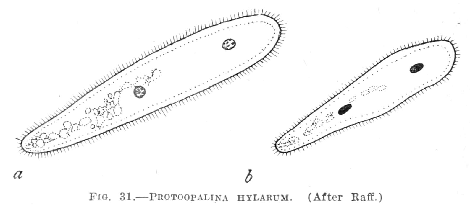
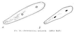
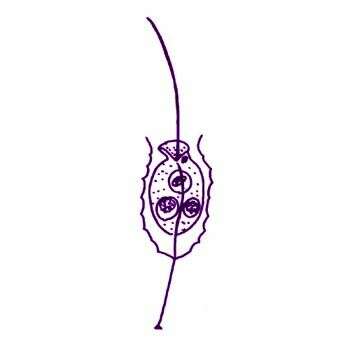
Summary.mw-parser-output table.commons-file-information-table,.mw-parser-output.fileinfotpl-type-information{border:1px solid #a2a9b1;background-color:#f8f9fa;padding:5px;font-size:95%;border-spacing:2px;box-sizing:border-box;margin:0;width:100%}.mw-parser-output table.commons-file-information-table>tbody>tr,.mw-parser-output.fileinfotpl-type-information>tbody>tr{vertical-align:top}.mw-parser-output table.commons-file-information-table>tbody>tr>td,.mw-parser-output table.commons-file-information-table>tbody>tr>th,.mw-parser-output.fileinfotpl-type-information>tbody>tr>td,.mw-parser-output.fileinfotpl-type-information>tbody>tr>th{padding:4px}.mw-parser-output.fileinfo-paramfield{background:#ccf;text-align:right;padding-right:0.4em;width:15%;font-weight:bold}.mw-parser-output.commons-file-information-table+table.commons-file-information-table,.mw-parser-output.commons-file-information-table+div.commons-file-information-table>table{border-top:0;padding-top:0;margin-top:-8px}@media only screen and (max-width:719px){.mw-parser-output table.commons-file-information-table,.mw-parser-output.commons-file-information-table.fileinfotpl-type-information{border-spacing:0;padding:0;word-break:break-word;width:100%!important}.mw-parser-output.commons-file-information-table>tbody,.mw-parser-output.fileinfotpl-type-information>tbody{display:block}.mw-parser-output.commons-file-information-table>tbody>tr>td,.mw-parser-output.commons-file-information-table>tbody>tr>th,.mw-parser-output.fileinfotpl-type-information>tbody>tr>td,.mw-parser-output.fileinfotpl-type-information>tbody>tr>th{padding:0.2em 0.4em;text-align:left;text-align:start}.mw-parser-output.commons-file-information-table>tbody>tr,.mw-parser-output.fileinfotpl-type-information>tbody>tr{display:flex;flex-direction:column}.mw-parser-output.commons-file-information-table+table.commons-file-information-table,.mw-parser-output.commons-file-information-table+div.commons-file-information-table>table{margin-top:-1px}.mw-parser-output.fileinfo-paramfield{box-sizing:border-box;flex:1 0 100%;width:100%}} Description: English: « Protoopalina hylarum » =
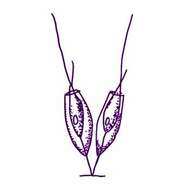
Protoopalina hylarumFrançais : « Protoopalina hylarum » =
Protoopalina hylarum. Date: 1923. Source:
Bulletin of the United States National Museum, vol. 120. Author: Maynard M. Metcalf (1868-1940), after Raff 1911.
-
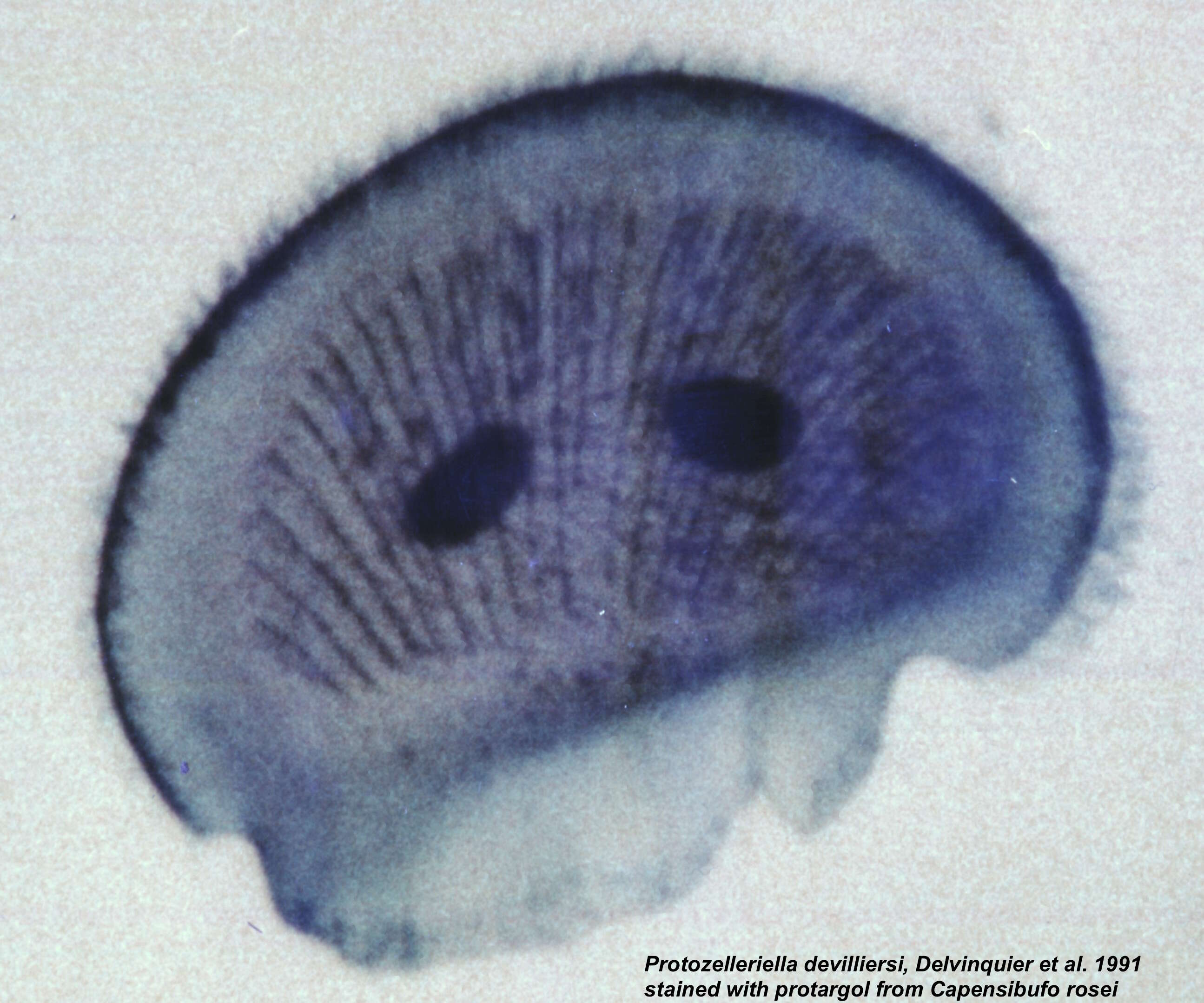
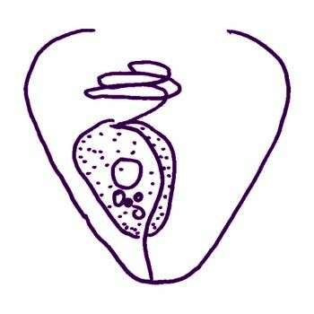
Description: English: Protargol stained specimen of Protozelleriella devilliersi. Date: 2 December 2019. Source: Own work. Author:
Ménidolcien. Camera location
34° 05′ 45″ S, 18° 27′ 00″ E View all coordinates using:
OpenStreetMap-34.095833; 18.450000. Holotypes and paratypes deposited in various museums (BM, USNM, AMNH, MCZ, TM and QM) Licensing[
edit] : This file is licensed under the
Creative Commons Attribution-Share Alike 4.0 International license. :. You are free: to share – to copy, distribute and transmit the work to remix – to adapt the work Under the following conditions: attribution – You must give appropriate credit, provide a link to the license, and indicate if changes were made. You may do so in any reasonable manner, but not in any way that suggests the licensor endorses you or your use. share alike – If you remix, transform, or build upon the material, you must distribute your contributions under the
same or compatible license as the original. https://creativecommons.org/licenses/by-sa/4.0 CC BY-SA 4.0 Creative Commons Attribution-Share Alike 4.0 truetrue.
-
San Martin De Castaneda, Castille and Leon, Spain
-
Rhizomonas setigera (Pavillard, 1916) Patterson et al., 1993. Cells found adhering to detritus, with one flagellum up to 25 microns long. The cell body is 6-20 microns in diameter and often embedded in mucus. Cells may produce elongate pseudopodia.
-
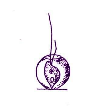
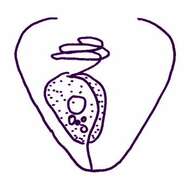
Hedraeophysa bulla Kent, 1880. Lorica subglobose, bubble-like, anterior aperture of small dimensions, organism occupying the greater portion of the cavity of the lorica, attached to its bottom, height of lorica 6.4 microns
-
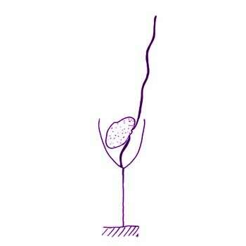
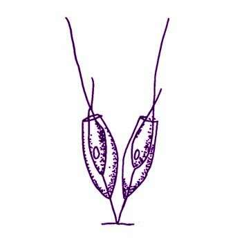
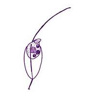
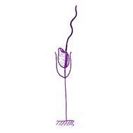
Bicosoeca conica Lemmermann, 1912. Bicosoeca cells with a body 2.5-5 microns in diameter, anterior flagellum 7-10 microns long, cell lives in a cone-shaped lorica with a stalk about 20 microns long that is slightly expanded at the point of attachment to the substrate.
-
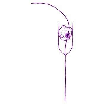
Bicosoeca lacustris James-Clark, 1867. Bicosoeca cells measuring 4-7 x 2.5-4 microns The lorica is an elongate goblet-shape with a rounded posterior end and a rather thick pedicel terminating in a distal button. The margin of the lorica curves inwards slightly when the cell is in its extended shape, and closes partially or completely when the cell retracts. The anterior flagellum tends to be held out at an oblique angle, and the posterior flagellum either narrows at the distal tip or else is connected to the lorica by a fine filament.
-
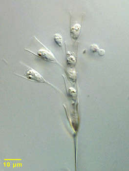
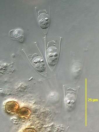
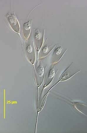
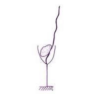
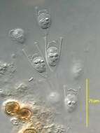
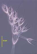
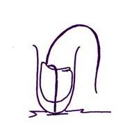
Portrait of Bicosoeca petiolata, a colorless loricate flagellate (synonymous with Poteriodendron petiolatum). This species is colonial with daughter cells forming stalks attached to the mother lorica after division. Most species of this genus are solitary. The cells of this colony are retracted with the long hair bearing flagellum tightly coiled on the anterior end of the cell (seen well in the cell on your left in this image). The second short smooth flagellum is directed posteriorly attaching the cell to its lorica. The lorica is organic. From fresh water marsh with Typha (Cattail) near Boise, Idaho. Differential interference contrast.
-
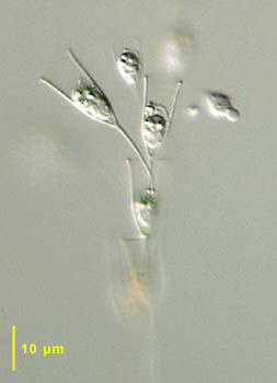
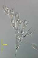
Portrait of Bicosoeca petiolata, a colorless loricate flagellate (synonymous with Poteriodendron petiolatum). This species is colonial with daughter cells forming stalks attached to the mother lorica after division. Most species of this genus are solitary. The cells of this colony are retracted. The second short smooth flagellum is directed posteriorly attaching the cell to its lorica. The lorica is organic. From fresh water marsh with Typha (Cattail) near Boise, Idaho. Differential interference contrast.
-
-
-
-
Bicosoeca epiphytica Hilliard, 1971. Bicosoeca cells with a body 3.5-6.5 x 2.5-4.5 microns long, lorica 8-10x3.5-5 microns The lorica of this species has an undulating surface. The lorica has about 8-10 ridges and is rounded at the posterior. It tapers slightly at the anterior, before a flared aperture. The lorica has a short pedicel, which is 0.5-1 times the lorica length, and terminates with a distal button as is characteristic in this genus.
-
Bicosoeca gracilipes James-Clark, 1867. Bicosoeca cells with a body that is oval, 3.5-7 x 3-4.5 microns Cells may be nearly the same size as the lorica or else only half as long. Two flagella insert at the base of the cytostome. The posterior flagellum lies in a conspicuous longitudinal groove and attaches directly to the base of the lorica. The anterior flagellum varies between 2 and 5 times the cell length, and is held in almost the same axis as the cell, but is slightly curved. The lorica is thin-walled and has almost parallel sides, but is slightly pointed at the posterior. The anterior margin of the lorica is closely adpressed to extended cells and can only be seen when the cell retracts. It usually curves outwards slightly, but may also be straight, or curve inwards slightly. The lorica has a fine pedicel of 2.5-4.5 times the cell length. The nucleus is posterior.
-
Bicosoeca maris Picken, 1941. Bicosoeca cells measuring 4.5-6.5 microns, D-shaped with an inconspicuous lip anteriorly. Anterior flagellum about 2-2.5 times cell length, held in a loose coil when cells are retracted to the posterior of the lorica. Posterior flagellum lies in a shallow longitudinal groove, which passes down the cell and attaches to the base of the lorica. The lorica has parallel sides, with a rounded posterior and a slightly everted anterior margin. It measures about 10 microns in length and is about half as wide.
-
Bicosoeca planctonica Kisselew 1931. Bicosoeca cells with lorica measuring 10-15 x 10-15 microns (11-15x13-17 microns), cell diameter about 5 microns Lorica shaped like a bowl with rounded bottom and wide anterior opening with more or less incurved margin. The protoplast is rounded or slightly triangular in outline.
-
Bicosoeca pocillum Kent, 1880. Bicosoeca cells in which the lorica is cup-shaped or subcylindrical, rounded posteriorly, the anterior margin abruptly truncate, neither everted or constricted, varying in height from one and a half to two or three times its greatest breadth, pedicel short, rarely half as high as the lorica, cell is rounded posteriorly, the anterior margin flattened, with on one side as a broad, flattened, lip-like process, occupying from one third to one half of the cavity of the lorica, posterior retractile ligament equalling the body in length, contractile vacuole posteriorly situated, nucleus spherical, subcentral. Length of lorica 10.2 to 17 microns, cell 7.8 microns
-
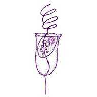
Bicosoeca pulchra Hilliard, 1971. Bicosoeca cells with a lorica that is 19.3-20.2 microns long, 9.7-10.6 microns wide. The lorica is delicately urn-shaped, posteriorly pointed, with a short strip appended to the base. There is a slight constriction just below a moderately flaring opening, while the lip at the oral rim may converge slightly. The lorica wall is thin, hyaline, and appears serrate in optical profile when observed in aqueous methylene blue. Staining reveals the presence of transverse bands, which number 10-12 in 10 microns The spherical protoplast (6.5-7 microns long, 4.5-6 microns wide) is colourless, granular, and has a central nucleus and a vacuole. A single flagellum bearing two rows of submicroscopic hairs extends dorsally from the cell and is about three times its length.
-
Bicosoeca tenuis Kent, 1880. Bicosoeca cells with a lorica that is elongate-ovate or subfusiforme, nearly three times as long as broad, tapering equally at each extremity, pedicel scarcely one-half the height of the lorica, protoplast with a lip-like projection, slightly exsert anteriorly. Length of lorica 8.5 to 10.2 microns The assignment of this species to Bicosoeca is incorrect, because in Bicosoeca, one flagellum attaches the cell to the lower end of the lorica, whereas here both flagella project from the opening of the lorica.
-
Bicosoeca vacillans Stole, 1888. Bicosoeca cells located in a stalked lorica, the chamber of which measured 17-25 microns long. The lorica chamber was approximately cylindrical with a slightly pointed posterior and a pedicel of 1-1.5 times the lorica length. Loricas may differ slightly in shape. The lorica wall was most frequently curved slightly outwards at the aperture to a greater or lesser extent and in some cases the lorica was waisted below the aperture. The lorica had fine horizontal bands spaced about 0.4 microns apart, with numerous fine perpendicular fibres in each band. Cells were sub-spherical and had a flattened indistinct peristome. The anterior flagellum was about 3 times the cell length and held at an angle to the longitudinal axis of the cell. The posterior flagellum was attached to the base of the lorica via a fine thread.


