-
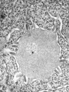
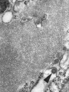
This transmission electron micrograph (TEM) revealed the presence of Lagos bat virus (LBV) virions, and an intracytoplasmic inclusion body in this tissue sample. LBV is a Rhabdoviridae family member, and a member of the genus, Lyssavirus.Created: 1975
-
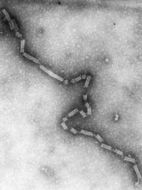
This negatively-stained transmission electron micrograph (TEM) revealed the presence of numerous negative-sense, single-stranded RNA ((-) ssRNA) Flanders virus virions. Note the bullet-like shape of these virions, which are very similar to other Rhabdoviruses, i.e., see PHIL 1876 depicting a TEM revealing the bullet-shaped rabies virus virions.Created: 1975
-
This negatively-stained transmission electron micrograph (TEM) revealed the presence of numerous bovine ephemeral fever virus virions, which are members of the Rhabdoviridae family of viruses, and the genus Ephemerovirus, infecting animals as well as plants.Created: 1975
-
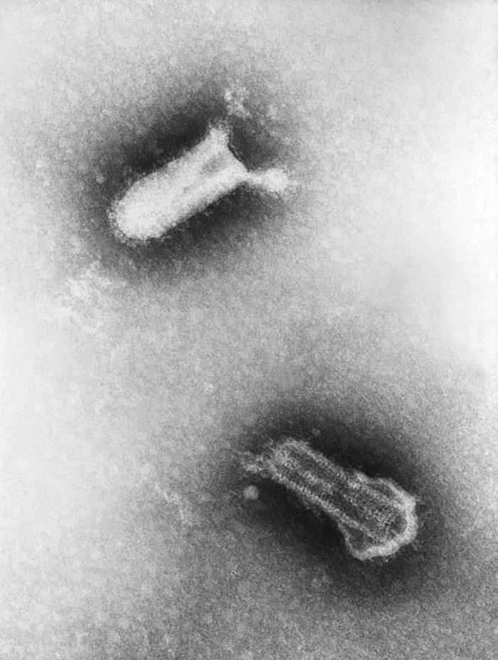
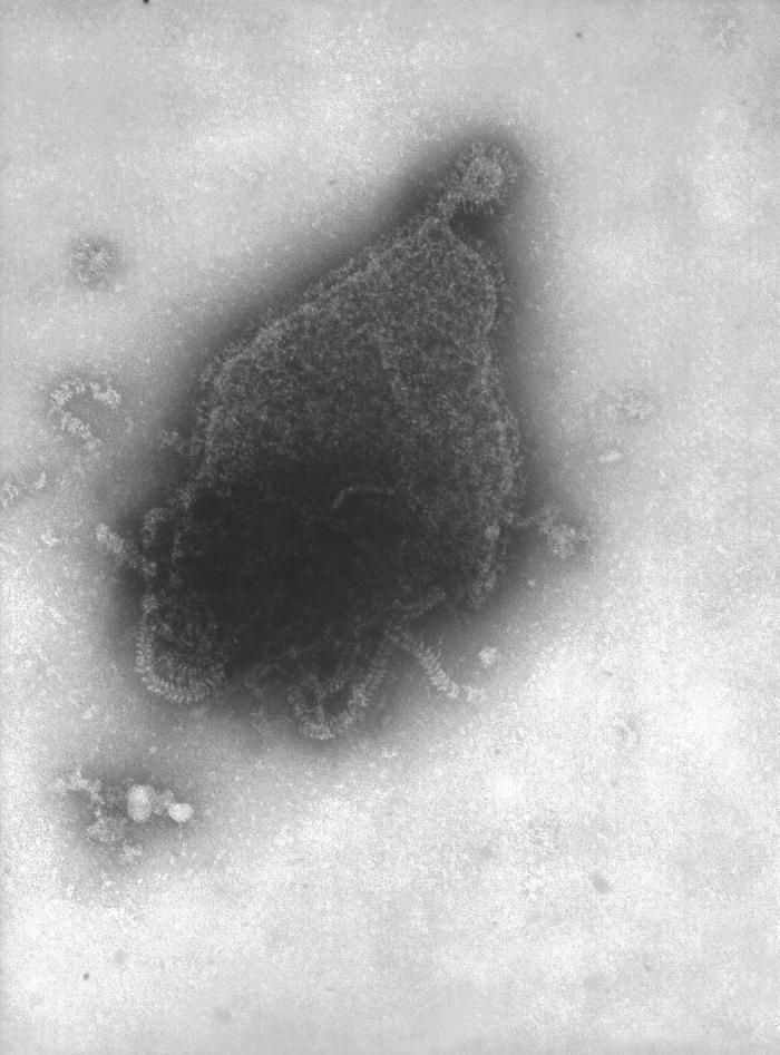
This negatively-stained transmission electron micrograph (TEM) revealed the presence of two Piry virus virions. Note the bullet-like shape of the small 155nm x 162nm virions. Normally, under electron microscopic examination, the virions are observed as being discoidal or spheroidal in shape, and only rarely as bullet-shaped, as was the case here.Created: 1975
-
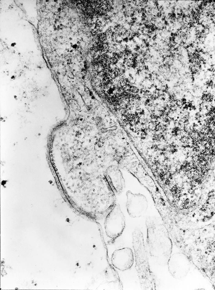
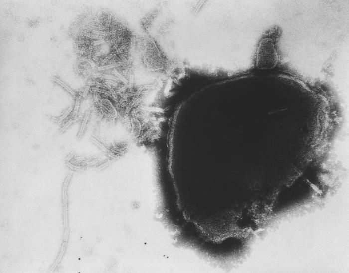
This negatively-stained transmission electron micrograph (TEM) revealed the presence of numerous Piry virus virions, many of which could be seen as they were budding from the host cell, thereby, becoming free to migrate throughout the hosts system. Note the bullet-like shape of the small 155nm x 162nm virions, as theyre freed from the host cell. Normally, under electron microscopic examination, the virions are observed as being discoidal or spheroidal in shape, and only rarely as bullet-shaped, as was the case here.Created: 1975
-
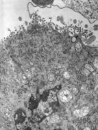
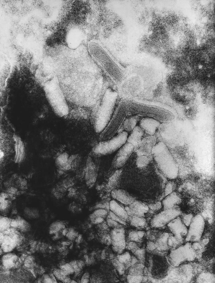
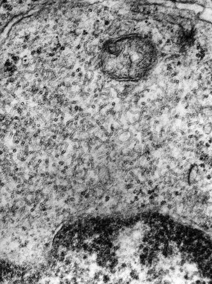
This transmission electron micrograph (TEM) revealed some of the internal cross-sectional structural morphology of a rabies virion (arrow) in this central nervous system tissue specimen. The virion is adjacent to a Negri body, which is pathognomonic in the positive diagnosis for Rabies. The virus infects the central nervous system, causing encephalopathy and ultimately death. Rabies virus belongs to the order Mononegavirales, viruses with a nonsegmented, negative-sense single-stranded RNA ((-) ssRNA) genomes. Within this group, viruses with a distinct "bullet" shape are classified in the Rhabdoviridae family, which includes at least three genera of animal viruses, Lyssavirus, Ephemerovirus, and Vesiculovirus. The genus Lyssavirus includes rabies virus, Lagos bat, Mokola virus, Duvenhage virus, European bat virus 1 & 2 and Australian bat virus.Created: 1975
-
This transmission electron micrograph (TEM) revealed some of the nucleocapsid morphologic features displayed by the human parainfluenza virus Type-4a (HPIV-4), a member of the Paramyxoviridae family. These viruses possess a genome consisting of negative-sense single-stranded RNA ((-) ssRNA).Each of the four HPIVs has different clinical and epidemiologic features. The most distinctive clinical feature of HPIV-1 and HPIV-2 is croup (i.e., laryngotracheobronchitis); HPIV-1 is the leading cause of croup in children, whereas HPIV-2 is less frequently detected. Both HPIV-1 and -2 can cause other upper and lower respiratory tract illnesses. HPIV-3 is more often associated with bronchiolitis and pneumonia. HPIV-4 is infrequently detected, possibly because it is less likely to cause severe disease. The incubation period for HPIVs is generally from 1 to 7 days.Created: 1975
-
This transmission electron micrograph (TEM) revealed the presence of the human parainfluenza type 4A virus (HPIV-4A), which like the mumps virus, is also a Paramyxoviridae family member, and a member of the genus, Rubulavirus.Created: 1975
-
This transmission electron micrograph (TEM) revealed the presence of the human parainfluenza type 4A virus (HPIV-4A), which like the mumps virus, is also a Paramyxoviridae family member, and a member of the genus, Rubulavirus.Created: 1975
-
This transmission electron micrograph (TEM) revealed the presence of numerous paramyxovirus virions, which in this instance, were responsible for a case of the mumps. Paramyxoviruses are members of the family, Paramyxoviridae, and those that cause mumps in humans belong to the genus, Rubulavirus. The virus itself can present itself in a number of morphologic shapes, including spherical, and stand-like, or filamentous, ranging from 150nm to 200nm in diameter, and 1000nm to 10000nm in length. At its core lies a non-segmented, negative-sense RNA genome.Created: 1975
-
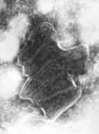
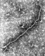
Under a highly magnified view of 168,000x this transmission electron micrographic (TEM) image revealed ultrastructural details of a Nipah virus nucleocapsid, a virus which was named for the location in Malaysia where it was first isolated.Created: 1999
-
Summary.mw-parser-output table.commons-file-information-table,.mw-parser-output.fileinfotpl-type-information{border:1px solid #a2a9b1;background-color:#f8f9fa;padding:5px;font-size:95%;border-spacing:2px;box-sizing:border-box;margin:0;width:100%}.mw-parser-output table.commons-file-information-table>tbody>tr,.mw-parser-output.fileinfotpl-type-information>tbody>tr{vertical-align:top}.mw-parser-output table.commons-file-information-table>tbody>tr>td,.mw-parser-output table.commons-file-information-table>tbody>tr>th,.mw-parser-output.fileinfotpl-type-information>tbody>tr>td,.mw-parser-output.fileinfotpl-type-information>tbody>tr>th{padding:4px}.mw-parser-output.fileinfo-paramfield{background:#ccf;text-align:right;padding-right:0.4em;width:15%;font-weight:bold}.mw-parser-output.commons-file-information-table+table.commons-file-information-table,.mw-parser-output.commons-file-information-table+div.commons-file-information-table>table{border-top:0;padding-top:0;margin-top:-8px}@media only screen and (max-width:719px){.mw-parser-output table.commons-file-information-table,.mw-parser-output.commons-file-information-table.fileinfotpl-type-information{border-spacing:0;padding:0;word-break:break-word;width:100%!important}.mw-parser-output.commons-file-information-table>tbody,.mw-parser-output.fileinfotpl-type-information>tbody{display:block}.mw-parser-output.commons-file-information-table>tbody>tr>td,.mw-parser-output.commons-file-information-table>tbody>tr>th,.mw-parser-output.fileinfotpl-type-information>tbody>tr>td,.mw-parser-output.fileinfotpl-type-information>tbody>tr>th{padding:0.2em 0.4em;text-align:left;text-align:start}.mw-parser-output.commons-file-information-table>tbody>tr,.mw-parser-output.fileinfotpl-type-information>tbody>tr{display:flex;flex-direction:column}.mw-parser-output.commons-file-information-table+table.commons-file-information-table,.mw-parser-output.commons-file-information-table+div.commons-file-information-table>table{margin-top:-1px}.mw-parser-output.fileinfo-paramfield{box-sizing:border-box;flex:1 0 100%;width:100%}} Description: English: Creative layout featuring 3D renderings of respiratory syncytial virus (RSV), a common contagious virus that infects the human respiratory tract. Credit: NIAID. Date: 20 September 2022, 04:45:06. Source:
https://www.flickr.com/photos/54591706@N02/52870645474/. Author: NIAID.
-
Swapnil Subhash Bawage, Pooja Munnilal Tiwari, Shreekumar Pillai, Vida Dennis, and Shree Ram Singh
Wikimedia Commons
Description: English: Structure and genome organization of respiratory syncytial virus (RSV). (a) Approximately 200 nm RSV virion particle and (b) single stranded negative RNA genome consisting of 10 genes. Date: 9 December 2013. Source:
https://www.hindawi.com/journals/av/2013/595768/. Author: Swapnil Subhash Bawage, Pooja Munnilal Tiwari, Shreekumar Pillai, Vida Dennis, and Shree Ram Singh.
-
Description: English: This electron micrograph depicts the Respiratory Syncytial Virus (RSV) pathogen. RSV is a negative-sense, enveloped RNA virus. The virion is variable in shape and size, with a diameter ranging between 120 and 300 nm, and is unstable in the environment surviving only a few hours on environmental surfaces. Polski: RSV w elektronowym mikroskopie transmisyjnym. Norsk bokmål: RS-virus. Date: 1981. Source: : This media comes from the
Centers for Disease Control and Prevention's
Public Health Image Library (PHIL), with identification number
#276. Note: Not all PHIL images are public domain; be sure to check copyright status and credit authors and content providers.
العربية |
Deutsch |
English |
македонски |
slovenščina |
+/−. Originally from
en.wikipedia; description page is/was
here. Author: Photo Credit: Content Providers(s): CDC/ Dr. Erskine Palmer. Permission(
Reusing this file): PD-USGov-HHS-CDC English: None - This image is in the public domain and thus free of any copyright restrictions. As a matter of courtesy we request that the content provider be credited and notified in any public or private usage of this image.
-
Summary.mw-parser-output table.commons-file-information-table,.mw-parser-output.fileinfotpl-type-information{border:1px solid #a2a9b1;background-color:#f8f9fa;padding:5px;font-size:95%;border-spacing:2px;box-sizing:border-box;margin:0;width:100%}.mw-parser-output table.commons-file-information-table>tbody>tr,.mw-parser-output.fileinfotpl-type-information>tbody>tr{vertical-align:top}.mw-parser-output table.commons-file-information-table>tbody>tr>td,.mw-parser-output table.commons-file-information-table>tbody>tr>th,.mw-parser-output.fileinfotpl-type-information>tbody>tr>td,.mw-parser-output.fileinfotpl-type-information>tbody>tr>th{padding:4px}.mw-parser-output.fileinfo-paramfield{background:#ccf;text-align:right;padding-right:0.4em;width:15%;font-weight:bold}.mw-parser-output.commons-file-information-table+table.commons-file-information-table,.mw-parser-output.commons-file-information-table+div.commons-file-information-table>table{border-top:0;padding-top:0;margin-top:-8px}@media only screen and (max-width:719px){.mw-parser-output table.commons-file-information-table,.mw-parser-output.commons-file-information-table.fileinfotpl-type-information{border-spacing:0;padding:0;word-break:break-word;width:100%!important}.mw-parser-output.commons-file-information-table>tbody,.mw-parser-output.fileinfotpl-type-information>tbody{display:block}.mw-parser-output.commons-file-information-table>tbody>tr>td,.mw-parser-output.commons-file-information-table>tbody>tr>th,.mw-parser-output.fileinfotpl-type-information>tbody>tr>td,.mw-parser-output.fileinfotpl-type-information>tbody>tr>th{padding:0.2em 0.4em;text-align:left;text-align:start}.mw-parser-output.commons-file-information-table>tbody>tr,.mw-parser-output.fileinfotpl-type-information>tbody>tr{display:flex;flex-direction:column}.mw-parser-output.commons-file-information-table+table.commons-file-information-table,.mw-parser-output.commons-file-information-table+div.commons-file-information-table>table{margin-top:-1px}.mw-parser-output.fileinfo-paramfield{box-sizing:border-box;flex:1 0 100%;width:100%}} Description: English: Creative work featuring 3D renditions of three dangerous respiratory viruses: RSV (top left), SARS-CoV-2 (bottom center), and flu (top right). Note: not to scale. Credit: NIAID. Date: 20 September 2022, 04:45:06. Source:
https://www.flickr.com/photos/54591706@N02/52566271818/. Author: NIAID.
-
Swapnil Subhash Bawage, Pooja Munnilal Tiwari, Shreekumar Pillai, Vida Dennis, and Shree Ram Singh
Wikimedia Commons
Description: English: A schematic representation of a simple DNA vaccine administered as a naked DNA vector or functionalized nanoparticle or as an encapsulation for controlled delivery. Date: 9 December 2013. Source:
https://www.hindawi.com/journals/av/2013/595768/. Author: Swapnil Subhash Bawage, Pooja Munnilal Tiwari, Shreekumar Pillai, Vida Dennis, and Shree Ram Singh.
-
Summary.mw-parser-output table.commons-file-information-table,.mw-parser-output.fileinfotpl-type-information{border:1px solid #a2a9b1;background-color:#f8f9fa;padding:5px;font-size:95%;border-spacing:2px;box-sizing:border-box;margin:0;width:100%}.mw-parser-output table.commons-file-information-table>tbody>tr,.mw-parser-output.fileinfotpl-type-information>tbody>tr{vertical-align:top}.mw-parser-output table.commons-file-information-table>tbody>tr>td,.mw-parser-output table.commons-file-information-table>tbody>tr>th,.mw-parser-output.fileinfotpl-type-information>tbody>tr>td,.mw-parser-output.fileinfotpl-type-information>tbody>tr>th{padding:4px}.mw-parser-output.fileinfo-paramfield{background:#ccf;text-align:right;padding-right:0.4em;width:15%;font-weight:bold}.mw-parser-output.commons-file-information-table+table.commons-file-information-table,.mw-parser-output.commons-file-information-table+div.commons-file-information-table>table{border-top:0;padding-top:0;margin-top:-8px}@media only screen and (max-width:719px){.mw-parser-output table.commons-file-information-table,.mw-parser-output.commons-file-information-table.fileinfotpl-type-information{border-spacing:0;padding:0;word-break:break-word;width:100%!important}.mw-parser-output.commons-file-information-table>tbody,.mw-parser-output.fileinfotpl-type-information>tbody{display:block}.mw-parser-output.commons-file-information-table>tbody>tr>td,.mw-parser-output.commons-file-information-table>tbody>tr>th,.mw-parser-output.fileinfotpl-type-information>tbody>tr>td,.mw-parser-output.fileinfotpl-type-information>tbody>tr>th{padding:0.2em 0.4em;text-align:left;text-align:start}.mw-parser-output.commons-file-information-table>tbody>tr,.mw-parser-output.fileinfotpl-type-information>tbody>tr{display:flex;flex-direction:column}.mw-parser-output.commons-file-information-table+table.commons-file-information-table,.mw-parser-output.commons-file-information-table+div.commons-file-information-table>table{margin-top:-1px}.mw-parser-output.fileinfo-paramfield{box-sizing:border-box;flex:1 0 100%;width:100%}} Description: English: Creative layout featuring 3D renderings of respiratory syncytial virus (RSV), a common contagious virus that infects the human respiratory tract. Credit: NIAID See May 4, 2023 press release, "NIH Celebrates FDA Approval of RSV Vaccine for People 60 Years of Age and Older," at www.niaid.nih.gov/news-events/nih-celebrates-fda-approval.. Date: 20 September 2022, 04:45:06. Source:
https://www.flickr.com/photos/54591706@N02/52869900362/. Author: NIAID.
-
Summary.mw-parser-output table.commons-file-information-table,.mw-parser-output.fileinfotpl-type-information{border:1px solid #a2a9b1;background-color:#f8f9fa;padding:5px;font-size:95%;border-spacing:2px;box-sizing:border-box;margin:0;width:100%}.mw-parser-output table.commons-file-information-table>tbody>tr,.mw-parser-output.fileinfotpl-type-information>tbody>tr{vertical-align:top}.mw-parser-output table.commons-file-information-table>tbody>tr>td,.mw-parser-output table.commons-file-information-table>tbody>tr>th,.mw-parser-output.fileinfotpl-type-information>tbody>tr>td,.mw-parser-output.fileinfotpl-type-information>tbody>tr>th{padding:4px}.mw-parser-output.fileinfo-paramfield{background:#ccf;text-align:right;padding-right:0.4em;width:15%;font-weight:bold}.mw-parser-output.commons-file-information-table+table.commons-file-information-table,.mw-parser-output.commons-file-information-table+div.commons-file-information-table>table{border-top:0;padding-top:0;margin-top:-8px}@media only screen and (max-width:719px){.mw-parser-output table.commons-file-information-table,.mw-parser-output.commons-file-information-table.fileinfotpl-type-information{border-spacing:0;padding:0;word-break:break-word;width:100%!important}.mw-parser-output.commons-file-information-table>tbody,.mw-parser-output.fileinfotpl-type-information>tbody{display:block}.mw-parser-output.commons-file-information-table>tbody>tr>td,.mw-parser-output.commons-file-information-table>tbody>tr>th,.mw-parser-output.fileinfotpl-type-information>tbody>tr>td,.mw-parser-output.fileinfotpl-type-information>tbody>tr>th{padding:0.2em 0.4em;text-align:left;text-align:start}.mw-parser-output.commons-file-information-table>tbody>tr,.mw-parser-output.fileinfotpl-type-information>tbody>tr{display:flex;flex-direction:column}.mw-parser-output.commons-file-information-table+table.commons-file-information-table,.mw-parser-output.commons-file-information-table+div.commons-file-information-table>table{margin-top:-1px}.mw-parser-output.fileinfo-paramfield{box-sizing:border-box;flex:1 0 100%;width:100%}} Description: English: Creative layout featuring 3D renderings of respiratory syncytial virus (RSV), a common contagious virus that infects the human respiratory tract. Credit: NIAID. Date: 20 September 2022, 04:45:06. Source:
https://www.flickr.com/photos/54591706@N02/52870479521/. Author: NIAID.
-
Summary.mw-parser-output table.commons-file-information-table,.mw-parser-output.fileinfotpl-type-information{border:1px solid #a2a9b1;background-color:#f8f9fa;padding:5px;font-size:95%;border-spacing:2px;box-sizing:border-box;margin:0;width:100%}.mw-parser-output table.commons-file-information-table>tbody>tr,.mw-parser-output.fileinfotpl-type-information>tbody>tr{vertical-align:top}.mw-parser-output table.commons-file-information-table>tbody>tr>td,.mw-parser-output table.commons-file-information-table>tbody>tr>th,.mw-parser-output.fileinfotpl-type-information>tbody>tr>td,.mw-parser-output.fileinfotpl-type-information>tbody>tr>th{padding:4px}.mw-parser-output.fileinfo-paramfield{background:#ccf;text-align:right;padding-right:0.4em;width:15%;font-weight:bold}.mw-parser-output.commons-file-information-table+table.commons-file-information-table,.mw-parser-output.commons-file-information-table+div.commons-file-information-table>table{border-top:0;padding-top:0;margin-top:-8px}@media only screen and (max-width:719px){.mw-parser-output table.commons-file-information-table,.mw-parser-output.commons-file-information-table.fileinfotpl-type-information{border-spacing:0;padding:0;word-break:break-word;width:100%!important}.mw-parser-output.commons-file-information-table>tbody,.mw-parser-output.fileinfotpl-type-information>tbody{display:block}.mw-parser-output.commons-file-information-table>tbody>tr>td,.mw-parser-output.commons-file-information-table>tbody>tr>th,.mw-parser-output.fileinfotpl-type-information>tbody>tr>td,.mw-parser-output.fileinfotpl-type-information>tbody>tr>th{padding:0.2em 0.4em;text-align:left;text-align:start}.mw-parser-output.commons-file-information-table>tbody>tr,.mw-parser-output.fileinfotpl-type-information>tbody>tr{display:flex;flex-direction:column}.mw-parser-output.commons-file-information-table+table.commons-file-information-table,.mw-parser-output.commons-file-information-table+div.commons-file-information-table>table{margin-top:-1px}.mw-parser-output.fileinfo-paramfield{box-sizing:border-box;flex:1 0 100%;width:100%}} Description: English: Creative layout featuring 3D renderings of respiratory syncytial virus (RSV), a common contagious virus that infects the human respiratory tract. Credit: NIAID. Date: 20 September 2022, 04:45:06. Source:
https://www.flickr.com/photos/54591706@N02/52870646799/. Author: NIAID.
-
Summary.mw-parser-output table.commons-file-information-table,.mw-parser-output.fileinfotpl-type-information{border:1px solid #a2a9b1;background-color:#f8f9fa;padding:5px;font-size:95%;border-spacing:2px;box-sizing:border-box;margin:0;width:100%}.mw-parser-output table.commons-file-information-table>tbody>tr,.mw-parser-output.fileinfotpl-type-information>tbody>tr{vertical-align:top}.mw-parser-output table.commons-file-information-table>tbody>tr>td,.mw-parser-output table.commons-file-information-table>tbody>tr>th,.mw-parser-output.fileinfotpl-type-information>tbody>tr>td,.mw-parser-output.fileinfotpl-type-information>tbody>tr>th{padding:4px}.mw-parser-output.fileinfo-paramfield{background:#ccf;text-align:right;padding-right:0.4em;width:15%;font-weight:bold}.mw-parser-output.commons-file-information-table+table.commons-file-information-table,.mw-parser-output.commons-file-information-table+div.commons-file-information-table>table{border-top:0;padding-top:0;margin-top:-8px}@media only screen and (max-width:719px){.mw-parser-output table.commons-file-information-table,.mw-parser-output.commons-file-information-table.fileinfotpl-type-information{border-spacing:0;padding:0;word-break:break-word;width:100%!important}.mw-parser-output.commons-file-information-table>tbody,.mw-parser-output.fileinfotpl-type-information>tbody{display:block}.mw-parser-output.commons-file-information-table>tbody>tr>td,.mw-parser-output.commons-file-information-table>tbody>tr>th,.mw-parser-output.fileinfotpl-type-information>tbody>tr>td,.mw-parser-output.fileinfotpl-type-information>tbody>tr>th{padding:0.2em 0.4em;text-align:left;text-align:start}.mw-parser-output.commons-file-information-table>tbody>tr,.mw-parser-output.fileinfotpl-type-information>tbody>tr{display:flex;flex-direction:column}.mw-parser-output.commons-file-information-table+table.commons-file-information-table,.mw-parser-output.commons-file-information-table+div.commons-file-information-table>table{margin-top:-1px}.mw-parser-output.fileinfo-paramfield{box-sizing:border-box;flex:1 0 100%;width:100%}} Description: English: Creative work featuring 3D renditions of three dangerous respiratory viruses: RSV (top left), SARS-CoV-2 (bottom center), and flu (top right). Note: not to scale. Credit: NIAID. Date: 20 September 2022, 04:45:06. Source:
https://www.flickr.com/photos/54591706@N02/52573302046/. Author: NIAID.
-
Swapnil Subhash Bawage, Pooja Munnilal Tiwari, Shreekumar Pillai, Vida Dennis, and Shree Ram Singh
Wikimedia Commons
Description: English: A schematic representation of biophysical method of respiratory syncytial virus (RSV) detection. Surface enhanced Raman spectroscopy. Date: 9 December 2013. Source:
https://www.hindawi.com/journals/av/2013/595768/. Author: Swapnil Subhash Bawage, Pooja Munnilal Tiwari, Shreekumar Pillai, Vida Dennis, and Shree Ram Singh.
-
Swapnil Subhash Bawage, Pooja Munnilal Tiwari, Shreekumar Pillai, Vida Dennis, and Shree Ram Singh
Wikimedia Commons
Description: English: A schematic representation of respiratory syncytial virus (RSV) management through coordinated diagnosis, prevention, and treatment. Date: 9 December 2013. Source:
https://www.hindawi.com/journals/av/2013/595768/. Author: Swapnil Subhash Bawage, Pooja Munnilal Tiwari, Shreekumar Pillai, Vida Dennis, and Shree Ram Singh.
-
Summary.mw-parser-output table.commons-file-information-table,.mw-parser-output.fileinfotpl-type-information{border:1px solid #a2a9b1;background-color:#f8f9fa;padding:5px;font-size:95%;border-spacing:2px;box-sizing:border-box;margin:0;width:100%}.mw-parser-output table.commons-file-information-table>tbody>tr,.mw-parser-output.fileinfotpl-type-information>tbody>tr{vertical-align:top}.mw-parser-output table.commons-file-information-table>tbody>tr>td,.mw-parser-output table.commons-file-information-table>tbody>tr>th,.mw-parser-output.fileinfotpl-type-information>tbody>tr>td,.mw-parser-output.fileinfotpl-type-information>tbody>tr>th{padding:4px}.mw-parser-output.fileinfo-paramfield{background:#ccf;text-align:right;padding-right:0.4em;width:15%;font-weight:bold}.mw-parser-output.commons-file-information-table+table.commons-file-information-table,.mw-parser-output.commons-file-information-table+div.commons-file-information-table>table{border-top:0;padding-top:0;margin-top:-8px}@media only screen and (max-width:719px){.mw-parser-output table.commons-file-information-table,.mw-parser-output.commons-file-information-table.fileinfotpl-type-information{border-spacing:0;padding:0;word-break:break-word;width:100%!important}.mw-parser-output.commons-file-information-table>tbody,.mw-parser-output.fileinfotpl-type-information>tbody{display:block}.mw-parser-output.commons-file-information-table>tbody>tr>td,.mw-parser-output.commons-file-information-table>tbody>tr>th,.mw-parser-output.fileinfotpl-type-information>tbody>tr>td,.mw-parser-output.fileinfotpl-type-information>tbody>tr>th{padding:0.2em 0.4em;text-align:left;text-align:start}.mw-parser-output.commons-file-information-table>tbody>tr,.mw-parser-output.fileinfotpl-type-information>tbody>tr{display:flex;flex-direction:column}.mw-parser-output.commons-file-information-table+table.commons-file-information-table,.mw-parser-output.commons-file-information-table+div.commons-file-information-table>table{margin-top:-1px}.mw-parser-output.fileinfo-paramfield{box-sizing:border-box;flex:1 0 100%;width:100%}} Description: English: Creative layout featuring 3D renderings of respiratory syncytial virus (RSV), a common contagious virus that infects the human respiratory tract. Credit: NIAID. Date: 20 September 2022, 04:45:06. Source:
https://www.flickr.com/photos/54591706@N02/52870647004/. Author: NIAID.