-
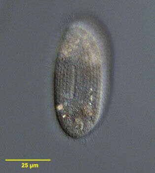
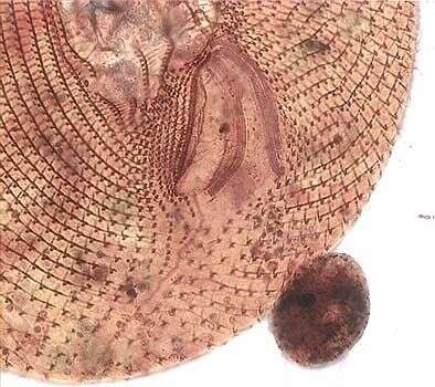
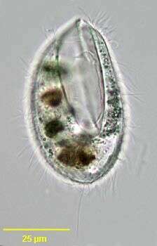
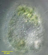
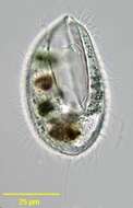
Right dorsolateral surface view of the hymenostome ciliate, Frontonia angusta (Kahl, 1931). Very similar in overall apppearance to F. acuminata (Ehrenberg,1833)Buetschli,1889. F. angusta lacks the anterior apical collection of pigmented granules seen in F. acuminata and its contractile vacuole has 3-4 excretory pores (4 in this case).The approximately 6 µm long extrusomes are clearly visible. Ingested diatoms and green algae are present. Collected from a freshwater pond near Boise, Idaho.DIC.
-
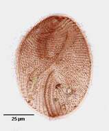
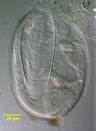
Ventral infraciliature of the hymenostome ciliate, Frontonia angusta (Kahl, 1931). Very similar in overall apppearance to F. acuminata (Ehrenberg,1833)Buetschli,1889. F. angusta lacks the anterior apical collection of pigmented granules seen in F. acuminata and its contractile vacuole has 3-4 excretory pores (not visible here).The prominent preoral and postoral sutures are visible. The 3 curved adoral membranelles are seen on the viewer's right of the oral apparatus. The vestibular ciliary rows are seen to the viewer's left of the the oral apparatus.The postoral ciliary field is seen abutting the posterior margin of the peristome to the viewer's right of the postoral suture.Stained by the silver carbonate technique (see Foissner, W. Europ. J. Protistol., 27:313-330;1991).Collected from a freshwater pond near Boise, Idaho.Brightfield.
-
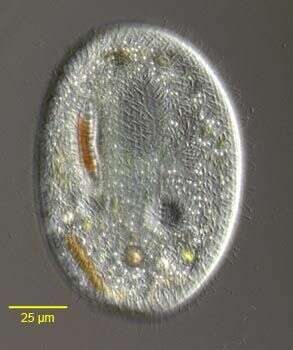
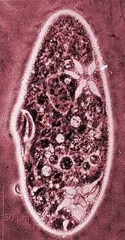
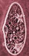
Optical section of the marine frontoniid ciliate, Schistophrya aplanata (Kahl,1933). Schistophrya is a monotypic genus. The cell outline is elongate and bluntly rounded anteriorly and posteriorly. The somatic ciliature is uniform. The pellicle is areolate (marked by uniform rectangular depressions). The slit-like oral aperture is located in mid-body and is bordered by thin slightly serrate lips (not seen in this image). The cytopharyngeal basket of fine trichites is not seen well in these images. A single contractile vacuole is located in the anterior half of the cell. There is a single ovoid macronucleus. A large aggregate of refractile dark granules is present at the anterior end. Fusiform subcortical extrusomes are present (seen in this image). S. aplanata is similar in appearance to the freshwater frontoniid ciliate, Clathrostoma viminale. Collected from a commercial saltwater aquarium in Boise, Idaho February 2004. DIC optics.
-
Portrait of the marine frontoniid ciliate, Schistophrya aplanata (Kahl, 1933). Schistophrya is a monotypic genus. The cell outline is elongate and bluntly rounded anteriorly and posteriorly. The somatic ciliature is uniform. The pellicle is areolate (marked by uniform rectangular depressions). The slit-like oral aperture is located in mid-body and is bordered by thin slightly serrate lips (seen well in this image). The cytopharyngeal basket of fine trichites is not seen well in these images. A single contractile vacuole is located in the anterior half of the cell. There is a single ovoid macronucleus. A large aggregate of refractile dark granules is present at the anterior end. Fusiform subcortical extrusomes are present. S. aplanata is similar in appearance to the freshwater frontoniid ciliate, Clathrostoma viminale. Collected from a commercial saltwater aquarium in Boise, Idaho February 2004. DIC optics.
-
Portrait of the marine frontoniid ciliate, Schistophrya aplanata (Kahl,1933). Schistophrya is a monotypic genus. The cell outline is elongate and bluntly rounded anteriorly and posteriorly. The somatic ciliature is uniform. The pellicle is areolate (marked by uniform rectangular depressions). The slit-like oral aperture is located in mid-body and is bordered by thin slightly serrate lips (seen well in this image). The cytopharyngeal basket of fine trichites is not seen well in these images. A single contractile vacuole is located in the anterior half of the cell. There is a single ovoid macronucleus. A large aggregate of refractile dark granules is present at the anterior end. Fusiform subcortical extrusomes are present. S. aplanata is similar in appearance to the freshwater frontoniid ciliate, Clathrostoma viminale. Collected from a commercial saltwater aquarium in Boise, Idaho February 2004. DIC optics.
-
Optical section of the marine frontoniid ciliate, Schistophrya aplanata (Kahl,1933). Schistophrya is a monotypic genus. The cell outline is elongate and bluntly rounded anteriorly and posteriorly. The somatic ciliature is uniform. The pellicle is areolate (marked by uniform rectangular depressions). The slit-like oral aperture is located in mid-body and is bordered by thin slightly serrate lips (not seen in this image). The cytopharyngeal basket of fine trichites is not seen well in these images. A single contractile vacuole is located in the anterior half of the cell. There is a single ovoid macronucleus. A large aggregate of refractile dark granules is present at the anterior end. Fusiform subcortical extrusomes are present (seen in this image). S. aplanata is similar in appearance to the freshwater frontoniid ciliate, Clathrostoma viminale. Collected from a commercial saltwater aquarium in Boise, Idaho February 2004. DIC optics.
-
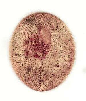
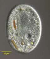
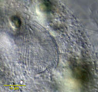
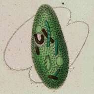
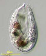
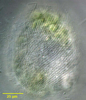
.Portrait of the frontoniid ciliate, Disematostoma buetschlii LAUTERBORN, 1894. D. buetschlii contains endosymbiotic algae but may lose (or digest) them during fall and winter (Ulrike-G.Berninger, Bland J.Finlay, and Hilda M.Canter; The Spatial Distribution and Ecology of Zoochlorellae-Bearing Ciliates in a Productive Pond. J.Protozool. 33(4):557-563, 1986). Although this specimen is slightly smaller (80 microns) than what is commonly reported for D. buetschlii (110 microns) it is otherwise indistinguishable. Kahl describes a smaller species without endosymbiotic algae (D. minor) (A.Kahl; [Urtiere oder Protozoa I: Wimpertiere oder Ciliata (Infusoria) 2. Holotricha]. Die Tierwelt Deutschlands und der angrenzenden Meeresteile. Germany:Verlag von Gustav Fischer. (2)-398). However it is unclear whether this is simply a small variety of D. buetschlii with algal endosymbionts. The cell shape is obovoid tapering to a blunt slightly curved point posteriorly. Dorsal surface convex with a flattened ventral surface. The cytostome (seen well in this image) is located in the anterior 1/3 with 3 left adoral membranelles and an inconspicuous undulating membrane on the right. 4-5 dense vestibular ciliary rows are found on the right of the cytostome. The reniform macronucleus is seen well here. The contractile vacuole is in the posterior half. The longitudinal somatic kineties terminate on prominent ladder-like preoral and postoral suture (the polar band). The preoral suture is seen well in this image. D. buetschlii is primarily algivorous and some of the green algae seen in the cytoplasm in this image may be in food vacuoles. Collected from freshwater pond near Boise, Idaho September 2003. DIC.
-
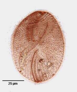
Dorsal surface of the frontoniid ciliate, Disematostoma buetschlii LAUTERBORN, 1894. D. buetschlii contains endosymbiotic algae but may lose (or digest) them during fall and winter (Ulrike-G.Berninger, Bland J.Finlay, and Hilda M.Canter; The Spatial Distribution and Ecology of Zoochlorellae-Bearing Ciliates in a Productive Pond. J.Protozool. 33(4):557-563, 1986). Although this specimen is slightly smaller (80 microns) than what is commonly reported for D. buetschlii (about 110 microns) it is otherwise indistinguishable. Kahl describes a smaller species without endosymbiotic algae (D. minor) (A.Kahl; [Urtiere oder Protozoa I: Wimpertiere oder Ciliata (Infusoria) 2. Holotricha]. Die Tierwelt Deutschlands und der angrenzenden Meeresteile. Germany:Verlag von Gustav Fischer. (2)-398). However it is unclear whether this is simply a small variety of D. buetschlii without algal endosymbionts. The cell shape is obovoid tapering to a blunt slightly curved point posteriorly. Dorsal surface convex with a flattened ventral surface. The cytostome is located in the anterior 1/3 with 3 left adoral membranelles and an inconspicuous undulating membrane on the right. 4-5 dense vestibular ciliary rows are found on the right of the cytostome. The reniform macronucleus is seen well here. The longitudinal somatic kineties terminate on prominent ladder-like preoral and postoral suture (the polar band). The polar band can be seen curving to the viewer's right in this image. The solitary dorsal excretory pore of the contractile vacuole is seen here to the viewer's right of the anterior part of the polar band.D. buetschlii is primarily algivorous and some of the green algae seen in the cytoplasm in this image may be in food vacuoles. Collected from freshwater pond near Boise, Idaho September 2003. DIC.
-

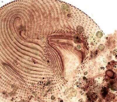
Detail view of the oral apparatus of the frontoniid ciliate, Disematostoma buetschlii LAUTERBORN, 1894. D. buetschlii contains endosymbiotic algae but may lose (or digest) them during fall and winter (Ulrike-G.Berninger, Bland J.Finlay, and Hilda M.Canter; The Spatial Distribution and Ecology of Zoochlorellae-Bearing Ciliates in a Productive Pond. J.Protozool. 33(4):557-563, 1986). Although this specimen is slightly smaller (80 microns) than what is commonly reported for D. buetschlii (? 110 microns) it is otherwise indistinguishable. A smaller species without endosymbiotic algae (D. minor) is described by Kahl (A.Kahl; [Urtiere oder Protozoa I: Wimpertiere oder Ciliata (Infusoria) 2. Holotricha]. Die Tierwelt Deutschlands und der angrenzenden Meeresteile. Germany:Verlag von Gustav Fischer. (2)-398). However it is unclear whether this is simply a small variety of D. buetschlii with algal endosymbionts. The cell shape is obovoid tapering to a blunt slightly curved point posteriorly. Dorsal surface convex with a flattened ventral surface. The cytostome is located in the anterior 1/3 with 3 left adoral membranelles and an inconspicuous undulating membrane on the right. 4-5 dense vestibular ciliary rows are found on the right of the cytostome (seen well in this image). The reniform macronucleus is seen well here. The contractile vacuole is in the posterior half. The longitudinal somatic kineties terminate on prominent ladder-like preoral and postoral suture (the polar band). D. buetschlii is primarily algivorous and some of the green algae seen in the cytoplasm in this image may be in food vacuoles. Collected from freshwater pond near Boise, Idaho September 2003. DIC.
-
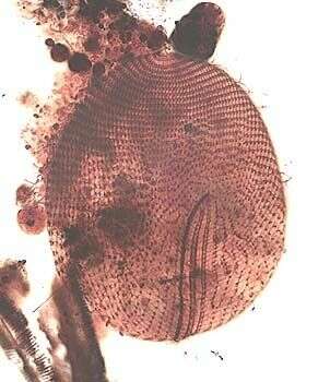
Right ventrolateral view of the infraciliature of Disematostoma buetschlii LAUTERBORN, 1894.Stained by the silver carbonate technique (see Foissner, W. Europ. J. Protistol., 27:313-330;1991).Brightfield.
-
Right dorsolateral view of the infraciliature of Disematostoma buetschlii LAUTERBORN, 1894.Stained by the silver carbonate technique (see Foissner, W. Europ. J. Protistol., 27:313-330;1991).Brightfield.
-
Dorsal view of the infraciliature of Disematostoma buetschlii LAUTERBORN, 1894.Stained by the silver carbonate technique (see Foissner, W. Europ. J. Protistol., 27:313-330;1991).Brightfield.
-
Dorsal view of the infraciliature of Disematostoma buetschlii LAUTERBORN, 1894.Stained by the silver carbonate technique (see Foissner, W. Europ. J. Protistol., 27:313-330;1991).Brightfield.
-
Detail of the oral infraciliature of Disematostoma buetschlii LAUTERBORN, 1894.Stained by the silver carbonate technique (see Foissner, W. Europ. J. Protistol., 27:313-330;1991).Brightfield.
-
Detail of the oral infraciliature of Disematostoma buetschlii LAUTERBORN, 1894.Stained by the silver carbonate technique (see Foissner, W. Europ. J. Protistol., 27:313-330;1991).Brightfield.
-
Phase contrast micrograph showing clearly one contractile vacuole with five filled channels.
-
-
-
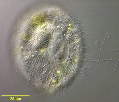
Lembadion (lem-bad-ee-on) is a freshwater planktonic ciliate. It has a large scoop to one side of the body, moves through the water in a rotating motion. In this action it scoops up small planktonic algae - its food. Phase contrast.
-
Lembadion (lem-bad-ee-on) is a freshwater planktonic ciliate. It has a large scoop to one side of the body, moves through the water in a rotating motion. In this action it scoops up small planktonic algae - its food. This one has been eating Cyclidium, which can be seen in the food vacuole. Differential interference contrast.
-
-
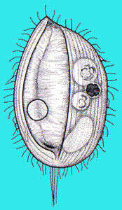
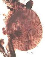
Portrait of Lembadion. Convex dorsum with concave ventral surface mostly occupied by large scoop-like buccal cavity. Buccal ciliature forms prominent "membranes" on right and left of buccal cavity. Swims rapidly rotating on long axis. Often with long tuft of caudal cilia. Dorsal contractile vacuole with collecting canals. From freshwater pond near Boise, Idaho. Brightfield.
-
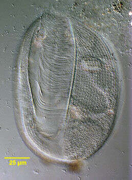
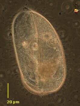
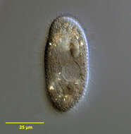
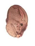
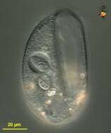
Portrait (ventral view) of the oligohymenophorean ciliate, Lembadion bullinum (Müller,1786;Perty,1849). Cell outline is oval. The ventral surface is concave and the dorsum convex. The very large scoop-like peristome occupies most of the ventral surface (seen well here). The cytostome is at the posterior end of the peristome. There is a small undulating membrane on the right margin of the peristome. A large sheet-like adoral membranelle arises from the left margin of the peristome (seen well here). There are 40-60 evenly spaced longitudinal somatic kineties. The pellicle of L. bullinum, divided into small roughly rectangular depressions, has a distinct cribriform pattern (seen clearly to the left of the peristome). This feature distinguishes L. bullinum from L. magnum which has a striate pellicular pattern. There is usually a tuft of longer caudal cilia. The contractile vacuole connects with its excretory pore by a long curved canal (not seen here). The single ovoid macronucleus and micronucleus are posterior (not seen here). Collected from a freshwater pond near Boise, Idaho May 2004. DIC optics.