-
Villoslada de Cameros, La Rioja, Spain
-
Castille and Leon, Spain
-
Ribadelago, Castille and Leon, Spain
-
Canencia, Comunidad de Madrid, Espaa
-
Peniscola, Valencia, Spain
-
Matute, La Rioja, Spain
-
Gravalos, La Rioja, Spain
-
Caada del Hoyo, Castilla-La Mancha, Espaa
-
Matalebreras, Castille and Leon, Spain
-
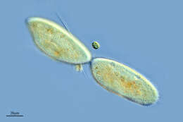
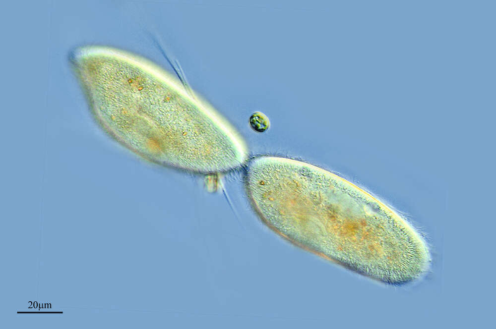
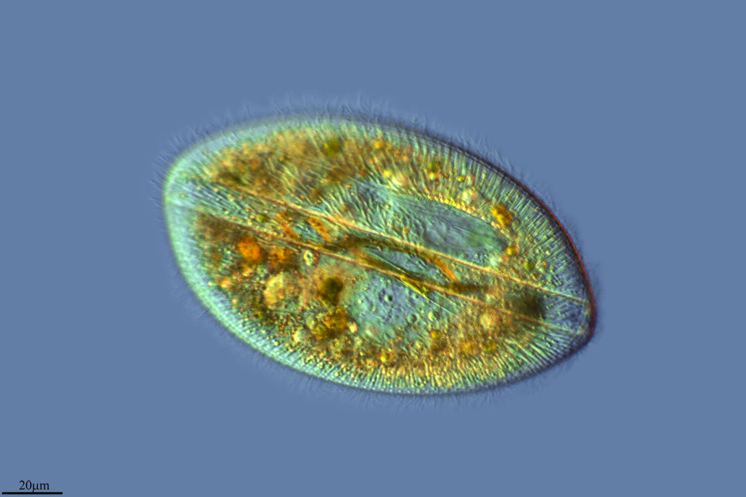
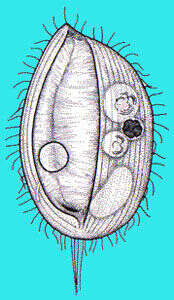
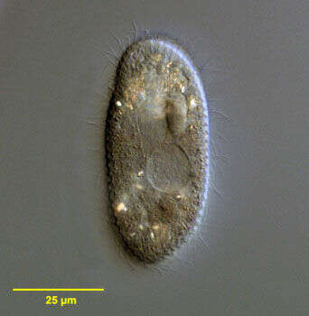
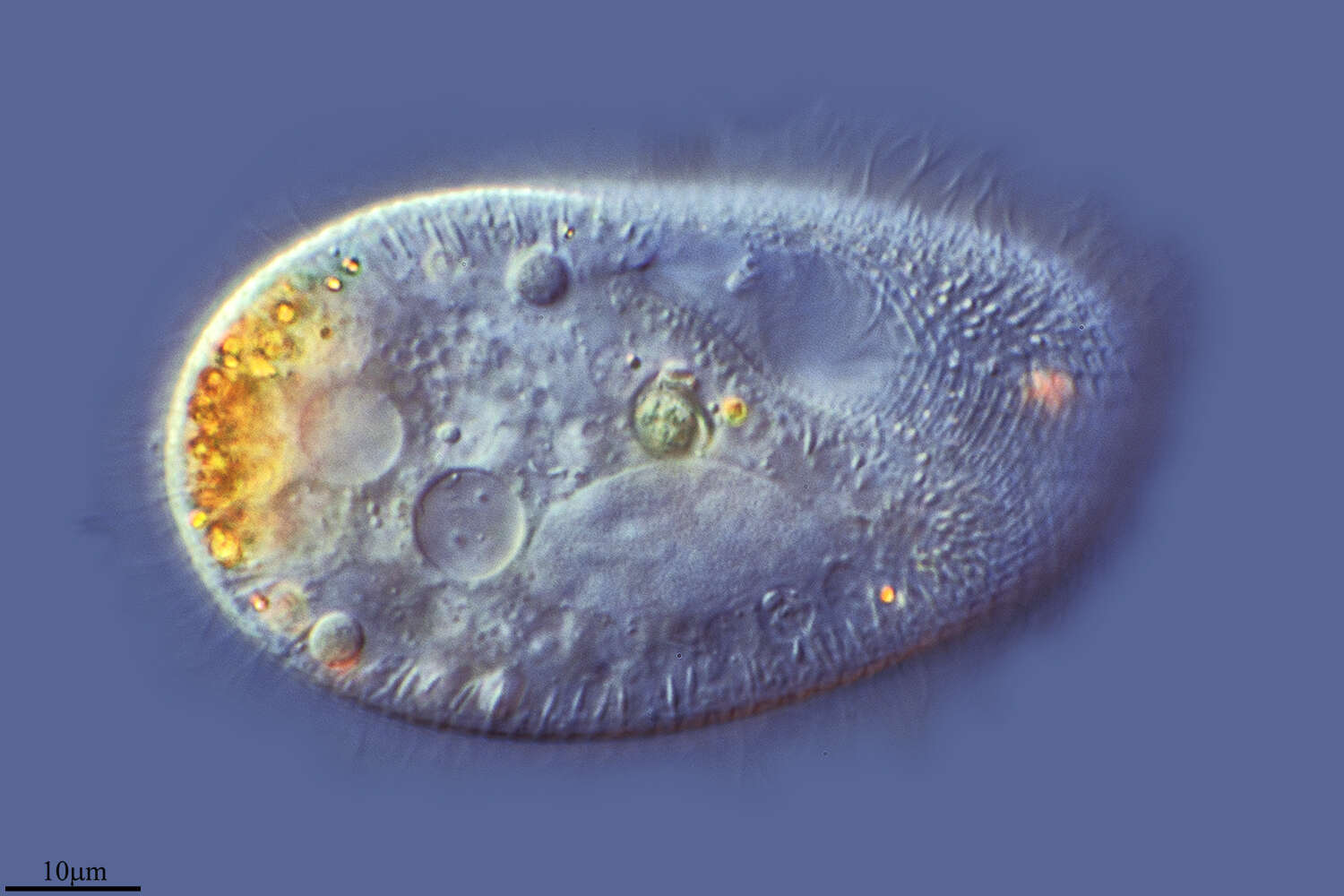
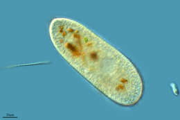
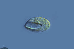
Portrait of the marine frontoniid ciliate, Schistophrya aplanata (Kahl,1933). Schistophrya is a monotypic genus. The cell outline is elongate and bluntly rounded anteriorly and posteriorly. The somatic ciliature is uniform. The pellicle is areolate (marked by uniform rectangular depressions). The slit-like oral aperture is located in mid-body and is bordered by thin slightly serrate lips (seen well in this image). The cytopharyngeal basket of fine trichites is not seen well in these images. A single contractile vacuole is located in the anterior half of the cell. There is a single ovoid macronucleus. A large aggregate of refractile dark granules is present at the anterior end. Fusiform subcortical extrusomes are present. S. aplanata is similar in appearance to the freshwater frontoniid ciliate, Clathrostoma viminale. Collected from a commercial saltwater aquarium in Boise, Idaho February 2004. DIC optics.
-
-
-
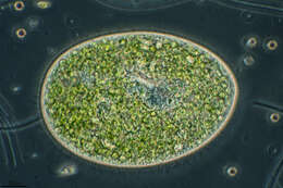
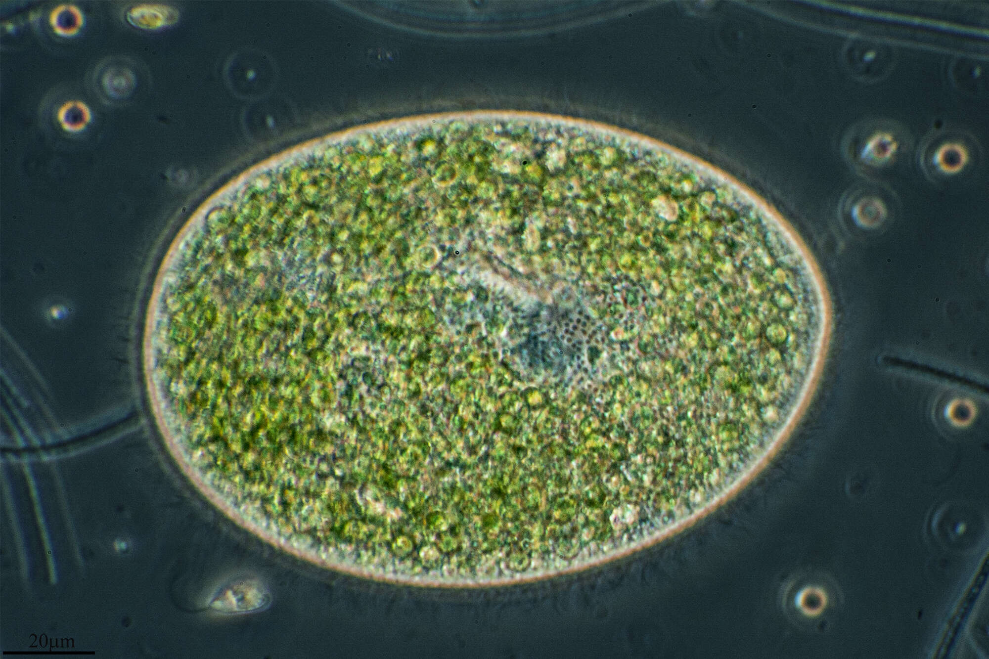
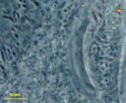
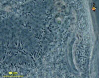
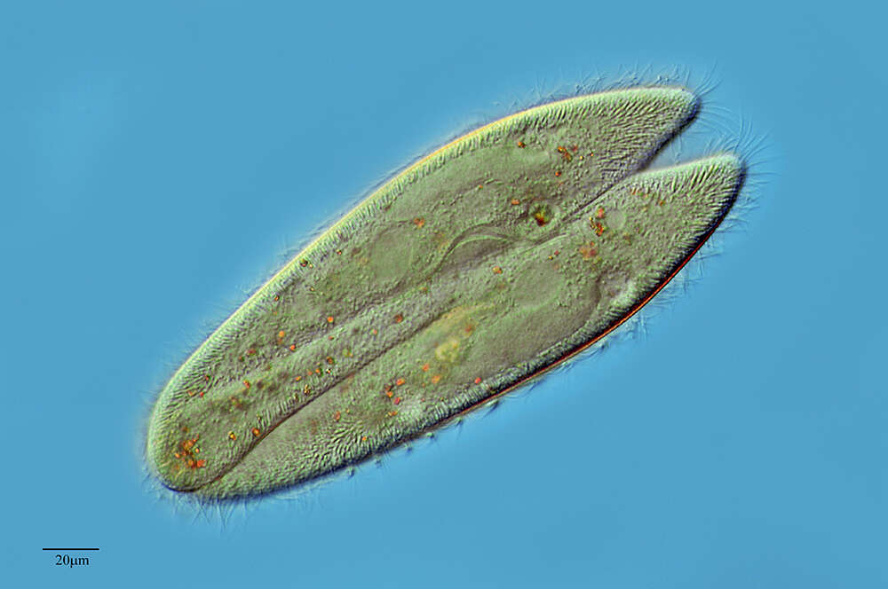
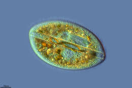
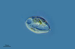
Paramecium (aurelia) (par-a-mee-see-um) is a very familiar genus of ciliates and this (morpho) species is best distinguished by the presence of two small micronuclei pressed up against the macronucleus. This image shows the peniculi or compound ciliary organelles in the mouth. Phase contrast.
-
-
Herrera de Soria, Castille and Leon, Spain
-
Mahide, Castilla y Len, Espaa
-
Rumoroso, Cantabria, Spain
-
-
Canencia, Madrid, Spain
-
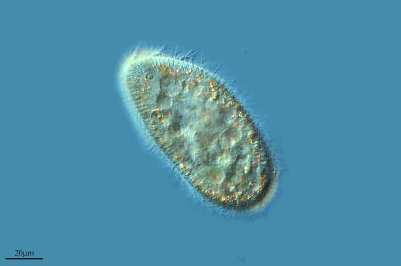
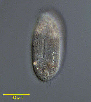
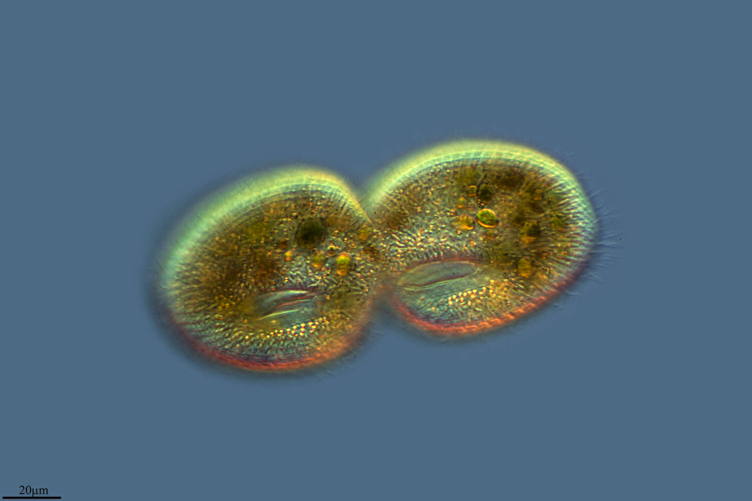
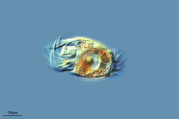
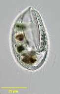
Optical section of the marine frontoniid ciliate, Schistophrya aplanata (Kahl,1933). Schistophrya is a monotypic genus. The cell outline is elongate and bluntly rounded anteriorly and posteriorly. The somatic ciliature is uniform. The pellicle is areolate (marked by uniform rectangular depressions). The slit-like oral aperture is located in mid-body and is bordered by thin slightly serrate lips (not seen in this image). The cytopharyngeal basket of fine trichites is not seen well in these images. A single contractile vacuole is located in the anterior half of the cell. There is a single ovoid macronucleus. A large aggregate of refractile dark granules is present at the anterior end. Fusiform subcortical extrusomes are present (seen in this image). S. aplanata is similar in appearance to the freshwater frontoniid ciliate, Clathrostoma viminale. Collected from a commercial saltwater aquarium in Boise, Idaho February 2004. DIC optics.
-
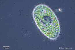
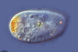
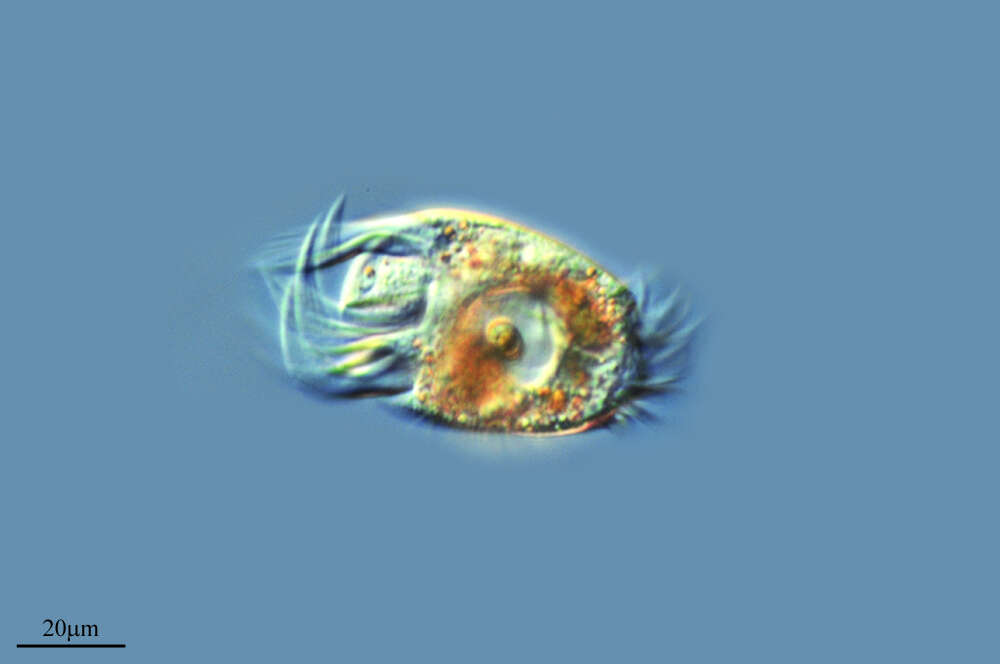
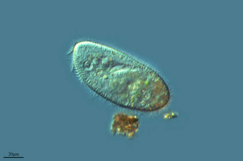
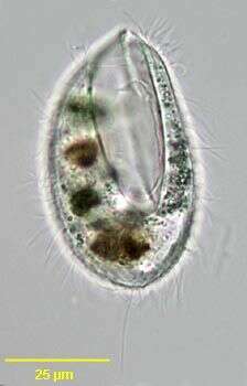
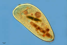
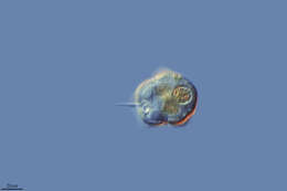
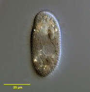
Portrait of Lembadion. Convex dorsum with concave ventral surface mostly occupied by large scoop-like buccal cavity. Buccal ciliature forms prominent "membranes" on right and left of buccal cavity. Swims rapidly rotating on long axis. Often with long tuft of caudal cilia. Dorsal contractile vacuole with collecting canals. From freshwater pond near Boise, Idaho. Brightfield.
-
Paramecium (aurelia) (par-a-mee-see-um) is a very familiar genus of ciliates and this (morpho) species is best distinguished by the presence of two small micronuclei pressed up against the macronucleus. They can be seen here to the north of the nucleus. Phase contrast.
-
Castille and Leon, Spain
-
Ribadelago de Franco, Castilla y Len, Espaa