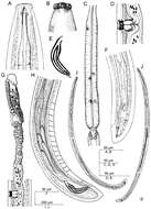
Figure 1.Crassolabium persicum sp. n. (all images are in lateral view) A Anterior region B Lip region and amphid fovea in surface C Pharyngeal expansion D Vagina E Spicules and lateral guiding piece F Female, posterior body region G Female, anterior genital branch H Male, posterior body region I Male, entire J Female, entire.
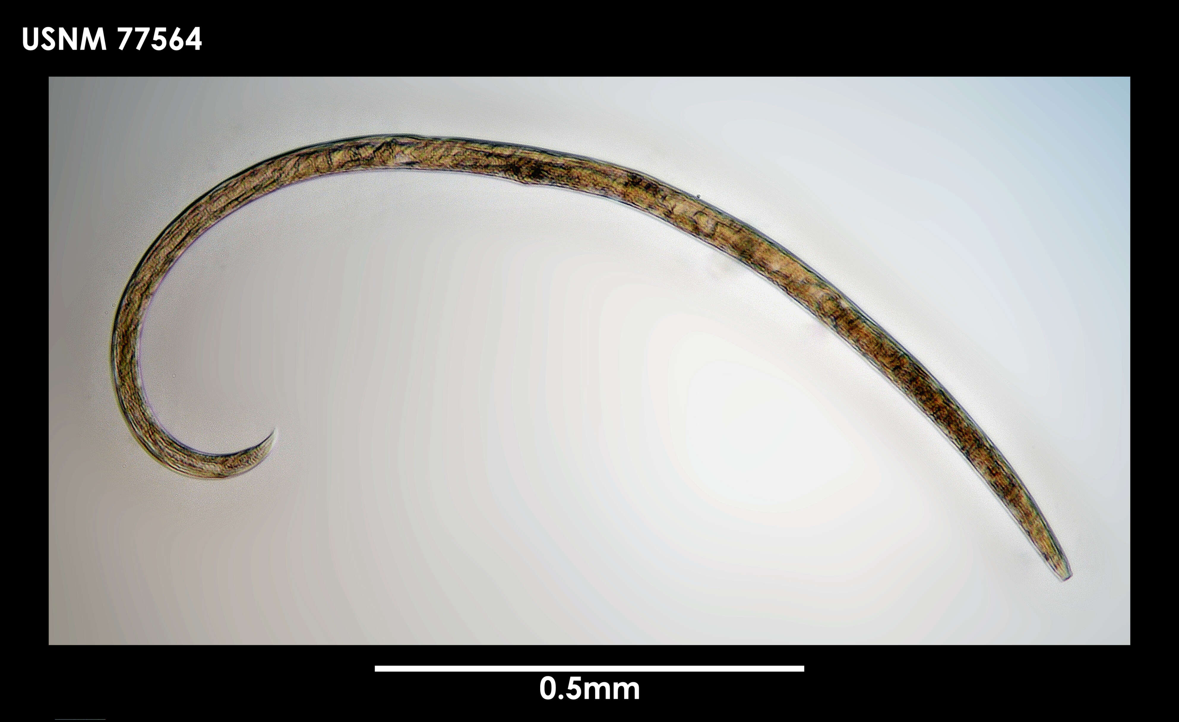
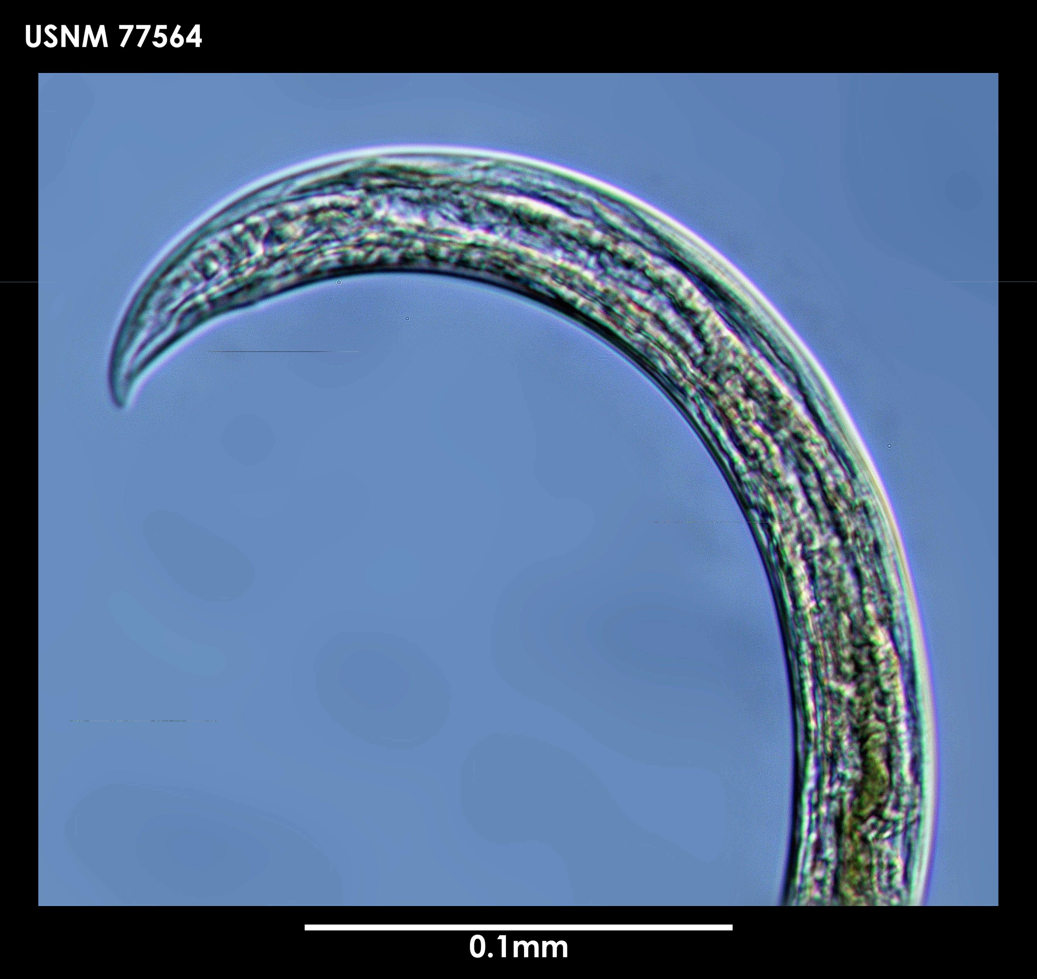
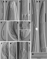
Figure 2.Crassolabium persicum sp. n. (light micrographs all in lateral view).A Female, entire B Anterior region C Pharyngo–intestinal junction D Female, genital system E Neck region F Male, entire G Male, posterior region H Vagina I Female, caudal region J Spicules K Lateral guiding piece L Oviduct–uterus junction M Lateral chord and pores. (Scale bars: A, F – 500 µm; B, H, K – 10 µm; C, G – 50 µm; D, E – 100 µm; I, J, L, M – 20 µm).
Instituto Nacional de Biodiversidad - INBio, Costa Rica.
INBio
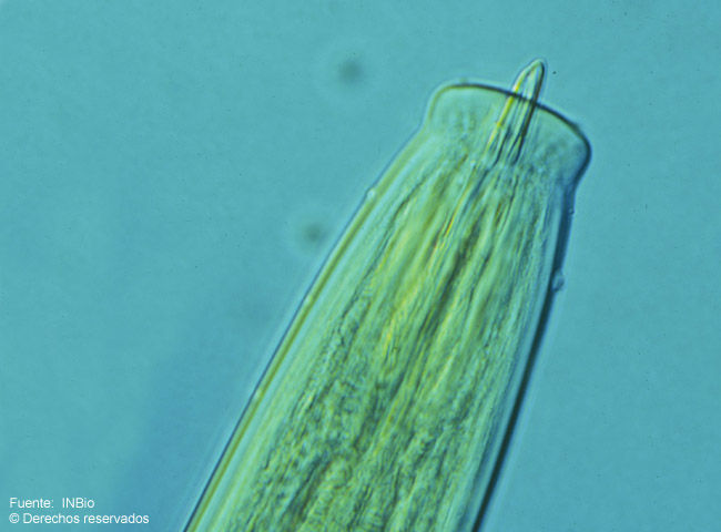
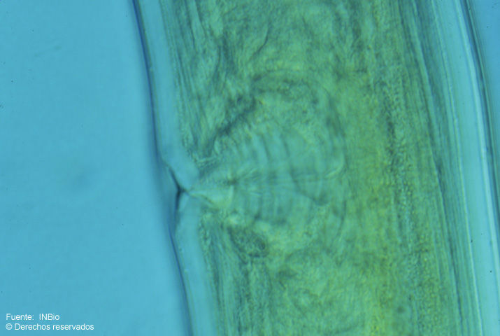
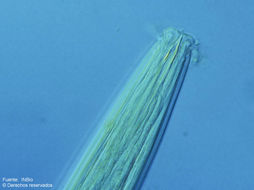
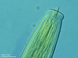
Fig.1 Fotomicrografía de la región anterior de la hembra. Nótense los labios fuertemente separados y la longitud del odontoestilete. Foto: A. Esquivel.
Instituto Nacional de Biodiversidad - INBio, Costa Rica.
INBio
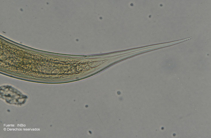
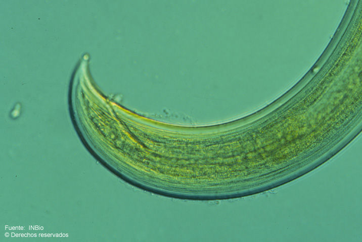
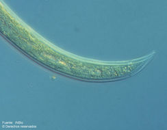
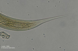
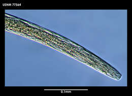
Fig.3 Fotomicrografía de la región posterior de la hembra. Se distingue claramente el recto. La cola es corta, conoide y arqueada en ambos sexos. Foto: A.Esquivel